
Recomendados
Más contenido relacionado
La actualidad más candente
La actualidad más candente (20)
Destacado
Destacado (20)
Similar a Clinical Assessment and Diagnosis of Respiratory Diseases
Similar a Clinical Assessment and Diagnosis of Respiratory Diseases (20)
Más de Yaser Ammar
Más de Yaser Ammar (15)
Último
Último (20)
Clinical Assessment and Diagnosis of Respiratory Diseases
- 1. • Clinical Assessment: - History: o Complaint (C/O): (Dyspnoea – cough – haemoptysis – chest pain) o Present History. o Past History. o Other History: Family, Occupational, Drug, Operative, Obstetric… - General Examination: o General look. o Gait, decubitus, posture. o Vital signs. o Systemic review. - Diagnosis of Respiratory System Diseases
- 2. - Chest Examination: o Inspection. o Palpation. o Percussion. o Auscultation. • Point of Care (POC) (Bed- Side or Hand- Held) Devices: - Glucometer. - Peak flowmeter. - Pulse Oximeter. • Respiratory Function Tests. • Other Investigations: - Chest X-Ray (CXR). - Electrocardiography (ECG).
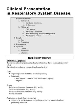
- 3. Dyspnoea: subjective feeling of difficulty in breathing due to increased respiratory effort. Exertional Dyspnoea: provoked or increased by physical activity. Types • Physiologic: with more than usual daily activity. • Pathologic: - Psychogenic: mainly at rest, with frequent sighing - Organic Grades • Provoked by more than usual daily activity • Provoked by usual daily activity • Provoked by less than usual daily activity • Present at rest Dyspnoea
- 4. New York Hear Association (NYHA) Functional Classification Of Heart Failure What produces symptoms (dyspnoea, palpitations, fatigue) and limitation of physical activity? Class I: more than usual physical activity. Class II: usual physical activity Class III: less than usual physical activity Class IV: without physical activity (symptoms at rest)
- 5. Progression to Grade 4 may occur over: • Minutes (Acute Dyspnoea): foreign body aspiration, bronchial asthma, pulmonary embolism • Days: rapidly accumulating pleural effusion • Months: interstitial lung disease • Years: emphysema • Orthopnoea It is dyspnoea produced or aggravated on lying down, relieved (partially or completely) in the upright position. The flat position may impair ventilation of lower lung segments. Orthopnoea may occur in heart failure and many chest disorders. It may also result from abdominal distension pushing the diaphragm upwards.
- 6. It is forced expiratory effort against a closed glottis which then suddenly opens with a jet of air expelled out, possibly along with secretions. Types • Dry (Irritant) Cough: The irritant stimulus may be obvious (smoke, dust, pharyngitis) and may not. Two common "concealed" causes of resistant dry cough are post- nasal discharge and GERD (gastro-oesophageal reflux disease). Cough
- 7. • Wet (Productive) Cough: Frothy Pink: pulmonary oedema Mucoid: bronchial asthma, chronic bronchitis Mucopurulent, Purulent (yellowish, thick): infections Rusty: lobar pneumonia Greenish/Bluish: Gram negative infection Foul Smelling: anaerobic infection Blackish: smoker, coal workers
- 8. It is coughing of blood or blood tinged sputum due to bleeding from the respiratory tract below the vocal cords. Bleeding originating above vocal cords (nose, mouth, larynx) may produce false haemoptysis. HaemoptysisHaemoptysis HaematemesisHaematemesis It isIt is Coughing of bloodCoughing of blood Vomiting ofVomiting of bloodblood ColorColor Bright redBright red Dark redDark red ReactionReaction alkalinealkaline acidicacidic MixedMixed withwith Sputum, air (frothy)Sputum, air (frothy) foodfood SputumSputum Blood tinged for 12 – 24Blood tinged for 12 – 24 h after the attackh after the attack normalnormal StoolStool normalnormal melenamelena Hemoptysis
- 9. Non- Cardiac Cardiac • Lung: pleurisy • Oesophagus: GERD • Stomach: gastritis, ulcer • GB: cholecystitis • Skin: Herpes Zoster • Sickle cell crisis Non- Ischemic Ischemic (Angina Pectoris) • Expanding aortic aneurysm • Dissecting aortic aneurysm • Pericarditis Non- Coronary Coronary • Aortic stenosis • Anaemia • Fever Not due to Coronary atherosclerosis due to Coronary atherosclerosis • Coronary arteritis • Coronary embolism Chest Pain
- 10. General Look • Complexion: Pallor – Jaundice - Cyanosis • Nutritional State: Malnutrition / Cachexia – Obesity • Features related to systemic disease: Diabetes mellitus – rheumatoid arthritis • Features related to drug toxicity: cushinoid facies Cyanosis bluish discoloration of skin, mucous membranes due to presence of: - > 5 gm deoxy Hb /100 mL blood Or - abnormal Hb (met or sulph Hb) in surface capillaries. Normal level of deoxy Hb /100 mL blood: - Arterial: 0.75 gm - Capillary 2.25 gm - Venous 3.75 gm
- 11. Consider average Hb: 15 gm/dL Arterial Blood Capillary Blood Venous Blood Cyanosis is NOT a reliable sign to monitor the degree of blood deoxygenation: • Effect of Hb level. • Effect of Hb type (MetHb – SulphHb) • Effect of skin color. Cyanosis is difficult to recognize in people with dark skin. Anemia Polycythemia
- 12. ↑ O2 Extraction ↓ O2 Loading Minor (Pulmonary) Circulation Major (Systemic) Circulation Central Cyanosis This traditional clinical sign of hypoxemia is insensitive bec. apperas at < 80% O2 saturation. Peripheral Cyanosis This is a sign of poor peripheral circulation.
- 13. Central CyanosisCentral Cyanosis 1 2 3 4 1) Hypoventilation - ↓ alveolar PO2 : eg, high altitude - Obstructive lung diseases - Restrictive lung diseases 2) Shunt - Cardiac Rt to Lt shunt (congenital cyanotic heart disease): eg, Fallot tetralogy - Pulmonary: pulmonary AV fistula 3) Diffusion Defect (Alveolo-Capillary Block) - Pulmonary fibrosis - Pulmonary oedema 4) Ventilation Perfusion (V/Q) Mismatch - Pulmonary embolism (ventilation > perfusion = dead space effect) - Atelectasis/collapse (perfusion > ventilation = shunt effect) - Most pulmonary disorders produce hypoxia by more than one mechanism. V/Q mismatch is the most common. Requisites for O2 Loading of Blood in Pulmonary Circulation
- 14. Peripheral CyanosisPeripheral Cyanosis 1) Generalized stagnation (low COP) i. HF ii. Shock 2) Localized stagnation i. Arterial • Lumen: thrombosis, embolism • Wall: Spasm: Raynaud`s disease, ergotism • Outside: Compression by a SOL i. Venous Chemical CyanosisChemical Cyanosis 1) Met-haemoglobin 2) Sulph-haemoglobin The bnormal Hb causes: - Dark color by itself. - Reducing the normal Hb, bec it has much higher affinity for O2
- 15. Central Cyanosis Peripheral Cyanosis Cause ↓ O2 saturation of core arterial blood (↓ O2 loading by cardiopulmonary circulation) Stagnant circulation → - ↓ peripheral arterial flow - ↑ O2 extraction by tissues Distribution Warm areas (tongue, interior of lips), all over Cold areas: tip of nose, lobule of ear, fingers, toes Temperature Warm Cold Clubbing + - Polycythaemia + - Effect of - Exercise - Warming - Oxygen May ↑ No effect ↓ to variable extent, maximal in low atmospheric PO2 and ↓ ↓ No effect
- 16. 1 4 3 2 O2 delivered to tissues in amounts matching with metabolic needs 4 Prerequisites: 1) Load 2) Vehicle 3) Route 4) Unload 1) Loading of blood with O2 in alveoli. 2) Carrying of O2 by blood: Hb in intact RBCs. 3) Blood flow: intact circulation. 4) Extraction of O2 from blood by tissues.
- 17. 1 4 3 2 1) Hypoxaemia Impaired blood oxygenation in alveolar capillaries → Low O2 tension in blood 2) Anaemia Deficient Hb in intact RBCs → Low O2 carrying capacity of blood 3) Ischaemia Circulatory insufficiency (local or general) → blood supply not matching with metabolic needs 4) Tissue Toxins Tissue inability to extract O2 (cyanide poisoning) 1) Hypoxic 2) Anaemic 3) Stagnant 4) Histotoxic HYPOXIA (Low O2 Tension in tissues)
- 18. Hypoxia Low Oxygen Tension in tissues Hypoxia Type Cyanosis Hypoxic Hypoxia Central Stagnant Hypoxia Peripheral Anaemic Hypoxia --- Histotoxic Hypoxia ---
- 19. Pulse Oximetry is a simple, relatively cheap, non- invasive technique to monitor blood oxygenation. It is a common POC device in ER, ICU, ambulance and even GP kit.
- 20. Principle Two LEDs are positioned on one side of the part examined, with their respective photodetectors on the opposite side, with 5 – 10 mm of tissues intervening. One LED emits red light (absorbed by deoxyHb). The other LED emits infrared light (absorbed by oxyHb). Relative absorption at each wave length is measured (spectrophotometry) by the photodetector several times per second and sent to a microprocessor that calculates the percent of oxyHb to total Hb (which represents O2 saturation). Q2 saturation readings are updated every 0.5 – 1 second so that they average the readings over the last 3 seconds. That is why the oxymeter reading should be taken 5 – 10 seconds after it is operated. Oxygen Saturation of Blood = Oxygen Content divided by Oxygen carrying capacity (2) (2)
- 21. Indications for Oximetry • Anaesthesia, sedation and perioperative use. • Acute and chronic chest conditions (BA, COPD, pneumonia). • Acute cardiac conditions (AMI, PE). • CVS (cerebrovascular stroke). • Acute metabolic derangements (DKA). • Assessment of cardiorespiratory fitness (walking tests). • Monitoring of oxygen therapy. • Monitoring of mechanical ventilation. • As alternative to ABG testing. • Neonatal care.
- 22. Confounding Factors that may Influence Oximetry Reading Medical Conditions • Anemia or polycythemia → O2 Saturation does not reflect O2 content. • Hypercapnoea (as in smokers): CarboxyHb is registered as 90% oxyHb and 10% deoxyHb → overestimation of O2 saturation. • Low peripheral perfusion → underestimation of O2 saturation, as in shock, heart failure, cold weather. Technical Factors • Excessive movement. • Strong ambient light (sun, infrared heater, phototherapy). • Unopposing LED and detector. • Automatic BP cuff → intermittent low reading.
- 23. Technique of Oximetry • The pt should stay still during measurement. If oxymeter is used to monitor effect of exercise, the pt should stay still a while whenever the oxymeter reading is being taken. • The probe may be placed on finger tip (usually index or middle finger) or ear lobe. The latter is less accurate and less commonly used. • The hand should be cleaned and dried; any nail polish removed. • If hands were cold, warm them by rubbing against each other. • The finger should fit snugly inside the oxymeter which should be properly fixed (either clip or wrap style). • The hand should be rested on the chest wall at the level of the heart, rather than the digit is held in air (as patients commonly do) to minimize motion artefact.
- 24. Technique of Oximetry (cont.) • Most oxymeters record the pulse rate and waveform as well. This can be used to confirm proper O2 saturation reading: o Hear rate displayed by the oxymeter should conform with that manually calculated (to indicate no motion artefact). o Pulse waveform should be evident with dicrotic notch. A flattened wave indicates poor perfusion. • Avoid strong ambient light. • Take the reading after it stabilizes after 5 – 10 seconds. Maintenance of Oximeter • Use a lightly damp cloth or paper towel to wipe away dust. • Us an alcohol swab for disinfection.
- 25. General Recommendations Based on Oximetry Reading 95 – 100% Normal 90 – 95% • More frequent monitoring. • Exclusion of low perfusion and technical factors causing low reading. 80 – 90% Supplemental oxygen to ↑ saturation above 90%. In most machines, the default low oxygen saturation alarm is 90%. < 80% • Check ABG. • Consider ventilatory support.
- 26. Muscle wasting / CachexiaMuscle wasting / Cachexia Obesity Hypoventilation Syndrome • Complexion: Pallor – Jaundice - Cyanosis • Nutritional State: Malnutrition / Cachexia – Obesity • Features related to systemic disease: Diabetes mellitus – rheumatoid arthritis • Features related to drug toxicity: cushinoid facies
- 27. Acanthosis Nigricans In diabetes mellitus A band of thickened darkened skin on the back of the neck.
- 28. Rheumatoid Arthritis May be associated with Interstitial pulmonary fibrosis Sublaxation of MP joints with ulnar deviation Hyperextension PIP joint, Flexion DIP joint
- 29. Cushinoid Facies Indicating Excess Adrenocortical Function Face is puffy, plethoric and somewhat rounded (moon face), with acne. Iatrogenic Cushing`s syndrome results from long standing treatment with systemic corticosteroids, as in severe cases of bronchial asthma and interstitial pulmonary fibrosis. Systemic exposure to absorbed inhaled steroids is rarely significant. Lomg term exposure to systemic steroids blunts the hypothalamic- pituitary-adrenal axis. So, reduction of oral steroid dose or conversion to inhaled steroids in cases with apparent cushinoid features should be gradual to avoid precipitation of adrenocortical insufficiency.
- 30. Gait, Decubitus, Posture • Gait: How the patient walks. • Decubitus: How the patient lies in bed. • Posture: particular part position. Antalgic Gait Orthopnic Decubitus Pursed-Lip Breathing
- 31. Vital Signs Pulse - Blood Pressure – Respiration - Temperature Normal Breathing Cheyne Stokes Breathing Acidotic (Kussmaul) Breathing
- 32. Bilateral OLL Unilateral OLL DVT → PE• Pulmonary hypertension → HF • Hypoalbuminaemia: o Malnutrition in cystic fibrosis o Copious sputum in bronchiectasis o Repeated Pleural tapping SystemicReview
- 34. Infrascapular region Interscapular region Suprascapular region Scapular region
- 35. Normal chest Barrel chest (Hyperinflated Chest) AP to Transverse diameter = 5:7 • AP to Transverse diameter = 1:1 • More transverse ribs and interspaces • Obtuse subcostal angle • Dorsal kyphosis Characteristic of COPD
- 37. Suprasternal, Supraclavicular Retractions Epigastric Retractions Inetrcostal Retractions
- 38. Active Accessory Muscles of Respiration
- 39. Palpation Trachea is markedly shifted to the left It is either pushed away from Rt side (pleural effusion or pneumothorax) or pulled to the Lt side (pulmonary atelectasis or fibrosis).
- 42. Adventitious Sounds Wheezes (Rhonchi) Crepitations (Rales) Pleural Friction Rub Stridor
- 43. Clinical Features ofClinical Features of RESPIRATORY DISTRESSRESPIRATORY DISTRESS SubjectiveSubjective (Symptoms)(Symptoms) ObjectiveObjective (Signs)(Signs) • Exertional dyspnoea • Orthopnoea • Tachypnoea (> 20 /min) • Inspiratory Retractions • Active accessory muscles of respiration • Wheezes
Notas del editor
- Complaint is the problem(s) that directed the patient to seek medical advice. Complaints should be arranged either by: Sequence (chronological order). Importance. Relevance to system concerned. Present History entails analysis of the complaints: onset, progression, precipitating and relieving factors. Past History usually includes comment on common chronic diseases as diabetes and hypertension, and previous treatments
- The differentiation between hemoptysis and hematemesis is sometimes misleading for the patient and even the physician. Blood in haematemesis may become darkened by the effect of gastric juice (formation of acid haematin). However, if bleeding is quite fresh or massive, blood would still be bright red. Melena means passage of dark altered (tarry) stool. Again, if haematemesis is massive and intestinal transit time is short, the blood passed in stool may not be altered completely. It should be differentiated from haematochazia (bleeding per rectum): passage of fresh unaltered blood in stool, a condition that occurs when bleeding originates more distally in the GIT.
- There are many non ischemic causes for chest pain. However, ischemic pain (angina pectoris) should be kept in mind, particularly in patients with risk factors. The lung parenchyma itself is insensitive to pain. Many pulmonary conditions do not give rise to chest pain until the overlying pleura is affected (pleurisy).
- During its passage through the blood vessels, the blood undergoes progressive reduction in its oxygen content. Therefore, oxyHb content of blood decreases, while deoxyHb increases. The bluish color can be noticed if the deoxyHb (reduced Hb) content of blood becomes &gt; 5 gm 100 mL blood. When Hb content as a whole of blood decreases (anaemia), cyanosis may not be apparent
- In anemia, the total Hb concentration is reduced. Even if the ratio of deoxyHb is increased due to marked hypoxaemia, cyanosis may not occur. In polycythemia, the total Hb concentration is increased. DeoxyHb is increased accordingly and may lead to cyanosis in absence of hypoxemia. Cyanosis occurs at lower levels of deoxyHb in presence of abnormal hemoglobins.
- Central cy
- Central cyanosis results from O2 loading by cardiopulmonary circulation. There are 4 requisites for proper oxygen loading of blood: Alveolar Ventilation Pulmonary Blood Flow Gas Exchange through respiratory (aveolo-capillary) membrane Ventilation Perfusion (V/Q) Matching Accordingly, there are 4 possible mechanisms for central cyanosis (for hypoxic hypoxia)
- SOL: space occupying lesion. Venous stagnation results from venous obstruction, eg Superior vena cava obstruction peripheral cyanosis in tongue and lips (exceptional) Causes of Methaemoglobinaemia: Congenital Nitrite producing intestinal flora (enterogenous cyanosis) Drugs: nitrates, sulphonamides
- Oxygen saturation can never exceed 100% (whereas Oxygen tension may under certain circumstances). Oxygen saturation may be a little below 100% in a perfectly normal person. We have some normal minor right to left shunts.
- The chest is an important part of the body to reveal signs of muscle wasting and cachexia. Cachexia indicates protein energy malnutrition (loss of both proteins and fats) in addition to toxaemia. Chest causes of cachexia include bronchial carcinoma, cystic fibrosis and chronic infections as bronchectasis, TB, chronic lung abscess, chronic empyema.
- DM is an important predisposing factor for chest infections, including TB.
- MP: Metacarpophalngeal PIP: proximal interphalangeal DIP: Distal interphalangeal
- Patients with possible adrenocortical insufficiency may require supplemental systemic steroids in stress situations as surgery and trauma.
- In antalgic gait (as in unilateral DVT), the patient tries to avoid weight bearing on the diseases limb. Pursed-lip breathing occurs in COPD and is intended to increase expiratory pressure to prevent premature closure of airways.
- Normally, inspiration is an active process while expiration is passive. Normally, expiration occupies a longer portion of respiratory cycle than inspiration. Acidotic breathing (rapid and deep) occurs in metabolic acidosis to compensate by inducing respiratory alkalosis (CO2 wash). Causes include renal failure, DKA and salicylate poisoning. Cheyne Stokes breathing occurs in cerebrovascular strokes and advanced heart, liver or renal failure.
- Sternal angle may be felt as a transverse bony ridge between the manubrium above and body of sternum below.
- Respiratory retractions indicate respiratory distress due to airway obstruction.
- This muscles do normally share in quiet respiration.
- Wheezes are continuous musical sounds caused by passage of air through narrowed bronchi (partial bronchial obstruction) Crepitations are interrupted non- musical sounds thought to result from passage of air through alveolar fluid or sudden tensing of alveolar walls. Pleural friction rub is a scratchy to and fro sound that results from friction of the 2 pleural layers against each other. Stridor is a high pitched inspiratory sound that results from upper airway obstruction (trachea and above)
- Tachypnoea Normal adult rate: 12 – 20/min Tachypnoea is a common sign of respiratory distress.