SlideShare una empresa de Scribd logo
1 de 58
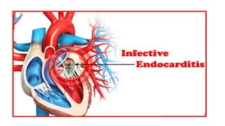
GUIDELINES FOR INFECTIVE
ENDOCARDITIS
• Clinical Practice Guidelines for the Prevention, Diagnosis &
Management of Infective Endocarditis 2017.
Contents
• Introduction
• Risk factors
• Classification
• Common Organism
• Clinical presentation
• Investigations
• Modified Duke’s criteria
• Management
• Summary
Introduction
• Infection of the endocardial surface of the heart (heart valves and
mural endocardium)
• Increasing in prevalence due to
• introduction of conduits (tubes),
• prosthetic materials and intracardiac devices
makes more susceptible
to infection in view of
the presence of these
foreign materials.
Classification
IE according to localisation of infection and presence of intracardiac material
• Left-sided native valve IE
• Left-sided PVE
>> Early PVE < 1 year after valve surgery
>> Late PVE > 1 year after valve surgery
• Right-sided native valve IE
• Device related IE
IE according to the mode of acquisition
• Healthcare associated IE
• Community acquired IE
• Intravenous drug abuseassociated IE
Acute endocarditis
• High grade and persistant fever
• Rapidldamages cardiac to structures,
• Seeds extracardiac sites
IF untreated progresses to death within weeks.
Subacute endocarditis
• Follows an slower course(slow cardiac damage)
• Rarely metastasizes; and
• Gradually progressive unless complicated by a major embolic event or a ruptured mycotic aneurysm.
Common Organisms
• Native Valve and late prosthetic valve : Strep Viridans
• Early Prosthetic valve : Staph Epidermidis and Staph Aureus
• IVDU esp tricuspid valve is : S. aureus
• Non-infective:
• systemic lupus erythematosus (Libman-Sacks)
• malignancy: marantic endocarditis
• Culture negative causes
• Brucella
• Coxiella burnetii
• HACEK: Hemophilus, Actinobacillus, Cardiobacterium, Eikenella, Kingella)
• Bartonella
• Prior antibiotic therapy
Pre-existing risk factors
• Previous history of IE.
• Pre-existing cardiac disease.
• Presence of prosthetic valves or prosthetic cardiac material.
• Presence of intracardiac devices.
• History of IVDU.
• Presence of chronic intravenous access (e.g. haemodialysis catheters,chemoports
and neonate/paediatric patients with indwelling central venous catheters).
• Presence of CHD Elderly or immunocompromised patients.
• Co-existing conditions such as diabetes, human immunodeficiency virus (HIV)
• infection and malignancy.
General Clinical Presentation
• Fever (87%) with chills, poor appetite and weight loss.
• Heart failure may be present at admission (up to 58%)
• New or altered cardiac murmur (50-85%)
• Embolic events may also cause presenting symptoms (27-30%)
Fever + New/Altered murmur : IE until proven otherwise
• Pt do not often present with classic textbook manifestations of subacute or chronic endocarditis.
Peripheral sign and
symptom
Due to septic emboli or immune complex deposition
Peripheral sign and
symptom
SKIN
Splinter
hemorhage
Janeway lesion
>> Non-tender
lesions
>> 1-4 mm in
diameter
>>Often
haemorrhagic
Osler’s
nodes
Painful
subcutane
ous
nodules
Clubbing
Peripheral sign and symptom
EYES
Conjuctival
hemorhage
Roth’s spot
White centred
retinal
haemorrhages)
Clinical features
Neurological
Mycotic
aneurysm
vessel wall s
infected with
bacteria, is
digested and a
false aneurysm
forms, which is
unstable and
highly prone to
rupture.
Clinical features
Renal
• Hematuria : due to glomerulonephritis
Differential diagnosis..
1) Acute Rhematic heart fever
2) Endocarditis due SLE (Libman-Sacks)
3) Atrial myxoma
Investigation
• FBC : Raised white cell count and Low hb
• UFEME : Ery positive
• Inflammatory marker
• Elevated CRP and ESR.
• Renal profile
• LFT
• ECG
• Chest X-ray
• Blood cultures : three set from three different sites prior to
antibiotics
If blood cultures negative after 5 days of incubation and no history of prior antimicrobial use, consider
blood culture negative infective endocarditis (BCNIE), which can be caused by fungi or fastidious
microorganisms, and perform the appropriate microbiological tests.
Definite IE Pathological criteria:
Microorganisms demonstrated by culture or HPE of a vegetation, a vegetation
that has embolised, or an intracardiac abscess specimen;
or pathological lesions; vegetation or intracardiac abscess confirmed by HPE
showing active endocarditis
Clinical criteria:
2 major criteria
or
1 major criterion and 3 minor criteria
or
5 minor criteria
Possible IE 1 major criterion and 1 minor criterion
or
3 minor criteria
Rejected IE Firm alternative diagnosis explaining evidence of IE
or resolution of IE syndrome with antimicrobial therapy for ≤ 4 days
or no pathological evidence of IE at surgery or autopsy with antimicrobial
therapy for ≤ 4 days
or does not meet criteria for possible IE as above
Major Criteria
Blood culture positive for IE Evidence of endocardial involvement
Typical microorganisms consistent with IE from 2
separate blood cultures:
• VGS, Streptococcus bovis, HACEK group, S. aureus
Or community-acquired enterococci in the absence
of a primary focus
Or microorganisms consistent with IE from
persistently positive blood cultures defined as follows:
>> At least 2 positive cultures of blood samples drawn
> 12 hours apart
>>Or all of 3 or a majority of ≥ 4 separate cultures of
blood (with first and last sample drawn at least 1
hour apart)
• Single positive blood culture from Coxiella burnetii
or phase 1 IgG antibody titres > 1:800
Echocardiogram positive for IE defined as follows:
• Oscillating intracardiac mass on valve or supporting
structures, in the path of regurgitant jets, or on
implanted material in the absence of an alternative
anatomic explanation
• Abscess
Or new partial dehiscence of prosthetic valve
Or new valvular regurgitation (worsening or changing
or pre-existing murmur not sufficient)
Minor
criteria
Predisposition: predisposing heart condition or IVDU
Fever: temperature > 38°C
Vascular phenomena: major arterial emboli, septic pulmonary infarcts, mycotic
aneurysm, intracranial haemorrhage, conjunctival haemorrhages and Janeway
lesions
Immunological phenomena: glomerulonephritis, Osler nodes, Roth spots and
rheumatoid factor
Microbiological evidence: positive blood cultures but does not meet a major
criterion as noted above (excludes single positive cultures for coagulase
negative staphylococci and microorganisms that do not cause endocarditis) or
serological evidence of active infection with microorganism consistent with IE
Transthoracic echocardiography
(TTE)
Transoesophageal echocardiography
(TEE)
Surgical or autopsy findings Echocardiography findings
Vegetation Infected mass attached to an
endocardial structure or on
implanted intracardiac material
Oscillating or non-oscillating
intracardiac mass on valve or
other endocardial structures, or on
implanted intracardiac material
Abscess Perivalvular cavity
with necrosis and purulent material
not communicating with the
cardiovascular lumen
Thickened, non-homogeneous
perivalvular area with echodense or
echolucent appearance
Pseudoaneurysms Perivalvular cavity communicating
with the cardiovascular lumen
Pulsatile perivalvular
echocardiographic-free space, with
colour-Doppler detected
Perforation Interruption of endocardial tissue
continuity
Interruption of endocardial tissue
continuity traversed by colour-
Doppler
Fistula Communication between two
neighbouring cavities through a
perforation
Colour-Doppler communication
between two neighbouring cavities
through a perforation
Valve aneurysm Saccular outpouching of valvular
tissue
Saccular bulging of valvular leaflet
tissue
Dehiscence of a
prosthetic valve
Dehiscence of the prosthesis Paravalvular regurgitation identified
by TTE/TEE, with or without rocking
motion of the prosthesis
Video Vegetation
• 2 chamber view
Video – Aortic abscess
Mercedes benz sign – aortic valve
Management of Infective
Endocarditis
The major goals of management of IE
are:
•To eradicate the infectious agent from
the endocardium.
•To address the complications of the
infection, both intra and extracardiac.
Things to monitor in Ward(1)
•General well being – septic looking, GCS
•Temperature : Fever usually resolves in a
few days after appropriate antibiotics
started
•Daily ECG – PR interval (conduction delay)
Things to monitor in Ward(2)
• Examined regularly for ssx of the following complications
• Heart failure: symptoms should be treated with standard medical therapy
and its severity regularly assessed. Heart failure may persist despite
microbiological resolution.
• Embolic events. - related to the migration of cardiac vegetations : liver,
spleen, kidneys and abdominal mesenteric vessels
• Common sites
• left-sided IE are the brain and spleen
• Right-sided IE – Pulmonary embolism
• Ongoing sepsis.
• Neurological sequelae. –ischemic and hemorhagic stroke, mycotic aneuryms,
cerebral abscess.
Things to monitor in Ward (3)
• Blood investigations
• Regular Inflammatory markers.
• Regular FBC to monitor WBCs, Hb and Plt
• Blood cultures
• BUSE for sign of AKI and it’s resolution.
• Echocardiography
• Monitor vegetation
• Resolution of IE: vegetations gradually reduce in size, mobility and increase in
echogenicity. In the long-term, these vegetations may not disappear or even change in
size, even with clinical treatment success.
• Risk of embolisation: vegetations increase in sizeand mobility.
Antimicrobial
management
1) Empirical antibiotic – based on clinical suspicion.
2) Definitive treatment – based on C+S.
Empirical
In our setting we use IV Ceftriaxone as empirical antibiotic
Add Cloxacillin if suspected Staph Aureus(IVDU or PVE)
Or
Vancomycin for suspected MRSA
1. Community-acquired native valves or
late prosthetic valves (≥ 12 months
post-surgery) endocarditis
Usually strep viridans
Ampicillin + Gentamicin (Low dose) +/-
Cloxacillin**
Or
Vancomycin + Gentamicin (Low dose)
2. Early PVE (< 12 months post-surgery) or
nosocomial and non-nosocomial
healthcare associated endocarditis.
• Usually MRSA
Vancomycin + Gentamicin (Low dose) +/-
Rifampicin** +/- Cefepime*
Definitive
Based on the culture
Ideally ,minimum inhibitory concentration(MIC) level is needed to
determine the number/dosage of the antimicrobial.
Native Valves
Methicillin-
Susceptible
Staphylococci (MSSA)
Complicated right sided
endocarditis: renal failure, extra
pulmonary metastatic infections
such as osteomyelitis, aortic or
mitral valve involvement,
meningitis, or infection by MRSA
Left sided endocarditis and
complicated right sided :
Cloxacillin 2gm IV in q4h for 6
weeks +/- Gentamicin 1mg/kg
IV/IM q8h for 3-5 days
Right sided endocarditis (tricuspid
valve) in uncomplicated
endocarditis :
Cloxacillin 2gm IV q4h for 2 weeks
+
Gentamicin 1mg/kg IM/IV q8h for
2 weeks
Prosthetic Valves
Methicillin-Susceptible
Staphylococci
Cloxacillin
2gm IV in q4h for
> 6 weeks
PLUS
Rifampicin 300mg PO q8h
for
> 6 weeks
PLUS
Gentamicin 1mg/kg IM/IV
q8h for 2 weeks
Native Valves
MRSA
left-sided and right-sided
Vancomycin
15-20 mg/kg/ dose (actual body weight)
IV every 8-12 hourly; not to
Exceed 2 g/dose
For 4-6 weeks
Prosthetic Valves
MRSA
Vancomycin 25-30mg/kg loading dose then
15mg/kg IV q12h for > 6 weeks, not to exceed
2gm/24h
PLUS
Rifampicin 300mg PO q8h for > 6 weeks
PLUS
Gentamicin 1mg/kg IM/IV q8h for 2 weeks
Surgical Intervention
Indications
• Severe valvular incompetence, haemodynamic instability or heart
failure.
• Uncontrolled sepsis and paravalvular extension of infection.
• Fungal or multiresistant endocarditis.
• Large vegetations (> 10 mm for left-sided IE) and recurrent systemic
embolisation.
Summary
Thank you
Appendices
The modified Duke criteria
Atrial myxoma
GUIDELINES FOR DIAGNOSIS AND MANAGEMENT OF INFECTIVE ENDOCARDITIS

Más contenido relacionado

La actualidad más candente

La actualidad más candente (20)

Restrictive cardiomyopathy
Restrictive cardiomyopathyRestrictive cardiomyopathy
Restrictive cardiomyopathy
 
Ventricular arrhythmias
Ventricular arrhythmiasVentricular arrhythmias
Ventricular arrhythmias
 
Infective endocarditis
Infective endocarditisInfective endocarditis
Infective endocarditis
 
Infective endocarditis
Infective endocarditisInfective endocarditis
Infective endocarditis
 
AORTIC STENOSIS
AORTIC STENOSISAORTIC STENOSIS
AORTIC STENOSIS
 
Complications of acute mi
Complications of acute miComplications of acute mi
Complications of acute mi
 
Infective Endocarditis
Infective EndocarditisInfective Endocarditis
Infective Endocarditis
 
Infective endocarditis
Infective endocarditisInfective endocarditis
Infective endocarditis
 
Complications of Myocardial Infarction (MI)
Complications of Myocardial Infarction (MI)Complications of Myocardial Infarction (MI)
Complications of Myocardial Infarction (MI)
 
Infective endocarditis
Infective endocarditisInfective endocarditis
Infective endocarditis
 
Ventricular arrhythmias
Ventricular arrhythmias Ventricular arrhythmias
Ventricular arrhythmias
 
Right and left ventricular hypertrophy
Right and left ventricular hypertrophyRight and left ventricular hypertrophy
Right and left ventricular hypertrophy
 
Aortic regurgitation
Aortic regurgitationAortic regurgitation
Aortic regurgitation
 
Rheumatic heart disease
Rheumatic heart diseaseRheumatic heart disease
Rheumatic heart disease
 
Myocarditis
MyocarditisMyocarditis
Myocarditis
 
Rheumatic heart disease
Rheumatic heart diseaseRheumatic heart disease
Rheumatic heart disease
 
Acute Coronary Syndrome - Overview
Acute Coronary Syndrome - OverviewAcute Coronary Syndrome - Overview
Acute Coronary Syndrome - Overview
 
HOCM Hypertrophic cardiomyopathy
HOCM Hypertrophic cardiomyopathyHOCM Hypertrophic cardiomyopathy
HOCM Hypertrophic cardiomyopathy
 
Aortic regurgitation
Aortic regurgitationAortic regurgitation
Aortic regurgitation
 
Myocarditis
MyocarditisMyocarditis
Myocarditis
 

Similar a GUIDELINES FOR DIAGNOSIS AND MANAGEMENT OF INFECTIVE ENDOCARDITIS

Similar a GUIDELINES FOR DIAGNOSIS AND MANAGEMENT OF INFECTIVE ENDOCARDITIS (20)

Infective endocarditis
Infective endocarditisInfective endocarditis
Infective endocarditis
 
4. Infective endocarditis.pptx
4. Infective endocarditis.pptx4. Infective endocarditis.pptx
4. Infective endocarditis.pptx
 
Infective endocarditis
Infective endocarditisInfective endocarditis
Infective endocarditis
 
Infective endocarditis updated
Infective endocarditis updatedInfective endocarditis updated
Infective endocarditis updated
 
Infective endocarditis by Dr. Basil Tumaini and Dr. Shamsherali Ebrahim
Infective endocarditis by Dr. Basil Tumaini and Dr. Shamsherali EbrahimInfective endocarditis by Dr. Basil Tumaini and Dr. Shamsherali Ebrahim
Infective endocarditis by Dr. Basil Tumaini and Dr. Shamsherali Ebrahim
 
Infective endocarditis
Infective endocarditisInfective endocarditis
Infective endocarditis
 
2.3. IE.ppt
2.3. IE.ppt2.3. IE.ppt
2.3. IE.ppt
 
INFECTIVE ENDOCARDITIS FMCJ.pptx
INFECTIVE ENDOCARDITIS FMCJ.pptxINFECTIVE ENDOCARDITIS FMCJ.pptx
INFECTIVE ENDOCARDITIS FMCJ.pptx
 
Endocarditis
EndocarditisEndocarditis
Endocarditis
 
Hypovolemic & septic shock
Hypovolemic & septic shockHypovolemic & septic shock
Hypovolemic & septic shock
 
Infective Endocarditis
Infective EndocarditisInfective Endocarditis
Infective Endocarditis
 
Infective endocarditis
Infective endocarditisInfective endocarditis
Infective endocarditis
 
Infective endocarditis ESC guidelines Ahmed Yehia. MD
Infective endocarditis ESC guidelines Ahmed Yehia. MDInfective endocarditis ESC guidelines Ahmed Yehia. MD
Infective endocarditis ESC guidelines Ahmed Yehia. MD
 
Infective endocarditis
Infective endocarditisInfective endocarditis
Infective endocarditis
 
INFECTIVE ENDOCARDITITS
INFECTIVE ENDOCARDITITSINFECTIVE ENDOCARDITITS
INFECTIVE ENDOCARDITITS
 
Infective endocarditis
Infective endocarditisInfective endocarditis
Infective endocarditis
 
Endocarditis and its management
Endocarditis and its managementEndocarditis and its management
Endocarditis and its management
 
Infective Endocarditis
Infective EndocarditisInfective Endocarditis
Infective Endocarditis
 
Endocarditis 2015
Endocarditis  2015Endocarditis  2015
Endocarditis 2015
 
Endocarditis 2015
Endocarditis  2015Endocarditis  2015
Endocarditis 2015
 

Último

Glomerular Filtration rate and its determinants.pptx
Glomerular Filtration rate and its determinants.pptxGlomerular Filtration rate and its determinants.pptx
Glomerular Filtration rate and its determinants.pptxDr.Nusrat Tariq
 
METHODS OF ACQUIRING KNOWLEDGE IN NURSING.pptx by navdeep kaur
METHODS OF ACQUIRING KNOWLEDGE IN NURSING.pptx by navdeep kaurMETHODS OF ACQUIRING KNOWLEDGE IN NURSING.pptx by navdeep kaur
METHODS OF ACQUIRING KNOWLEDGE IN NURSING.pptx by navdeep kaurNavdeep Kaur
 
world health day presentation ppt download
world health day presentation ppt downloadworld health day presentation ppt download
world health day presentation ppt downloadAnkitKumar311566
 
Statistical modeling in pharmaceutical research and development.
Statistical modeling in pharmaceutical research and development.Statistical modeling in pharmaceutical research and development.
Statistical modeling in pharmaceutical research and development.ANJALI
 
History and Development of Pharmacovigilence.pdf
History and Development of Pharmacovigilence.pdfHistory and Development of Pharmacovigilence.pdf
History and Development of Pharmacovigilence.pdfSasikiranMarri
 
Presentation on Parasympathetic Nervous System
Presentation on Parasympathetic Nervous SystemPresentation on Parasympathetic Nervous System
Presentation on Parasympathetic Nervous SystemPrerana Jadhav
 
PULMONARY EMBOLISM AND ITS MANAGEMENTS.pdf
PULMONARY EMBOLISM AND ITS MANAGEMENTS.pdfPULMONARY EMBOLISM AND ITS MANAGEMENTS.pdf
PULMONARY EMBOLISM AND ITS MANAGEMENTS.pdfDolisha Warbi
 
call girls in Dwarka Sector 21 Metro DELHI 🔝 >༒9540349809 🔝 genuine Escort Se...
call girls in Dwarka Sector 21 Metro DELHI 🔝 >༒9540349809 🔝 genuine Escort Se...call girls in Dwarka Sector 21 Metro DELHI 🔝 >༒9540349809 🔝 genuine Escort Se...
call girls in Dwarka Sector 21 Metro DELHI 🔝 >༒9540349809 🔝 genuine Escort Se...saminamagar
 
SYNDESMOTIC INJURY- ANATOMICAL REPAIR.pptx
SYNDESMOTIC INJURY- ANATOMICAL REPAIR.pptxSYNDESMOTIC INJURY- ANATOMICAL REPAIR.pptx
SYNDESMOTIC INJURY- ANATOMICAL REPAIR.pptxdrashraf369
 
VarSeq 2.6.0: Advancing Pharmacogenomics and Genomic Analysis
VarSeq 2.6.0: Advancing Pharmacogenomics and Genomic AnalysisVarSeq 2.6.0: Advancing Pharmacogenomics and Genomic Analysis
VarSeq 2.6.0: Advancing Pharmacogenomics and Genomic AnalysisGolden Helix
 
Wessex Health Partners Wessex Integrated Care, Population Health, Research & ...
Wessex Health Partners Wessex Integrated Care, Population Health, Research & ...Wessex Health Partners Wessex Integrated Care, Population Health, Research & ...
Wessex Health Partners Wessex Integrated Care, Population Health, Research & ...Wessex Health Partners
 
Pharmaceutical Marketting: Unit-5, Pricing
Pharmaceutical Marketting: Unit-5, PricingPharmaceutical Marketting: Unit-5, Pricing
Pharmaceutical Marketting: Unit-5, PricingArunagarwal328757
 
The next social challenge to public health: the information environment.pptx
The next social challenge to public health:  the information environment.pptxThe next social challenge to public health:  the information environment.pptx
The next social challenge to public health: the information environment.pptxTina Purnat
 
SWD (Short wave diathermy)- Physiotherapy.ppt
SWD (Short wave diathermy)- Physiotherapy.pptSWD (Short wave diathermy)- Physiotherapy.ppt
SWD (Short wave diathermy)- Physiotherapy.pptMumux Mirani
 
Presentation on General Anesthetics pdf.
Presentation on General Anesthetics pdf.Presentation on General Anesthetics pdf.
Presentation on General Anesthetics pdf.Prerana Jadhav
 
POST NATAL EXERCISES AND ITS IMPACT.pptx
POST NATAL EXERCISES AND ITS IMPACT.pptxPOST NATAL EXERCISES AND ITS IMPACT.pptx
POST NATAL EXERCISES AND ITS IMPACT.pptxvirengeeta
 
Radiation Dosimetry Parameters and Isodose Curves.pptx
Radiation Dosimetry Parameters and Isodose Curves.pptxRadiation Dosimetry Parameters and Isodose Curves.pptx
Radiation Dosimetry Parameters and Isodose Curves.pptxDr. Dheeraj Kumar
 
PULMONARY EDEMA AND ITS MANAGEMENT.pdf
PULMONARY EDEMA AND  ITS  MANAGEMENT.pdfPULMONARY EDEMA AND  ITS  MANAGEMENT.pdf
PULMONARY EDEMA AND ITS MANAGEMENT.pdfDolisha Warbi
 
April 2024 ONCOLOGY CARTOON by DR KANHU CHARAN PATRO
April 2024 ONCOLOGY CARTOON by  DR KANHU CHARAN PATROApril 2024 ONCOLOGY CARTOON by  DR KANHU CHARAN PATRO
April 2024 ONCOLOGY CARTOON by DR KANHU CHARAN PATROKanhu Charan
 

Último (20)

Glomerular Filtration rate and its determinants.pptx
Glomerular Filtration rate and its determinants.pptxGlomerular Filtration rate and its determinants.pptx
Glomerular Filtration rate and its determinants.pptx
 
METHODS OF ACQUIRING KNOWLEDGE IN NURSING.pptx by navdeep kaur
METHODS OF ACQUIRING KNOWLEDGE IN NURSING.pptx by navdeep kaurMETHODS OF ACQUIRING KNOWLEDGE IN NURSING.pptx by navdeep kaur
METHODS OF ACQUIRING KNOWLEDGE IN NURSING.pptx by navdeep kaur
 
world health day presentation ppt download
world health day presentation ppt downloadworld health day presentation ppt download
world health day presentation ppt download
 
Statistical modeling in pharmaceutical research and development.
Statistical modeling in pharmaceutical research and development.Statistical modeling in pharmaceutical research and development.
Statistical modeling in pharmaceutical research and development.
 
History and Development of Pharmacovigilence.pdf
History and Development of Pharmacovigilence.pdfHistory and Development of Pharmacovigilence.pdf
History and Development of Pharmacovigilence.pdf
 
Presentation on Parasympathetic Nervous System
Presentation on Parasympathetic Nervous SystemPresentation on Parasympathetic Nervous System
Presentation on Parasympathetic Nervous System
 
PULMONARY EMBOLISM AND ITS MANAGEMENTS.pdf
PULMONARY EMBOLISM AND ITS MANAGEMENTS.pdfPULMONARY EMBOLISM AND ITS MANAGEMENTS.pdf
PULMONARY EMBOLISM AND ITS MANAGEMENTS.pdf
 
call girls in Dwarka Sector 21 Metro DELHI 🔝 >༒9540349809 🔝 genuine Escort Se...
call girls in Dwarka Sector 21 Metro DELHI 🔝 >༒9540349809 🔝 genuine Escort Se...call girls in Dwarka Sector 21 Metro DELHI 🔝 >༒9540349809 🔝 genuine Escort Se...
call girls in Dwarka Sector 21 Metro DELHI 🔝 >༒9540349809 🔝 genuine Escort Se...
 
SYNDESMOTIC INJURY- ANATOMICAL REPAIR.pptx
SYNDESMOTIC INJURY- ANATOMICAL REPAIR.pptxSYNDESMOTIC INJURY- ANATOMICAL REPAIR.pptx
SYNDESMOTIC INJURY- ANATOMICAL REPAIR.pptx
 
VarSeq 2.6.0: Advancing Pharmacogenomics and Genomic Analysis
VarSeq 2.6.0: Advancing Pharmacogenomics and Genomic AnalysisVarSeq 2.6.0: Advancing Pharmacogenomics and Genomic Analysis
VarSeq 2.6.0: Advancing Pharmacogenomics and Genomic Analysis
 
Wessex Health Partners Wessex Integrated Care, Population Health, Research & ...
Wessex Health Partners Wessex Integrated Care, Population Health, Research & ...Wessex Health Partners Wessex Integrated Care, Population Health, Research & ...
Wessex Health Partners Wessex Integrated Care, Population Health, Research & ...
 
Epilepsy
EpilepsyEpilepsy
Epilepsy
 
Pharmaceutical Marketting: Unit-5, Pricing
Pharmaceutical Marketting: Unit-5, PricingPharmaceutical Marketting: Unit-5, Pricing
Pharmaceutical Marketting: Unit-5, Pricing
 
The next social challenge to public health: the information environment.pptx
The next social challenge to public health:  the information environment.pptxThe next social challenge to public health:  the information environment.pptx
The next social challenge to public health: the information environment.pptx
 
SWD (Short wave diathermy)- Physiotherapy.ppt
SWD (Short wave diathermy)- Physiotherapy.pptSWD (Short wave diathermy)- Physiotherapy.ppt
SWD (Short wave diathermy)- Physiotherapy.ppt
 
Presentation on General Anesthetics pdf.
Presentation on General Anesthetics pdf.Presentation on General Anesthetics pdf.
Presentation on General Anesthetics pdf.
 
POST NATAL EXERCISES AND ITS IMPACT.pptx
POST NATAL EXERCISES AND ITS IMPACT.pptxPOST NATAL EXERCISES AND ITS IMPACT.pptx
POST NATAL EXERCISES AND ITS IMPACT.pptx
 
Radiation Dosimetry Parameters and Isodose Curves.pptx
Radiation Dosimetry Parameters and Isodose Curves.pptxRadiation Dosimetry Parameters and Isodose Curves.pptx
Radiation Dosimetry Parameters and Isodose Curves.pptx
 
PULMONARY EDEMA AND ITS MANAGEMENT.pdf
PULMONARY EDEMA AND  ITS  MANAGEMENT.pdfPULMONARY EDEMA AND  ITS  MANAGEMENT.pdf
PULMONARY EDEMA AND ITS MANAGEMENT.pdf
 
April 2024 ONCOLOGY CARTOON by DR KANHU CHARAN PATRO
April 2024 ONCOLOGY CARTOON by  DR KANHU CHARAN PATROApril 2024 ONCOLOGY CARTOON by  DR KANHU CHARAN PATRO
April 2024 ONCOLOGY CARTOON by DR KANHU CHARAN PATRO
 

GUIDELINES FOR DIAGNOSIS AND MANAGEMENT OF INFECTIVE ENDOCARDITIS

  • 1.
  • 2. GUIDELINES FOR INFECTIVE ENDOCARDITIS • Clinical Practice Guidelines for the Prevention, Diagnosis & Management of Infective Endocarditis 2017.
  • 3. Contents • Introduction • Risk factors • Classification • Common Organism • Clinical presentation • Investigations • Modified Duke’s criteria • Management • Summary
  • 4. Introduction • Infection of the endocardial surface of the heart (heart valves and mural endocardium) • Increasing in prevalence due to • introduction of conduits (tubes), • prosthetic materials and intracardiac devices makes more susceptible to infection in view of the presence of these foreign materials.
  • 6. IE according to localisation of infection and presence of intracardiac material • Left-sided native valve IE • Left-sided PVE >> Early PVE < 1 year after valve surgery >> Late PVE > 1 year after valve surgery • Right-sided native valve IE • Device related IE IE according to the mode of acquisition • Healthcare associated IE • Community acquired IE • Intravenous drug abuseassociated IE Acute endocarditis • High grade and persistant fever • Rapidldamages cardiac to structures, • Seeds extracardiac sites IF untreated progresses to death within weeks. Subacute endocarditis • Follows an slower course(slow cardiac damage) • Rarely metastasizes; and • Gradually progressive unless complicated by a major embolic event or a ruptured mycotic aneurysm.
  • 7.
  • 8. Common Organisms • Native Valve and late prosthetic valve : Strep Viridans • Early Prosthetic valve : Staph Epidermidis and Staph Aureus • IVDU esp tricuspid valve is : S. aureus • Non-infective: • systemic lupus erythematosus (Libman-Sacks) • malignancy: marantic endocarditis • Culture negative causes • Brucella • Coxiella burnetii • HACEK: Hemophilus, Actinobacillus, Cardiobacterium, Eikenella, Kingella) • Bartonella • Prior antibiotic therapy
  • 9.
  • 10. Pre-existing risk factors • Previous history of IE. • Pre-existing cardiac disease. • Presence of prosthetic valves or prosthetic cardiac material. • Presence of intracardiac devices. • History of IVDU. • Presence of chronic intravenous access (e.g. haemodialysis catheters,chemoports and neonate/paediatric patients with indwelling central venous catheters). • Presence of CHD Elderly or immunocompromised patients. • Co-existing conditions such as diabetes, human immunodeficiency virus (HIV) • infection and malignancy.
  • 11. General Clinical Presentation • Fever (87%) with chills, poor appetite and weight loss. • Heart failure may be present at admission (up to 58%) • New or altered cardiac murmur (50-85%) • Embolic events may also cause presenting symptoms (27-30%) Fever + New/Altered murmur : IE until proven otherwise • Pt do not often present with classic textbook manifestations of subacute or chronic endocarditis.
  • 12. Peripheral sign and symptom Due to septic emboli or immune complex deposition
  • 13. Peripheral sign and symptom SKIN Splinter hemorhage Janeway lesion >> Non-tender lesions >> 1-4 mm in diameter >>Often haemorrhagic Osler’s nodes Painful subcutane ous nodules Clubbing
  • 14. Peripheral sign and symptom EYES Conjuctival hemorhage Roth’s spot White centred retinal haemorrhages)
  • 15. Clinical features Neurological Mycotic aneurysm vessel wall s infected with bacteria, is digested and a false aneurysm forms, which is unstable and highly prone to rupture.
  • 16. Clinical features Renal • Hematuria : due to glomerulonephritis
  • 17.
  • 18. Differential diagnosis.. 1) Acute Rhematic heart fever 2) Endocarditis due SLE (Libman-Sacks) 3) Atrial myxoma
  • 19. Investigation • FBC : Raised white cell count and Low hb • UFEME : Ery positive • Inflammatory marker • Elevated CRP and ESR. • Renal profile • LFT • ECG • Chest X-ray • Blood cultures : three set from three different sites prior to antibiotics If blood cultures negative after 5 days of incubation and no history of prior antimicrobial use, consider blood culture negative infective endocarditis (BCNIE), which can be caused by fungi or fastidious microorganisms, and perform the appropriate microbiological tests.
  • 20. Definite IE Pathological criteria: Microorganisms demonstrated by culture or HPE of a vegetation, a vegetation that has embolised, or an intracardiac abscess specimen; or pathological lesions; vegetation or intracardiac abscess confirmed by HPE showing active endocarditis Clinical criteria: 2 major criteria or 1 major criterion and 3 minor criteria or 5 minor criteria Possible IE 1 major criterion and 1 minor criterion or 3 minor criteria Rejected IE Firm alternative diagnosis explaining evidence of IE or resolution of IE syndrome with antimicrobial therapy for ≤ 4 days or no pathological evidence of IE at surgery or autopsy with antimicrobial therapy for ≤ 4 days or does not meet criteria for possible IE as above
  • 21. Major Criteria Blood culture positive for IE Evidence of endocardial involvement Typical microorganisms consistent with IE from 2 separate blood cultures: • VGS, Streptococcus bovis, HACEK group, S. aureus Or community-acquired enterococci in the absence of a primary focus Or microorganisms consistent with IE from persistently positive blood cultures defined as follows: >> At least 2 positive cultures of blood samples drawn > 12 hours apart >>Or all of 3 or a majority of ≥ 4 separate cultures of blood (with first and last sample drawn at least 1 hour apart) • Single positive blood culture from Coxiella burnetii or phase 1 IgG antibody titres > 1:800 Echocardiogram positive for IE defined as follows: • Oscillating intracardiac mass on valve or supporting structures, in the path of regurgitant jets, or on implanted material in the absence of an alternative anatomic explanation • Abscess Or new partial dehiscence of prosthetic valve Or new valvular regurgitation (worsening or changing or pre-existing murmur not sufficient)
  • 22. Minor criteria Predisposition: predisposing heart condition or IVDU Fever: temperature > 38°C Vascular phenomena: major arterial emboli, septic pulmonary infarcts, mycotic aneurysm, intracranial haemorrhage, conjunctival haemorrhages and Janeway lesions Immunological phenomena: glomerulonephritis, Osler nodes, Roth spots and rheumatoid factor Microbiological evidence: positive blood cultures but does not meet a major criterion as noted above (excludes single positive cultures for coagulase negative staphylococci and microorganisms that do not cause endocarditis) or serological evidence of active infection with microorganism consistent with IE
  • 24. Surgical or autopsy findings Echocardiography findings Vegetation Infected mass attached to an endocardial structure or on implanted intracardiac material Oscillating or non-oscillating intracardiac mass on valve or other endocardial structures, or on implanted intracardiac material Abscess Perivalvular cavity with necrosis and purulent material not communicating with the cardiovascular lumen Thickened, non-homogeneous perivalvular area with echodense or echolucent appearance Pseudoaneurysms Perivalvular cavity communicating with the cardiovascular lumen Pulsatile perivalvular echocardiographic-free space, with colour-Doppler detected Perforation Interruption of endocardial tissue continuity Interruption of endocardial tissue continuity traversed by colour- Doppler Fistula Communication between two neighbouring cavities through a perforation Colour-Doppler communication between two neighbouring cavities through a perforation Valve aneurysm Saccular outpouching of valvular tissue Saccular bulging of valvular leaflet tissue Dehiscence of a prosthetic valve Dehiscence of the prosthesis Paravalvular regurgitation identified by TTE/TEE, with or without rocking motion of the prosthesis
  • 25. Video Vegetation • 2 chamber view
  • 26. Video – Aortic abscess Mercedes benz sign – aortic valve
  • 27.
  • 29. The major goals of management of IE are: •To eradicate the infectious agent from the endocardium. •To address the complications of the infection, both intra and extracardiac.
  • 30. Things to monitor in Ward(1) •General well being – septic looking, GCS •Temperature : Fever usually resolves in a few days after appropriate antibiotics started •Daily ECG – PR interval (conduction delay)
  • 31. Things to monitor in Ward(2) • Examined regularly for ssx of the following complications • Heart failure: symptoms should be treated with standard medical therapy and its severity regularly assessed. Heart failure may persist despite microbiological resolution. • Embolic events. - related to the migration of cardiac vegetations : liver, spleen, kidneys and abdominal mesenteric vessels • Common sites • left-sided IE are the brain and spleen • Right-sided IE – Pulmonary embolism • Ongoing sepsis. • Neurological sequelae. –ischemic and hemorhagic stroke, mycotic aneuryms, cerebral abscess.
  • 32. Things to monitor in Ward (3) • Blood investigations • Regular Inflammatory markers. • Regular FBC to monitor WBCs, Hb and Plt • Blood cultures • BUSE for sign of AKI and it’s resolution. • Echocardiography • Monitor vegetation • Resolution of IE: vegetations gradually reduce in size, mobility and increase in echogenicity. In the long-term, these vegetations may not disappear or even change in size, even with clinical treatment success. • Risk of embolisation: vegetations increase in sizeand mobility.
  • 33. Antimicrobial management 1) Empirical antibiotic – based on clinical suspicion. 2) Definitive treatment – based on C+S.
  • 34.
  • 35. Empirical In our setting we use IV Ceftriaxone as empirical antibiotic Add Cloxacillin if suspected Staph Aureus(IVDU or PVE) Or Vancomycin for suspected MRSA 1. Community-acquired native valves or late prosthetic valves (≥ 12 months post-surgery) endocarditis Usually strep viridans Ampicillin + Gentamicin (Low dose) +/- Cloxacillin** Or Vancomycin + Gentamicin (Low dose) 2. Early PVE (< 12 months post-surgery) or nosocomial and non-nosocomial healthcare associated endocarditis. • Usually MRSA Vancomycin + Gentamicin (Low dose) +/- Rifampicin** +/- Cefepime*
  • 36. Definitive Based on the culture Ideally ,minimum inhibitory concentration(MIC) level is needed to determine the number/dosage of the antimicrobial.
  • 37. Native Valves Methicillin- Susceptible Staphylococci (MSSA) Complicated right sided endocarditis: renal failure, extra pulmonary metastatic infections such as osteomyelitis, aortic or mitral valve involvement, meningitis, or infection by MRSA Left sided endocarditis and complicated right sided : Cloxacillin 2gm IV in q4h for 6 weeks +/- Gentamicin 1mg/kg IV/IM q8h for 3-5 days Right sided endocarditis (tricuspid valve) in uncomplicated endocarditis : Cloxacillin 2gm IV q4h for 2 weeks + Gentamicin 1mg/kg IM/IV q8h for 2 weeks
  • 38. Prosthetic Valves Methicillin-Susceptible Staphylococci Cloxacillin 2gm IV in q4h for > 6 weeks PLUS Rifampicin 300mg PO q8h for > 6 weeks PLUS Gentamicin 1mg/kg IM/IV q8h for 2 weeks
  • 39. Native Valves MRSA left-sided and right-sided Vancomycin 15-20 mg/kg/ dose (actual body weight) IV every 8-12 hourly; not to Exceed 2 g/dose For 4-6 weeks Prosthetic Valves MRSA Vancomycin 25-30mg/kg loading dose then 15mg/kg IV q12h for > 6 weeks, not to exceed 2gm/24h PLUS Rifampicin 300mg PO q8h for > 6 weeks PLUS Gentamicin 1mg/kg IM/IV q8h for 2 weeks
  • 40. Surgical Intervention Indications • Severe valvular incompetence, haemodynamic instability or heart failure. • Uncontrolled sepsis and paravalvular extension of infection. • Fungal or multiresistant endocarditis. • Large vegetations (> 10 mm for left-sided IE) and recurrent systemic embolisation.
  • 41.
  • 43.
  • 46. The modified Duke criteria
  • 47.
  • 48.
  • 49.
  • 50.
  • 51.
  • 52.
  • 53.
  • 54.
  • 55.
  • 56.

Notas del editor

  1. NBTE also arises as a result of a hypercoagulable state; this gives rise to mara n tic endocarditis (uninfected vegetations seen in patients with malignancy and chronic diseases) and to bland vegetations complicating systemic lupus erythematosus and the antiphosphoIipid antibody syndrome.
  2. ECG: &amp;gt;&amp;gt; Should be done daily to monitor heart rhythm and to look for conduction defects. This is especially important in cases of prosthetic valve IE or native valve IE as they are at higher risk for extension of infection to the conduction pathways which can occur very abruptly. &amp;gt;&amp;gt; Conduction defects may be a sign of perivalvular extension of infection especially in cases involving the aortic valve.
  3. lowest concentration of an antimicrobial (like an antifungal, antibiotic or bacteriostatic) drug that will inhibit the visible growth of a microorganism.
  4. **For patients with suspected S. aureus infections (such as IVDU or patients with prosthesis) and acute presentation **Rifampicin is only recommended for PVE and it should be started 3-5 days later than vancomycin and gentamicin ^Cefepime is indicated if local Epidemiology suggests for non-HACEK Gram- negative rod infections (such as Pseudomonas)
  5. lowest concentration of an antimicrobial (like an antifungal, antibiotic or bacteriostatic) drug that will inhibit the visible growth of a microorganism.
  6. The undamaged endothelium is resistant to infection by most bacteria and to thrombus formation. Endothelial injury (e.g.at the site of impact of high relocity blood jets or on the low-pressure side of a cardiac structural lesion) allows either direct infection by virulent organisms or the development of a platelet-fibrin thrombus-acondition called nonbacterial thrombotic endocarditis (NBTE). This thrombus serves as a site of bacterial attachment during transient bacteremia. The cardiac conditions most commonly resulting in NBTE are mitral regurgitation, aortic stenosis, aortic regurgitation, ventricular septal defects, and complex congenital heart disease. NBTE also arises as a result of a hypercoagulable state; this gives rise to marantic endocarditis (uninfected vegetations seen in patients with malignancy and chronic diseases) and to bland vegetations.