Bangalore Call Girls Marathahalli 📞 9907093804 High Profile Service 100% Safe
x ray
1. P A G E | 1
INTRODUCTION
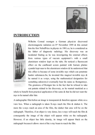
Wilhelm Conrad roentgen a German physicist discovered
electromagnetic radiation on 8th November 1895 & this earned
him the first NobelPrize in physics in 1901.so, he is considered as
the father of diagnostic radiology. His discovery was an
incidental finding as he was investigating the external effects
from various types of vaccum equipment’s with a small
aluminium window kept on the tube. He noticed a fluorescent
effect on the cardboard screen painted with barium platino
cyanide kept near to the aluminium window & he understood that
this effect is because of some invisible rays which can penetrate
harder substances.So, he invented this magical invisible rays &
he named it as x-rays, using the mathematical designation for
something unknown.it eventually bear his name as Rontgenrays.
The greatness of Rontgen lies in the fact that he refused to take
out patents related to his discovery, as he wanted mankind as a
whole to benefit from practical application of the same & that he did not want the
rays to be named after him
A radiographic film before an image is transparent & therefore appears white on a
view box. When a radiograph is taken X-rays reach the film & darken it. The
more the x-rays reach an area of the film, the darker that area will be on the
radiograph. Therefore, if an object is very dense, less x-rays will reach the film &
consequently the image of the object will appear white on the radiograph.
However, if an object has little density, its image will appear black on the
radiograph becauseit allows most of the x-ray beam to reach the film.
2. P A G E | 2
RADIOGRAPHIC
DENSITIES
Only five basic radiographic densities
exist. They are in order of increasing
brightness; gas, fat, fluid, bone& metal
densities .This is a key concept.
Anatomic structures seen on the
radiograph can be identified by their
characteristic density.
For e.g; the lungs are dark, or air density, because they
are filled with air. Organs such as the heart are largely
composed of water. Therefore, it is no surprise that they
appear lighter than the lungs, because they are fluid density.
Bones are brighter structures because they are composed of
calcium
PROPERTIESOF X-RAYS
Wavelength : 10 – 0.01nm
Frequency : 30 petahertz to 30 exahertz
Energy : 120 eV to 120 Kev
Shorter in wavelength than UV rays & larger than gamma rays
X-rays 0.12 to 12 Kev (10-0.10nm wavelength)are soft x-rays
12 to 120 Kev (0.10 – 0.010nm wavelength) are hard x-rays
[Due to their penetrating abilities, hard x-rays penetrate solid objects, to take
images of objects in diagnostic radiography & crystallography; soft x-rays can
hardly be said to penetrate matter]
3. P A G E | 3
UNDER EXPOSED&OVER EXPOSED FILM
A good quality film is necessary for a right diagnosis. The precision of a
diagnosis depends upon the proper exposure of the film. A film should not be too
dark (over exposed) or (under exposed). While reading the x-rays it is important
that we should see the name, number, left or right of the film. Making a right
diagnosis in a wrong patient will be very much embarrassing, so identification of
the x-ray in terms of right, left, patientsname, age& gender is very important for
properradiological diagnosis.
PURPOSEOF X-RAY EXAMINATION& TYPES
To confirm a diagnosis (e.g; pneumonia)
To exclude injury or disease(e.g; fracture)
To establish the extent of disease(e.g; metastatic invasion)
To assess the progress of disease or its responseto treatment
(e.g; osteomyelitis, fracture healing etc.)
TYPES: In x-rays again simple x-ray, miniature x-ray, computed x-ray, xero
radiography, contrast medium x-ray like barium meal, barium swallow, barium
enema, bronchography, sialography, phlebogram, arteriogram, intra cardiac
catheterization, cine radiography etc. are there
4. P A G E | 4
DIGITAL SUBTRACTION X-RAYis a newer modality of investigation. Here a
positive film & a negative film are superimposed on each other & a picture is
taken. After taking the first x-ray, contrast is injected & the second is taken. So
thesuperimposed film will show only the portion containing the dye. This is
useful for angiograms, barium studies & all other situations using contrast
CONTRAST MEDIUM X-RAY:To visualise radiolucent structures like
stomach, intestines, blood vessels, ducts etc., a radiopaque substance is passed or
injected into them & x-ray film is taken. Barium meal, intra venous pyelography
(IVP), myelogram, intra cardiac catheterization, etc., are called contrast medium
x-rays. In these, indirect viewing, of the required part with the help of injecting a
radio opaque substanceis possible.
MAMMOGRAPHY:A mammogram is an x-ray examination of the breast. It is
used to detect & diagnose breast disease in women who either have breast
problems such as a lump, pain, or nipple discharge, as well as for women who
have no breast complaints. A screening mammogram is an x-ray of the breast
changes in women who have no signs of breast cancer. It usually involves two x-
rays of each breast. Using a mammogram, it is possible to detect a tumour that
cannot be felt. A diagnostic mammogram is an x-ray of the breast used to
diagnose unusual breast changes, such as a lump, pain, nipple thickening or
discharge, or a change in breast size or shape.
A mammogram helps to identify the following conditions:
Calcification – tiny mineral deposits within the breast tissue. There are two categories
of calcifications:
i. Macro calcification- coarse calcium deposits that usually
indicates degenerative changes in the breasts, such as aging of
the breastarteries,old injuries, inflammations etc.
5. P A G E | 5
ii. Micro calcification- tiny(less than 1/50 of an inch) specks of
calcium. When many micro calcifications are seen in one area,
they are referred to as a cluster.
Masses – may occur with or without associated calcifications, & may be due to
different causes,includingcyst&benign breastconditions.
Cyst is a non-cancerous collection of fluid in the breast. Cysts cannot be diagnosed
by physical examination alone nor by mammography alone. Either breast ultrasound
or aspiration with a needle is required.
FLUOROSCOPY –fluoroscopy is a study of moving body structures – similar to
an x-ray “movie”. A continuous x-ray beam is passed through the body part being
examined, & is transmitted to a TV- like monitor so that the body part & its
motion can be seen in detail.
Fluoroscopy is used in many types of examinations & procedures, such as barium
x-rays, cardiac catheterization, & placement of intravenous (IV) catheters (hollow
tubes inserted into veins or arteries0. In barium x-rays, fluoroscopy allows the
physician to see the movement of the intestines as the barium moves through
them. In cardiac catheterization, fluoroscopy enables the physician to see the flow
of blood through the coronary arteries in order to evaluate the presence of arterial
blockages. For intravenous catheter insertion, fluoroscopy assists the physician in
guiding the catheter into a specific location inside the body.
INTRAVENOUS PYELOGRAM (IVP)– An IVP is a diagnostic x-ray of the
kidneys, ureters, & bladder. When a contrast agent is injected intravenously, the
urinary tract will show up very clearly, which is not seen on regular x-rays. An
IVP may be done for many reasons including: to detect kidney tumours, to
identify blockages or obstructions of the normal flow of urine, to detect kidney
stones or bladder stones, to establish if the prostate gland is enlarged, to detect
injuries to the urinary tract.
6. P A G E | 6
BARIUM X-RAY – pure barium is rarely used outside the laboratory. Barium
compounds are found primarily in two ores; the first is barite & the second is
witherite. Barite contains a sulphate compound while witherite contains barium
carbonate. While barium & all of its soluble compounds are poisonous, barium
sulphate is used in medical procedures because it does not dissolve in water or
other body fluids. Like other meals, barium is a good conductor of heat &
electricity. It is silver & white in colour & relatively flexible. Chemically it
resembles calcium & strontium fellow members of the alkaline earth family of
metals.
Barium’s many compounds have a number of practical applications. Perhaps the
most familiar is the one used in a medical procedure, the barium meal & enema.
When doctors need to examine a patient’s digestive system, a mixture containing
barium sulphate is used to coat the inner lining of the intestines. Similarly, to
enhance examination of the stomach & oesophagus; the patient drinks a chalky
barium sulphate liquid.
When the patient is x-rayed, the barium coating inside the digestive tract absorbs
a large proportion of the radiation. Barium sulphate is non-toxic insoluble salt.
This highlights the black & white contrast of the x-ray photograph, so that doctors
can better diagnose digestive problems.
For some patients it is necessary to inject contrast agents directly into arteries or
veins. In this case, iodine based molecules are suitable contrast agents. Iodine is
chosen because it has a relatively high atomic mass & so markedly attenuates x-
rays, but also, importantly, it is naturally excreted via the urinary system.
7. P A G E | 7
DIFFERENT VIEWS IN X-RAY
Skiagrams are taken in different positions of the subject in relation to the source
of x-rays & the photographic film. Some of the common position used are:
(1)ANTERO-POSTERIOR VIEW (AP VIEW)
In this the x-ray tube anterior to the subject & film is placed posteriorly. So
rays are passing antero- posteriorly. Posterior structures are better visualized in
this view.Taken at a distance of 100 cm. usually taken in supine position (lying
down). It is substituted for the PA VIEW in very sick patients, infants & in one
who is unable to stand or sit.
(2) POSTERO-ANTERIOR VIEW
In this the x-ray tube is posterior to the subject & the film is anterior, the x-rays
thus passing postero anteriorly through the subject. Anteriorly placed structures are
more clearly visible in this view. Routine PA VIEW is taken with patient upright
(standing or sitting) with the x-ray tube directed horizontally at a distance of 6 feet
from the film.
(3)OBLIQUE VIEW
In taking oblique views, the subject stands in front of upright film cassette holder
& is then turned 45 degree oblique (left or right). Oblique views are taken to
visualize certain special structures.
In the case of chest x-rays, oblique views could be
8. P A G E | 8
i. Right anterior oblique view (R.A.O)
ii. Left anterior oblique view (L.A.O)
(4)LATERAL VIEW
These are used to assess the depth of the structures. The position in this view is
at right angle to that in AP or PA VIEW. Lateral views are of two types;
i. Right lateral view- when the film is kept against the right side of the subject
ii. Left lateral view – when the film is kept against the left side of the subject
The fundamental rule of the radiography is to get the lesion as close to the film as
possible, hence if the lesion is on right side we should take right lateral view & if
the lesion on the left side the view to be taken will be left lateral view. This
reduces the magnification & enhances the sharpness.
(5)WATERS’S VIEW (occipito mental view)
This view is taken to see the paranasal sinuses. To take this view the head is
extended with chin touching the palate & nose raised from the plate (tragocanthal
line forming an angle of 37 degree with the film cassette). X-rays are passed from
posterior side.
(6)EXPIRATORY FILM
This view is used to demonstrate a small pneumothorax, vascular lesions,
azygos veins or the changing shape of pulmonary hydatid cyst. It should be
remembered that normally a chest PA VIEW is taken in full inspiration.
(7)TRENDELENBURG VIEW
9. P A G E | 9
This view is taken with the patient in the head –low position. This helps to
demonstrate movement of the fungal ball in the cavity. Shift of fluid in the pleura
or pericardium can also be noted, when the patient is put in trendelenburg view
(8)APICOGRAM/LORDOTIC VIEW
Compared to the PA VIEW, in the lordotic view the lung apices are seen
clearly & the x-ray is devoid of the overlapping clavicular shadows. Often small
nodules, calcifications, azygos lobe & even middle lobe collapse are clearly
demonstrated in this view.
(9)DECUBITUS VIEW
In this view, patient is lying on one side & x-ray beam is perpendicular to the
film. When the patient is lying on his right side & faces the tube, & the film is
placed behind his back, it is called right lateral decubitus view, similarly when he
is lying on the left side it will be a left lateral decubitus view. Best for
demonstrating minimal pleural effusion & to confirm air fluid levels in the lung.
For demonstrating small pneumothorax, affected side should be in non-dependent
position.
(10)AXIAL VIEW
Best used to assess the talocalcaneal joint & plantar aspects of the calcaneus. The
axial view has a diagnostic sensitivity of 87% for calcaneus fractures. Patient is
supine or seated with the affected limb extended, the posterior aspect of the ankle
is resting on the image receptor. Foot is dorsiflexed until the plantar surface is
running perpendicular to the image receptor
(11)SKYLINE VIEW
10. P A G E | 10
Looks between the kneecap & the thigh bone. It is taken with the knee bent (about
30 degrees) & is used to diagnose arthritis. It is much less common. Only about
30% of orthopaedic surgeons use this view during a routine examination. This
view is similar to the angle we see when sitting with our leg out in front of us,
slightly bent. Only two bones are visible in this skyline view x-ray; the patella &
the femur. In a healthy knee, the groove in the femur should match the shape of
the patella & there should be a nice gap between the two of a consistent width.
(12) MORTISE VIEW
Ankle AP mortise view is part of a three view series of the distal tibia, distal
fibula, proximal talus & proximal metatarsals. This view helps to assess the
mortis joint of the ankle. The patient may be supine or sitting upright with the legs
straighten on the table. The leg must be rotated internally 15-20 degree allowing
the intermalleolar line to be parallel to the detector. This usually results in the 5 th
toe directly in line with the centre of the calcaneum. Internal rotation must be
from the hip, rotation isolated at the ankle will cause a non-diagnostic image, and
foot is to be in slight dorsiflexion.
INDICATIONS&CONTRAINDICATIONS:X-RAY
INDICATIONS
Fractures
Dislocation
Determination of age by knowledge of ossification of bones
For diagnosis of
Deficiency disease (e.g; rickets, scurvy); Infections(e.g; osteomyelitis);
Osteoarthritis; Tumours; Non-fusion of bones (congenital deformity)
11. P A G E | 11
EXAMINATION OF X-RAY
When a radiograph is requested, in most cases the standard views comprising an
antero-posterior (AP) & a lateral projection will be provided
Someone experienced in looking at radiographs will recognise the main pathology
at a glance without following any analytical procedure, just as a familiar face is
identified without apparently paying conscious attention to the relevant position &
size of its main features. Until such skills are developed, it may be helpful for the
student to have some simple scheme follow. One such is to look at each
radiograph as though in the cinematic sequence of long shot, medium shot &
close up. In the long (wide angle) shot a scene is established & the overall
relationship of the important features is made clear. Start by looking at the
radiograph in a general, unfocusedway, as though you were standing well back
from it.
Looking a little more closely, note whether the bone texture appears normal or
disturbed, such as in osteoporosis, Paget’sdisease, avascular necrosis,etc. note if
there are any areas of new bone, such as exostoses, sub periosteal new bone
formation etc. note whether there are any areas of bone destruction, such as may
be found in the presence of many tumours.
Examination of the bone in close up may be done in two ways; either trace
methodically round the contours of the bone, noting any abnormality in route, or
go through a checklist of your own making: checklists can have different bases &
can be used in combination.
A list may be based on pathology ,you might then look for evidence of congenital
abnormality,infection,trauma,neoplasm,metabolic disturbances, degeneration ; a
list have an anatomical base; you might then assess ligamentous attachments,
jointmargins, the joint space& the cortical & cancellous bone elements.
12. P A G E | 12
Before asking for an x-ray, the following points should be kept in mind.
Both AP VIEW & LATERAL VIEWS should be requested in most cases.
X-ray requisition must specify the area of suspicion.
X-ray of the pelvis with both hips should be asked for in all cases of
suspected pelvic injury. Major injuries of the thigh are often associated
with.
Special views show fractures better in some cases
(e.g; oblique view wrist-scaphoid fracture, judet view-acetabular fracture,
merchant view-patello femoral joint, skyline view-calcaneous fracture)
In some situations radiographs additional to the standard AP & lateral projections
may be required.
These may include:
COMPARISON FILMS – here films of the other limb may be taken so that the
two sides may be compared; this may be indicated where there is some
difficulty in interpreting the radiographs.(e.g;elbow region in the children, where
the epiphyseal structures are continuously changing or where there is some
unexplained shadowor congenital abnormality)
LOCALISED VIEWS –where there is marked local tenderness , but routine
films are normal, coned down localised views may give sufficient gain in detail
to reveal, for e.g; hairline fracture
STRESS FILMS – stress films can be of value in certain situations, especially
when a substantial tear of a major ligament is suspected. For e.g; where the
lateral ligament of the ankle is thought to be torn, x-ray of the joint taken with
the foot in forced inversion may demonstrate instability of the talus in the ankle
mortice.
13. P A G E | 13
READING AN X-RAY
An x-ray view box should be used in all cases. If a fracture is obvious, one
must make a note of the following points.
Which bone is affected?
Which partof the bone is affected? E.g;shaft etc.
At whatlevel is the fracture? i.e.;whetherthe fracture is in the upper,middle orlowerthird.
Whatis the pattern of the fracture? i.e.;whether the fracture is transverse,oblique etc.
Is the fracture displaced? If yes, in what direction i.e.;whether it is a shift (anteroposterior,
sideways), atilt, or angulation in any direction, or a rotational displacement. Rotational
displacementis sometimes notvisible on x-rays,& can only be diagnosed clinically.
Is the fracture line extending into the nearby joint?
Does the underlying bone appear pathological? E.g;a cyst, abnormal texture of the whole
bone etc.
Is it fresh or an old fracture? An x-ray of a fresh fracture shows a soft tissue shadow
resulting from haematoma, & the fracture ends are sharp. An old fracture shows callus
formation & disuse osteoporosis &the fracture ends are smoothened.
If the fracture is not obvious immediately, all the bones & joints seen on the x- ray must be
examined systematically for a break in the cortex or loss of joint congruity. X-ray findings
should be co –related with clinical findings, so as to avoid error because of some artefacts
which may mimic a fracture. Also one must ensure that the part under question is visible
on x-ray. An x- ray of a bone must include the joints proximal & distal to the bone. Do look
for an associated injury to all the otherbones & joints visible on the x-ray.
One must be aware of some normal x-ray findings, which are often misinterpreted as
fracture. e.g; epiphyseal lines, vascular markings on bones, accessory bones etc. A
comparison with the x-ray of the opposite limb helps in clearing any doubt. There are some
injuries particularly liable to be missed by a novice. Before an x-ray is passed as normal,
one mustcarefully look forthese injuries.
14. P A G E | 14
USES OF X-RAY
X-rays are especially useful in the detection of the skeletal system, but are also
useful for detecting some disease processes in soft tissue. Some notable examples
are the very common chest x-ray, which can be used to identify lung diseases
such as pneumonia, lung cancer or pulmonary oedema, & the abdominal x-ray,
which can detect intestinal obstruction, free air(from visceral perforations)& free
fluid(in ascites)
X-rays may also be used to detect pathology such as gallstones (which are
rarely radiopaque) or kidney stones which are often visible. Traditional plain x-
rays are less useful in the imaging of soft tissues such as the brain or muscle.
Imaging alternatives for soft tissues are CT scanning, MRI, and ultrasound.
X-rays helps in determining the type & extent of a fracture, helps in detecting
pathological changes in the lungs,& with use of radio-opaque contrast media(such
as Barium),can visualise structures of the stomach & intestines. Helps in
diagnosing ulcers or certain types of colon ulcer. A general practitioner should
know about what type of x-ray or scanning is required in a particular condition or
part of the body.
Simple disorders like fractures, dislocations, bone tumours, congenital &
nutritional deformities of bone, arthritis, vertebral disc prolapse, peptic ulcer
through barium meal x-ray, pulmonary tuberculosis, enlargement of heart, aortic
aneurysm, intestinal perforation, intestinal obstruction, foreign bodies in the GI
tract, dental disorders etc. can easily be diagnosed by studying & observing a few
films of x-ray. Dental x-ray plays a major role in identifying root infection,
abnormal dentition, identification of the individual in medico-legal cases etc.
Since 2005, x-rays are listed as a carcinogen by many governments. The use
of x-rays as a treatment is known as Radiotherapy & is largely used for the
management (including palliation) of cancer; it requires higher radiation energies
than for imaging alone.
15. P A G E | 15
RISKS IN X-RAYS
X-rays are relatively safe method of investigation & the radiation exposure is
relatively low, depending upon the study. Experimental & epidemiological data,
however, do not support the proposition that there is a threshold dose of radiation
below which there is no increased risk of cancer. Diagnostic x-rays account for
14% of the total annual radiation exposure from man-made & natural sources
worldwide. It is estimated that the additional radiation will increase a person’s
cumulative risk of getting cancer by age 75 by 0.6-1.8%. The amount of absorbed
radiation depends upon the type of x-ray test & the body part involved. CT &
fluoroscopyentail higher doseof radiation than do plain x-rays.
To place the increased risk in perspective, a plain chest x-ray or dental x-ray
will expose a person to the same amount from background radiation that we are
exposed to (depending upon location)everyday over 10 days. Each such x-ray
would add less than 1 per 1,000,000 to the lifetime cancer risk. An abdominal or
chest CT would be the equivalent to 2-3 years of background radiation, increasing
the lifetime cancer risk between 1 per 10,000 & 1 per 1000. These numbers are
very small compared to the roughly 40% chance of developing any cancer during
over lifetime.
Father’s exposed to diagnostic x-rays are more likely to have infants who
contract leukaemia, especially if exposure is closer to conception or includes two
or more x-rays of the lower gastro-intestinal (G.I) tract or lower abdomen. This
side effect was misused by Hitler’s doctor in concentration camps to sterilize the
Jew prisoners to eliminate the generations. These prisoners were made to fill a
lengthy questionnaire at a special counter, taking few hours & for the same
duration, a heavy x-ray radiation from a hidden source was focused on their pelvic
areas. By the time they left the counter, irreversible damage had been done to the
testicles, turning most of them sterile. (The risk of radiation is greater to unborn babies,
so in pregnant patients, the benefits of the investigation (x-ray) should be balanced with the
potential hazards to the unborn foetus.)
16. P A G E | 16
SAFETY WITH X-RAY APPARATUS
Whenever a patient undergoes an x-ray or nuclear medicine investigation, a dose
of radiation is given. As a general principle it is expected that the dose given is as
low as reasonably possible for a diagnostic image to be obtained. Numerous laws
govern the amount of radiation exposure that a patient can undergo for a variety
of procedures, & these are monitored to prevent any excess or additional dosage.
Whenever a radiograph is booked, the clinician ordering the procedures must
appreciate its necessity & understand the dose given to the patient to ensure that
the benefits significantly outweigh the risks. Investigations must be carried out
judiciously, based on a sound clinical history & examination, for which an
understanding of anatomy is vital.
PRINCIPLES OF RADIATION SAFETY
1. MINIMISE EXPOSURE TIME
Ensure all personnel minimise time spentclose to machine.
2. MAXIMISE DISTANCE FROM SOURCE
Exposure falls at the square of distance from source. The longer the distance, the
lowerthe dose.
3. USE CORRECTSHIELDING
Most analytical x-ray machines have adequate shielding provided. It is important to
periodically verify that shielding is functioning properly. Never tamper with or alter
shielding.
4. FOLLOW MANUFACTURERS INSTRUCTIONS
5. FOLLOW THE “ALARA”PRINCIPLE
Keep dose As Low as Reasonably Achievable.

![P A G E | 2
RADIOGRAPHIC
DENSITIES
Only five basic radiographic densities
exist. They are in order of increasing
brightness; gas, fat, fluid, bone& metal
densities .This is a key concept.
Anatomic structures seen on the
radiograph can be identified by their
characteristic density.
For e.g; the lungs are dark, or air density, because they
are filled with air. Organs such as the heart are largely
composed of water. Therefore, it is no surprise that they
appear lighter than the lungs, because they are fluid density.
Bones are brighter structures because they are composed of
calcium
PROPERTIESOF X-RAYS
Wavelength : 10 – 0.01nm
Frequency : 30 petahertz to 30 exahertz
Energy : 120 eV to 120 Kev
Shorter in wavelength than UV rays & larger than gamma rays
X-rays 0.12 to 12 Kev (10-0.10nm wavelength)are soft x-rays
12 to 120 Kev (0.10 – 0.010nm wavelength) are hard x-rays
[Due to their penetrating abilities, hard x-rays penetrate solid objects, to take
images of objects in diagnostic radiography & crystallography; soft x-rays can
hardly be said to penetrate matter]](data:image/gif;base64,R0lGODlhAQABAIAAAAAAAP///yH5BAEAAAAALAAAAAABAAEAAAIBRAA7)