Meniscus injury
•Descargar como PPTX, PDF•
70 recomendaciones•16,881 vistas

Orthopedic Knee Injuries Meniscus tear Injury

Recomendados
Más contenido relacionado
La actualidad más candente
La actualidad más candente (20)
Similar a Meniscus injury
Similar a Meniscus injury (20)
Más de Rifhan Kamaruddin
Más de Rifhan Kamaruddin (14)
Último
Último (20)
Meniscus injury
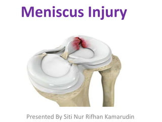
- 1. Meniscus Injury Presented By Siti Nur Rifhan Kamarudin
- 2. ANATOMY • Meniscus is a cushion structure made of cartilage which fits within the knee joint between tibia and femur. • Each Menisci has - Two ends - Two borders - Two surfaces
- 4. MEDIAL MENISCUS • C- Shaped structure and lateral meniscus is more circular. • Anterior horn : Attached to the tibia anterior to the intercondylar eminence to the ACL. • Posterior horn : Anchored immediately in front of the attachment of PCL posterior to the intercondylar eminence.
- 5. Medial Meniscus • Peripheral border attached to the medial capsule through the coronary ligament to the upper border of tibia. • Most of the weight borne on the posterior portion of meniscus
- 6. LATERAL MENISCUS • Circular shaped • The anterior and posterior horns are closer to each other & near insertion of ACL • Anterior Horn : Attached to the tibia in front of the intercondylar eminence. • Posterior Horn : Attached to the posterior aspect of the intercondylar eminence in front of posterior attachment of medial meniscus.
- 7. Lateral Meniscus • The lateral meniscus is mobile and medical meniscus is more fixed -> causing more tears to occurs in medical meniscus • Lateral meniscus is associated with discoid meniscus and meniscal cysts • Lateral meniscus is also assoc. with acute injury to ACL Medial Meniscus • Tears of medical meniscus occurs more with degenerative tears • Associated with a baker’s cyst.
- 8. BLOOD SUPPLY • The blood supply of meniscus decides the healing potential of the meniscus • The outer one-third of meniscus is vascular. It will heal if repaired • The inner one-third is not vascular and is nourished by synovial fluid. • The middle third is red/white and it is avascular. • The blood supply of meniscus originates from medial and lateral genicular arteries
- 9. FUNCTIONS OF MENISCUS • Shock Absorber: Provides load sharing across knee by increasing the contact area and decreasing the contact stress. • Act as joint filler : Compensates for the gross incongruity between tibial and femoral articulating surfaces. • Joint Lubrication: help to distribute Synovial fluid through the joint and aiding the nutrition of articular cartilage.
- 10. OVERVIEW of MENISCAL INJURY • Epidemiology: - Most common indication for knee surgery • Location: Medial Tears - More common - Degenerative tears in older patients usually occur in posterior horn of medial meniscus. Lateral Tears - More common in acute ACL tears
- 11. CLINICAL FEATURES • Pt is usually a young person who sustain twisting injury to the knee • Knee pain (often severe) • Swelling of the knee within 48hours • “Locking” : Sudden inability to extend the knee fully – suggest a ‘bucket-handle tear’. • Popping or clicking within the knee. • Limited motion of knee joint. • Tenderness when pressing on the meniscus (Knee joint line)
- 12. CLASSIFICATION OF MENISCAL TEAR • Based on Location Red Zone: Outer third, vascularized Red-White Zone : Middle Third White Zone : Inner third, Vascularized
- 13. Based On Pattern • Vertical/Longitudinal - Common, esp. with ACL tears • Bucket Handle - Vertical tear which may displace into notch • Horizontal - More common in older population - May be associated with meniscal cysts
- 14. PHYSICAL EXAMINATION • The joint may be held slightly flexed and there is often an effusion. • In late presentations, the quadriceps will be wasted. • Tenderness is localized to the joint line, particularly the medial line. • Flexion is usually full but extension is often limited.
- 15. SPECIAL TESTS 1) Thessaly Test • Standing at 20 degrees of knee flexion on affected limb • Patient twists with knee external and internal rotation. • Positive Test: Clicking, pain or discomfort on joint line.
- 17. 2) McMurrays Test • Principle: To trap the meniscus between the tibia and femur. • Pt needs to be relaxed. • One hand on knee joint line. Other hand holds the foot & ankle. • Flex the knee as far as possible (Hyperflexion) • Externally rotate(Medial Me.) or internally rotate (Lateral Me.) the tibia and then extend the knee. • Positive McMurray’s : Clicking or popping felt associated with pain.
- 19. 2) Apley’s Grinding test • Patient is in prone position • Knee flexed to 90 degrees • The leg is rotated from side to side • Compression force applied • A painful response signifies a torn or degenerate meniscus.
- 21. IMAGING Radiographs • Should be normal in young patient with acute meniscal injury MRI • Most sensitive diagnostic test • Findings - MRI Grade III signal is indicative of a tear - Parameniscal cyst indicates presence of meniscal tear - May see ‘Double PCL” sign that indicates bucket- handle meniscal tear.
- 23. MANAGEMENT NON-OPERATIVE TREATMENT Indication: First line of treatment for degenerative tears : Acute episode without locking but with acute synovitis • Immediate abstinence from weight bearing • Rest • Ice pack application • Compression dressing • NSAIDS • Rehabilitation exercises
- 24. SURGICAL MANAGEMENT 1)Meniscectomy 2)Meniscal Repair 3)Meniscal Transplantation
- 25. OPERATIVE TREATMENT 1) Partial Meniscectomy • Indication: Tears not amenable to repair (complex, degenerative, radial tear patterns) : Repair failure > 2 times • Objective: Remove the torn meniscal fragment and contour the peripheral rim, leaving a balanced, stable rim of meniscal tissue. • Outcomes - >80% satisfactory function • Partial is preferred over total meniscectomy - Shorter operating time, Faster recovery, better post-op function.
- 26. Anthroscopic Meniscal Repair 3 important steps: - Appropriate patient selection : should have documented tear that is able to heal - Tear debridement and local synovial, meniscal and capsular ablation to stimulate a proliferative fibroblastic response - Suture placement to reduce and stabilize the meniscus
- 27. Meniscal Repair Risks: – Saphenous Nerve and Vein damage – Peroneal Nerve – Popliteal Vessels
- 28. 3) Meniscal Transplantation • Attempts at meniscal replacement with - Allograft meniscus - Autograft fascial material - Synthetic meniscus
- 29. REFERENCES • Apley and Solomon’s Concise System of Orthopedic and Trauma, 4th Edition
Notas del editor
- if the meniscus is removed or injured, the pt will develop arthritis of knee joint.