Management of patient with burns
•Descargar como PPTX, PDF•
270 recomendaciones•141,539 vistas

Management of patient with burns

Recomendados
Más contenido relacionado
La actualidad más candente
La actualidad más candente (20)
Similar a Management of patient with burns
Similar a Management of patient with burns (20)
Más de salman habeeb
Más de salman habeeb (20)
Último
Último (20)
Management of patient with burns
- 1. MANAGEMENT OF PATIENT WITH BURNS
- 2. Definition • Injuries that result from direct contact or exposure to any physical, thermal, chemical, electrical, or radiation source are termed as Burns.
- 3. STATISTICS An estimated 265000 deaths every year are caused by burns. One of leading causes of disability-adjusted life-years (DALYs) lost in low- and middle-income countries.
- 4. Problem Statement : India 70 lakh burn injury cases annually Over 10,00,000 people are moderately or severely burnt every year 1.4 lakh people die of burn every year. Around 70% of all burn injuries occur in most productive age group (15-35 years). Majority are women & children. As many as 80% of cases admitted are a result of accidents at home (kitchen-related incidents)
- 6. Etiology Based on Cause o Thermal o Electrical o Chemical o Radiation o Inhalation
- 7. Thermal Injuries • Most common • Types : Dry & wet Contact • Direct contact with hot object (i.e. pan or iron) • Anything that sticks to skin (i.e. tar, grease or foods)
- 8. Flame oDirect contact with flame (dry heat) o structural fires / clothing catching on fire
- 9. Electrical Burns • Usually follows accidental contact with exposed object conducting electricity o Electrically powered devices o Electrical wiring o Power transmission lines • Can also result from Lightning • Damage depends on intensity of current
- 10. • Low-tension injuries(<1000 V) o Low energy burns Minimal damage to subcutaneous tissue o Entry & Exit points – fingers small deep burns o AC Tetany within muscles, cardiac arrest due to interference with normal cardiac pacing o High-tension injuries(>1000V) • Earthed high tension lines Arc over the patient Flash burn
- 11. • Severity depends upon: owhat tissue current passes through (Low voltage/ High voltage) owidth or extent of the current pathway oAC or DC oduration of current contact
- 13. • Lightning oHIGH VOLTAGE!!! oInjury may result from • Direct Strike • Side Flash
- 14. Chemical Burns • Usually associated with industrial exposure • Accidental mishandling of household cleaners Degree of tissue damage determined by - Chemical nature of the agent - Concentration of the agent - Duration of skin contact Acids- Eg- Formic acid,sulphuric acid Alkalis - Eg. Lime, potassium hydroxide
- 15. Radiation Exposure • Waves or particles of energy that are emitted from radioactive sources • Alpha radiation Large, travel a short distance, minimal penetrating ability Can harm internal organs if inhaled, ingested or absorbed • Beta radiation Small, more energy, more penetrating ability Usually enter through damaged skin, ingestion or inhalation
- 16. INHALATION • Smoke and inhalation injury carbon monoxide poisoning inhalation injury above glottis inhalation injury below glottis
- 17. According Depth of burn • Superficial Partial-Thickness (First Degree burn) cause-Sunburn Low-intensity flash Skin involvement- Epidermis Symptoms- Reddened, Tingling, Pain that is soothed by cooling
- 18. Deep Partial-Thickness (Second Degree) Cause • Scalds • Flash flame • Contact burns • chemical Skin involvement- Epidermis, upper dermis, portion of deeper dermis Manifestations- Blisters that are red, shiny. Severe pain caused by nerve injury ,mild to moderate edema • Recovery in 2 to 4 weeks, some scarring and depigmentation contractures
- 19. Full-Thickness (Third Degree) Cause- • Flame • Prolonged exposure to • hot liquids • Electric current • Chemical Skin involvement- Epidermis, entire dermis, and sometimes subcutaneous tissue; may involve connective tissue, muscle, and bone Manifestations- Dry; pale white, Leathery, visible thrombosed blood vessels • Pain free, all skin elements and local nerve endings are destroyed, surgical intervention required for healing
- 20. 4th Degree E+D+S+muscles, tendons & bone
- 22. Extent of Body Surface Area Injured • RULE OF NINES, • LUND AND BROWDER METHOD, • PALM METHOD.
- 23. RULE OF NINES • An estimation of the TBSA involved in a burn is simplified by using the rule of nines • The rule of nines is a quick way to calculate the extent of burns. The system assigns percentages in multiples of nine to major body surfaces
- 26. LUND AND BROWDER METHOD • A more precise method of estimating the extent of a burn is the Lund and Browder method, which recognizes that the percentage of TBSA of various anatomic parts • By dividing the body into very small areas and providing an estimate of the proportion of TBSA
- 28. PALM METHOD • In patients with scattered burns, a method to estimate the percentage • of burn is the palm method. The size of the patient’s palm is approximately 1% of TBSA.
- 29. Location of burn • Burns to face, neck ,chest and back may inhibit respiratory function due to mechanical obstruction secondary to edema, eschar formation • Burns to the ear, nose are susceptible to infection because of poor blood supply • Burns to buttocks, genitalia are susceptible to infection because of contamination • Burns on extremities cause circulatory compromise and neurologic impairment.
- 31. Zones of burn injury
- 32. Zones of burn injury • The inner zone (known as the zone of coagulation, where cellular death occurs) sustains the most damage o Necrotic area with cellular disruption o Irreversible tissue damage • The middle area, or zone of stasis, has a compromised blood supply, inflammation, and tissue injury, Can survive or go on to coagulative necrosis depending on wound environment • The outer zone—the zone of hyperemia—sustains the least damage
- 34. Pathophysiology Burns> 30% Cell lysis increased capillary loss of skin barrier permeability Hemolysis Hyperkalemia inflamatory altered Na,H20,Protien process thermoreglatn Haemo/myoglobinuria shift extravascular Acute tubular neccrosis intravascular volume vasodilation hypothermia ACUTE RENAL FAILURE BURNS SHOCK HYPOTENSION ARRYTHMIAS MODS
- 35. MANAGEMENT
- 36. Phases of burn management • 1. emergent phase/resuscitative phase • 2.Acute phase/ wound healing phase • 3. Rehabilitative phase/Restorative phase
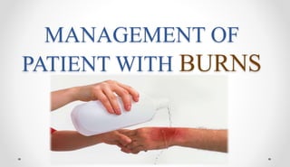
- 37. PRE HOSPITAL MANAGEMENT • Rescuer to avoid injuring himself • Remove patient from source of injury • Stop burn process • Burning clothing; jewelry, watches, belts to be removed • Pour ample water on burnt area (not ice/ ice packs – skin injury & hypothermia)
- 38. Chemical burns: Remove saturated clothing Brush skin if agent is powder Irrigation with copious amount water to be started and continued in hospital Electrical burns: Turn off the current Use non-conductor item to separate from source
- 39. • Small thermal burns (<10% TBSA ) may be covered with a clean, tap water-damped towel for patient comfort and protection until definite medical care instituted • Cooling of injured area within 1 minute helps minimize the depth of injury • If the burn injury is large (>10% TBSA) it is not advisable to immerse the body part in cool water since doing so might lead to extensive heat loss
- 40. Do not break blisters. Do not apply lotions, powders, grease, ghee, gentian violet, calamine lotion, toothpastes, butter and other sticky agents over the burn wound. Prevent contamination: Wrap burn part in clean dry sheet /cloth. Assess for life threatening injuries.
- 41. EMERGENT/RESUSCITATIVE PHASE • This phase may last 24-48 hours after injury Resuscitation phase characterized by: Life-threatening airway problems Cardiopulmonary instability Hypovolemia Goal: Maintain vital organ function and perfusion
- 42. • Assess A B C • ET intubation + assisted ventilation with 100% O2 if: oOvert signs and symptoms of airway obstruction (Progressive hoarseness) oSuspected inhalational injury (smoke/ carbon monoxide intoxication) oUnconscious patient/ rapidly deteriorating patient oAcute respiratory distress oBurns of face & neck oExtensive Burns (> 40% TBSA)
- 43. • Large gauge I.V catheter • Central line Insertion • Venesection • Foleys catheter and NG tube placement • Quick assessment of extent • Tetanus prophylaxis (the only IM administered inj) • Weigh the patient
- 44. • History o Mechanism of injury o Time of injury o Surroundings (closed space/ chemicals) • Physical examination o Head to toe assessment o Careful neurological examination (cerebral anoxia) o Labs: CBC, electrolytes, BUN o Pulmonary assessment: ABG, CXR, carboxyhemoglobin
- 45. • Pulse in extremities: manual/ doppler • Loss of distal circulation • Pallor/coolness/absent pulse/loss capillary refill/decreased oxygen saturation • Absent pulse: emergency escharotomy to release constrictive, unyielding eschar
- 46. ESCHAROTOMY • It is the surgical division of the nonviable skin and tissues , which allows the cutaneous envelope to become more compliant •Deep 2nd & 3rd degree circumferential burns o Chest: To allow respiratory movement o Limb: To restore circulation in limb with excess swelling under rigid eschar • Not in SC tissue Exposes SC fat
- 48. FLUID RESUCITATION • Parkland Formula • Evan’s formula: • Brooke formula
- 49. Parkland Formula Fluid of Choice Lactated Ringer’s (RL) NS can produce hyperchloremic acidosis 4 ml x % of burn x weight (Kg) in 24 hours First ½ of total volume given in the first 8 hours Remaining ½ of total volume given over following 16 hours NEXT 24 HRS Total volume ½ of first day Colloids ( 0.5 ml / kg / % ) 5 % glucose to make up the rest
- 50. Brooke formula( modified) 2 ml x % of burn x weight (Kg) in 24 hours First ½ of total volume given in the first 8 hours Remaining ½ of total volume given over following 16 hours NEXT 24 HRS Total volume ½ of first day Colloids (0 .3-0.5 ml / kg / % )
- 51. Evan’s formula Requirement for first 24 hrs Colloids : 1ml/kg/% burn Saline : 1ml/kg/% burn D5 : 2000ml Requirement for second 24 hrs ½ of first 24 hrs
- 52. Assessment of Adequacy of Fluid Resuscitation • Monitor o Urinary Output • Adult: > 1 ml/ kg/ hr o Daily Weight o Vital Signs • Heart rate and blood pressure • CVP • Level of Consciousness o Laboratory values
- 53. RESUSCITATION FAILURE • Delayed resuscitation • Electric burns • Inhalation injury • Escharotomy • Carbon monoxide poisoning • Elderly patients
- 54. Wound care • Wound care should be delayed until a patent airway, adequate circulation and adequate fluid replacement have been established. • 2 types of wound treatment used to control infection 1. open method 2. multiple dressing change method
- 55. Closed method
- 56. Antimicrobial Agent • Silvadene (silver sulfadiazine)1% cream- • Most bactericidal agent • Minimal penetration of eschar • Mafenide acetate 5% to 10% (Sulfamylon) hydrophilic-based cream • Effective against gram-negative and gram-positive organisms • Diffuses rapidly through eschar In 10% strength, it is the agent of choice for electrical burns because of its ability to penetrate thick eschar
- 57. • Silver nitrate 0.5% aqueous solution- • Bacteriostatic and fungicidal • Does not penetrate eschar
- 58. Analgesia • Morphine sulphate • Fentanyl • Methadone • Haloperidol • Lorazepam • Midazolam
- 59. ACUTE PHASE • Begins 48 to 72 hours after the burn injury. • In this phase the extracellular fluid start mobilize and start diuresis • This phase is completes when wound is covered by skin grafts or the wounds are healed • This may take weeks or many months
- 60. • Eschar begins to separate fairly after injury • Re epitheliazation begins at wound margin and appears as red/pink scar tissue • Hyponatremia/hypernatremia • Hypokalemia/hyperkalemia • Decreased hematocrit
- 61. Management GOALS • Prevention of infection and Wound care • Excision and grafting • Pain management • Nutritional therapy • Physical, psychosocial and occupational therapy
- 62. Prevention of infection and Wound care • Burn wounds are frequently monitored for bacterial colonization • Wound swab cultures and invasive biopsies • Cleanse and debride the area of necrotic tissue that would promote bacterial growth
- 63. Debridement of the wound • May be completed at the bedside or as a surgical procedure. Types of Debridement: Natural • Body & bacterial enzymes dissolve eschar; takes a long time Mechanical • Sharp (scissors), Wet-to-Dry Dressings or Enzymatic Agents Surgical
- 64. Wound/Skin Grafting • If wounds are deep (full-thickness) or extensive, spontaneous re-epithelialization is not possible. Therefore, coverage of the burn wound is necessary by using patients own skin or other methods.
- 65. TYPES • Permanent Skin Grafts Autografts Cultured Epithelial Autografts (CEA)
- 66. Types • TEMPORARY Biosynthetic- Homograft / allograft (cadaveric) heterograft/Xenograft (porcine) Artificial Skins (collagen based) Trancyte/ Integra Synthetic Biobrane/Opsite
- 67. Permanent skin graft Autograft •Harvested from pt •Non-antigenic •Less expensive •Decreased risk of infection •Can utilize meshing to cover large area •Disadvantage : lack of sites and painful
- 70. Permanent skin graft Cultured Epithelial Autografts (CEA) • A small piece of pt’s skin is harvested and grown in a culture medium (PDGF impregnated) • Takes 3 weeks to grow enough for the first graft • Very fragile; immobile for 10 days post grafting • Useful for limited donor sites • Disadvantage : very expensive; poor long term cosmetic results and skin remains fragile for years
- 72. Temporary Skin Grafts Biosynthetic • Homograft/Allograft • Live or cadaver human donors • Fairly expensive/ all the function of skin • Best infection control of all biologic coverings • Disadvantage : • Disease transmission (HBV & HIV) • Antigenic: body rejects in 2 weeks • Not always available • Storage problems
- 73. Temporary Skin Grafts BIOSYNTHETIC -Heterograft • Xenograft • Graft between 2 different species • Porcine most common • Fresh, frozen or freeze-dried (longer shelf life) • Amenable to meshing & antimicrobial impregnation • Antigenic: body rejects in 3-4 days • Fairly inexpensive • Disadvantage : Higher risk of infection
- 75. Temporary skin graft Artificial Skins • Transcyte: oA collagen based dressing impregnated with newborn fibroblasts. • Integra: oA collagen based product that helps to form a “neodermis” ono anti-microbial property Synthetic • Any non-biologic dressing that will help prevent fluid & heat loss oBiobrane, Xeroform, OpSite or Beta Glucan collagen matrix
- 77. Nutritional therapy o High-protein & high-calorie diet o Often requiring various supplements o Routes: • ORAL (BEST) • Enteral oGut is the preferred alternative route oG-tube or J-tube (Head injury/ surgery/ unconscious) • Parenteral oTPN and PPN oAssociated with an increased risk of infections
- 78. Physical and psychosocial care • Active and passive ROM excercises should be performed all joints • Support and counselling • Adjust with disabilities
- 79. Rehabilitation phase • It starts when the patients burn wounds are healed and patient is able to resume a level of selfcare activity • This occur from weeks to months • GOALS • resuming a functional role in society and to accomplish functional and cosmetic reconstructive surgery
- 80. • New skin starts to appear which is flat and pink • Mature healing is reached in 6 months to 2 years • Scarring can happen discolouration contour- skin is no longer flat or slightly elevate but become elevated and enlarged above original burned area • Apply water moisturisers and emolients to prevent dryness and itching • Protect from direct sunlight for 6 to 9 months
- 81. Complications • EMERGENT PHASE CVS- dysrhythmias and hypovolemic shock Resp- upper RT injury, pulmonary edema, ARDS, pneumonia urinary- Acute Tubular necrosis, ARF ACUTE PHASE infection – sepsis, septicemia ( pseudomonas) G.I- Paralytic ileus, curlings ulcer REHABILITATION PHASE Contracture- abnormal condition of a joint characterised by flexion and fixation
- 82. • Curling's ulcer Curling ulcer is an acute gastric erosion resulting as a complication from severe burns when reduced plasma volume leads to ischemia and cell necrosis (sloughing) of the gastric mucosa.
- 86. NURSING DIAGNOSIS • impaired gas exchange related to carbon monoxide poisoning, smoke inhalation, and upper airway obstruction • Ineffective airway clearance related to edema and effects of smoke inhalation • Fluid volume deficit related to increased capillary permeability and evaporative losses from the burn wound • Hypothermia related to loss of skin microcirculation and open wounds • Pain related to tissue and nerve injury and emotional impact of injury • Anxiety related to fear and the emotional impact of burn injury
- 87. • Fluid volume excess related to resumption of capillary integrity and fluid shift from interstitial to intravascular compartment • Risk for infection related to loss of skin barrier and impaired immune response • Altered nutrition, less than body requirements, related to hypermetabolism and wound healing • Impaired skin integrity related to open burn wounds • Impaired physical mobility related to burn wound edema, pain, and joint contractures • Ineffective individual coping related to fear and anxiety, grieving, and forced dependence on health care providers