SlideShare una empresa de Scribd logo
1 de 57
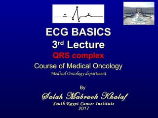
ECG BASICSECG BASICS
33rdrd
LectureLecture
QRS complexQRS complex
By
Salah Mabruok Khalaf
South Egypt Cancer Institute
2017
Course of Medical Oncology
Medical Oncology department
• Definitions of QRS:
• Q wave – first downward deflection after P
wave
• R wave – first upward deflection after Q
wave
• R` wave – any second upward deflection
• S wave – first downward deflection after the
R wave
• QRS duration :
0.06 to 0.12 msec= 3 ss
• Nomenclature of QRS
Ventricular Depolarization
Occurs in 2 stagesOccurs in 2 stages:
1- septal depolarization:
direction from stronger
left bundle to the right bundle
V1 +ve wave = septal r
V6 -ve wave = septal q
V1
V6
Ventricular Depolarization
2- wall depolarization:
directed mainly to the left
due to stronger left ventricle
V1 -ve wave = deep S
V6 +ve wave = Tall R
V1 V6
Lead Septal Wall Total
V1
V6
Are the QRS complexes normal?
• Voltage?
• Duration?
• Pathological Q waves?
• QRS axis?
Voltage of QRS
Low Voltage
• Diagnostic Criteria
1. Voltage of entire QRS complex in all limb leads <
5mm(≤ 1 Ls).
2. Voltage of entire QRS complex in all chest leads <
10mm(≤ 2 Ls).
3. Either criteria may be met to qualify as "low
voltage".
ECG on presentation with low QRS voltages compatible
with pericardial effusion
After two months of treatment and disappearance of
pericardial effusion and normalisation of ECG
Differential Diagnosis of low QRS voltages
1) Increased Distance
1) Pericardial effusion
2) Obesity
3) COPD with hyperinflation
4) Pleural effusion
5) Constrictive pericarditis
2) Infiltrative Heart and Diseased myocardium
1) Amyloidosis
2) Scleroderma
3) Hemachromatosis
4) Cardiomyopathy
5) Myocardial Ischemia or infarction
3) Decreased Metabolic activity
1) Myxoedema
2) Hypothermia
High voltage
Ventricular Hypertrophy
• Conditions that increase the load, pressure
or volume, on either the left or right ventricle,
cause a compensatory increase in the
ventricular muscle massmuscle mass.
• This increase in muscle mass is seen on the
surface electrocardiogram as an increase in
QRS voltage.
1- Left Ventricular Hypertrophy
V1 V6
1- Left Ventricular Hypertrophy:
ESTES Criteria for LVH
("diagnostic", ≥5 points; "probable", 4 points)
ECG Criteria Points
Voltage Criteria (any of):
Tall R in V5 or V6 >30 mm (≥ 6 Ls)
Deep S in V1 or V2 > 30 mm (≥ 6 Ls)
Tall R in V5 or V6 + Deep S in V1 > 35 mm (≥ 7 Ls)
3 points
Left Atrial Enlargement in V1 (p mitrale) 3 points
ST-T Abnormalities:
Without digitalis
With digitalis
3 points
1 point
Left axis deviation 2 points
QRS duration 0.09 sec 1 point
Delayed intrinsicoid deflection in V5 or V6 (>0.05 sec)
(i.e., time from QRS onset to peak R is >0.05 sec)
1 point
S in V1 or V2 > 30 mm (≥ 6 Ls)
R in V5 or V6 > 30 mm (≥ 6 Ls)
Left Atrial Enlargement in V1
ST-T Abnormalities
S in V1 + R in V5 or V6 > 35 mm (≥ 7 Ls)
• ESTES Criteria: 3 points for voltage in V5, 3
points for ST-T changes (more than 5 points
• Note
– LAD - P mitrale in V1
2-Right Ventricular Hypertrophy:
V1
V6
2-Right Ventricular Hypertrophy
11 ss
7 ss
RAD R in v1 > 7 mm
R/S ratio > 1 and
negative T wave
RAE
RVHRVH
3-Biventricular Hypertrophy:
• Biventricular Hypertrophy (difficult ECG
diagnosis to make)
In the presence of LAELAE any one of the following
suggests this diagnosis:
• R/S ratio in V5 or V6 < 1
• S in V5 or V6 > 6 mm
• RAD (>90 degrees)
Other suggestive ECG findings:
• Criteria for LVH and RVH both met
• LVH criteria met and RAD or RAE present
Tall R wave in lead V1
1. Right Ventricular Hypertrophy.
2. Posterior infarction
3. Muscular dystrophy.
4. Wolff-Parkinson-White syndrome.
5. Right bundle branch block.
• Poor R Wave Progression – defined as loss of, or no
R waves in leads V1-3 (R ≤ 2mm):
1. Normal variant (if the rest of the ECG is normal)
2. LVH (look for voltage criteria and ST-T changes of
LV "strain")
3. Complete or incomplete LBBB (increased QRS
duration)
4. Left anterior fascicular block (should see LAD in
frontal plane)
5. Anterior or anteroseptal MI
6. Emphysema and COPD (look for R/S ratio in V5-6
<1)
7. Diffuse infiltrative or myopathic processes
Poor R Wave ProgressionPoor R Wave Progression
QRS duration=3 ssQRS duration=3 ss
QRS duration=3 ss
• Causes of wide QRS:
I- Intrinsic intraventricular delay:
• Right BBB.
• Left BBB.
• pacemaker.
II- Extrinsic (toxic) intraventricular delay:
• Hyperkalemia.
• Drugs.
III- Ventricular ectopy: premature, escape, or paced
IV- Wolf-Parkinson-White syndrome
V- Myocardial infarction.
Right Bundle Branch Block (RBBB):
Ventricular Depolari Occurs in
3 stages:
1- septal depolarization:
direction from left bundle
to the right.
V1 +ve wave = septal r
V6 -ve wave = septal q
V1 V6
2- Lt vent. depolarization:
directed to the left
V1 deep -ve wave = S
V6 tall +ve wave = R
V1 V6
3- Rt vent. depolarization:
directed to the right from
the Lt vent
V1 deep -ve wave = R'
V6 tall +ve wave = S
V1 V6
Lead Septal Lt vent Rt vent Total
V1 R S R' RSR'
V6 q R S qRS
Character of RBBB
1. Terminal R' wave in lead V1 (rSR' complex)
2. Terminal S waves in leads I, aVL, V6 (qRS).
3. "Complete" RBBB has a QRS duration >0.12s
4. "Incomplete" RBBB has a QRS duration of
0.10 - 0.12s with the same terminal QRS
features. This is often a normal variant.
Character of RBBB
5. The ST-T waves in RBBB should be oriented
opposite to the direction of the terminal QRS
forces
– In leads with terminal R or R' forces the ST-T should be
negative or downwards
– In leads with terminal S forces the ST-T should be
positive or upwards.
5. If the ST-T waves are in the same direction as the
terminal QRS forces, they should be labeled
primary ST-T wave abnormalities.
RBBBRBBB
• The ECG below illustrates primary ST-T wave
abnormalities (leads I, II, aVR, V5, V6) in a
patient with RBBB.
Character of RBBB
6. The frontal plane QRS axis in RBBB should be in
the normal range (i.e., -30 to +90 degrees).
– If left axis deviation is present, think about left anterior
fascicular block in addition to the RBBB
– If right axis deviation is present, think about left
posterior fascicular block in addition to the RBBB.
Left Bundle Branch Block (LBBB):
Ventricular Depolari Occurs in
3 stages:
1- septal depolarization:
direction from right
to the left
V1 -ve wave = septal q
V6 +ve wave = septal r
V1 V6
Left Bundle Branch Block (LBBB):
2- Rt ventr depolarization:
direction to the right
V1 +ve wave
V6 -ve wave
V1 V6
Left Bundle Branch Block (LBBB):
3- Lt ventr depolarization:
direction to the left from
Rt vent
V1 -ve wave
V6 +ve wave
V1 V6
Lead Septal Lt vent Rt vent Total
V1
W-shaped
V6
M-shaped
Character of LBBB
1. Terminal R waves in lead I, aVL, V6 broad
usually M-shaped
2. Poor R progression from V1 to V3 is common.
3. "Complete" LBBB" has a QRS duration
>0.12s .
4. "Incomplete" LBBB looks like LBBB but QRS
duration = 0.10 to 0.12s, with less ST-T
change. This is often a progression of LVH.
5. The "normal" ST-T waves in LBBB should be
oriented opposite to the direction of the
terminal QRS forces;
LBBB
Left Anterior Fascicular Block
III II
AvF
rS
LAD
AvL qR
V1-3
Poor R wave
progression
V5-6
Tall R wave
Mimic LVH
• Criteria of left anterior fascicular block
• I. QRS duration < 0.10 sec.
• II. LAD.
• III. QRS morphology
• Limb leads
– qR in leads I and aVL.
– rS in leads II, III, and aVF.
– R peaks in aVL before aVR.
• Precordial leads
– Poor R wave progression.
– Persistent S wave in V5-V6
• In this ECG, note –
• 75 degree QRS axis
• rS complexes in II, III, aVF
• tiny q-wave in aVL
• poor R progression V1-3
• late S waves in leads V5-6.
• QRS duration is normal, and there is a slight slur to
the R wave downstroke in lead aVL.
Left posterior Fascicular Block
III II
AvF
rSI
RAD
qR
R in III > R in II
• Left Posterior Fascicular Block (LPFB)....
Very rare intraventricular defect!
1. Right axis deviation in the frontal plane (usually >
+100 degrees)
2. rS complex in lead I
3. qR complexes in leads II, III, aVF, with R in lead III
> R in lead II
4. QRS duration usually <0.12s unless coexisting
RBBB
5. Must first exclude (on clinical grounds) other
causes of right axis deviation such as cor
pulmonale, pulmonary heart disease,
pulmonary hypertension, etc., because these
conditions can result in the identical ECG
picture!
R in III > R in IIRAD rS
Bifascicular Blocks
• RBBB plus either LAFB (common) or LPFB (uncommon)
• Features of RBBB plus frontal plane features of the
fascicular block (axis deviation, etc.)
• The above ECG shows classic RBBB (note rSR' in V1)
plus LAFB (note QRS axis = -45 degrees, rS in II, III, aVF;
and small q in aVL).
• Electrocardiogram of a 59-year-old man
showing a bifascicular block (consisting of a
RBBB and LAFB).
Bifascicular block.
• Right bundle branch block and left anterior
fascicular block.
Causes of wide QRS:
II- Extrinsic (toxic) intraventricular delay:
• Hyperkalemia.
• Drugs.
Hyperkalemia
• The following changes may be seen in
hyperkalaemia
1. small or absent P waves
2. shortened or absent ST segment
3. atrial fibrillation
4. ventricular fibrillation
5. wide QRS
6. wide, tall and tented T waves
Potassium
•P wave small or absent
•Oscillation or fibrillation
•T wide and tall
•Absent or shortened ST
segment
Hyperkalemia and ECG changesHyperkalemia and ECG changes
according to its levelaccording to its level
Drugs
• Antiarrhythmic drugs
• Beta-blockers
• Calcium channel blockers
• Cardiac Digitalis
• Clonidine (Catapres)
• Cholinergic
• Central Tricyclic antidepressant
Causes of wide QRS:
III- Ventricular ectopy: see arrhythmia
lecture
IV- WPW syndrome: see 1st
lecture.
V- Myocardial infarction: see 6th
lecture.
3rd part ECG Basics  QRS complex Dr Salah Mabrouk Khallaf

Más contenido relacionado

La actualidad más candente

Bundle branch blocks by Dr Sujith Chadala
Bundle branch blocks by Dr Sujith ChadalaBundle branch blocks by Dr Sujith Chadala
Bundle branch blocks by Dr Sujith ChadalaDr Sujith Chadala
 
Aortic stenosis - Echocardiography
Aortic stenosis - EchocardiographyAortic stenosis - Echocardiography
Aortic stenosis - EchocardiographyAnkur Gupta
 
Echocardiographic assesment of systolic and diastolic dysfunction
Echocardiographic assesment of systolic and diastolic dysfunctionEchocardiographic assesment of systolic and diastolic dysfunction
Echocardiographic assesment of systolic and diastolic dysfunctionMalleswara rao Dangeti
 
Ecg localization of myocardial infarction
Ecg localization of myocardial infarctionEcg localization of myocardial infarction
Ecg localization of myocardial infarctionShleshaPradhan
 
Bundle branch blocks
Bundle branch blocksBundle branch blocks
Bundle branch blocksSonukurian
 
ventricular tachycardia (VT) Localisation
ventricular tachycardia (VT) Localisationventricular tachycardia (VT) Localisation
ventricular tachycardia (VT) LocalisationMalleswara rao Dangeti
 
Myocardial infarction (MI) ecg localisation
Myocardial infarction (MI) ecg localisationMyocardial infarction (MI) ecg localisation
Myocardial infarction (MI) ecg localisationMalleswara rao Dangeti
 
Idiopathic ventricular tachycardia
Idiopathic ventricular tachycardiaIdiopathic ventricular tachycardia
Idiopathic ventricular tachycardiaRamachandra Barik
 
Echocardiographic evaluation of Aortic stenosis
Echocardiographic evaluation of Aortic stenosisEchocardiographic evaluation of Aortic stenosis
Echocardiographic evaluation of Aortic stenosisAswin Rm
 
Echocardiography in ischemic heart disease
Echocardiography in ischemic heart diseaseEchocardiography in ischemic heart disease
Echocardiography in ischemic heart diseaseBhargav Kiran
 
The ecg in chmaber enlargement approach
The ecg in chmaber enlargement approachThe ecg in chmaber enlargement approach
The ecg in chmaber enlargement approachDheeraj kumar
 
Ecg criteria of chamber enlargement
Ecg criteria of chamber enlargementEcg criteria of chamber enlargement
Ecg criteria of chamber enlargementAdarsh
 
Left Bundle Branch Block (LBBB)
Left Bundle Branch Block (LBBB)Left Bundle Branch Block (LBBB)
Left Bundle Branch Block (LBBB)Kerolus Shehata
 

La actualidad más candente (20)

Bundle branch blocks by Dr Sujith Chadala
Bundle branch blocks by Dr Sujith ChadalaBundle branch blocks by Dr Sujith Chadala
Bundle branch blocks by Dr Sujith Chadala
 
Aortic stenosis - Echocardiography
Aortic stenosis - EchocardiographyAortic stenosis - Echocardiography
Aortic stenosis - Echocardiography
 
Echocardiographic assesment of systolic and diastolic dysfunction
Echocardiographic assesment of systolic and diastolic dysfunctionEchocardiographic assesment of systolic and diastolic dysfunction
Echocardiographic assesment of systolic and diastolic dysfunction
 
LBBB
LBBBLBBB
LBBB
 
IDIOPATHIC VT
IDIOPATHIC VTIDIOPATHIC VT
IDIOPATHIC VT
 
Ecg localization of myocardial infarction
Ecg localization of myocardial infarctionEcg localization of myocardial infarction
Ecg localization of myocardial infarction
 
Bundle branch blocks
Bundle branch blocksBundle branch blocks
Bundle branch blocks
 
Approach to svt
Approach to svt Approach to svt
Approach to svt
 
ventricular tachycardia (VT) Localisation
ventricular tachycardia (VT) Localisationventricular tachycardia (VT) Localisation
ventricular tachycardia (VT) Localisation
 
Right bundle branch block
Right bundle branch blockRight bundle branch block
Right bundle branch block
 
Myocardial infarction (MI) ecg localisation
Myocardial infarction (MI) ecg localisationMyocardial infarction (MI) ecg localisation
Myocardial infarction (MI) ecg localisation
 
Idiopathic ventricular tachycardia
Idiopathic ventricular tachycardiaIdiopathic ventricular tachycardia
Idiopathic ventricular tachycardia
 
Echocardiographic evaluation of Aortic stenosis
Echocardiographic evaluation of Aortic stenosisEchocardiographic evaluation of Aortic stenosis
Echocardiographic evaluation of Aortic stenosis
 
ECG: RBBB with LAFB
ECG: RBBB with LAFBECG: RBBB with LAFB
ECG: RBBB with LAFB
 
ECG: WPW Syndrome
ECG: WPW SyndromeECG: WPW Syndrome
ECG: WPW Syndrome
 
Echocardiography in ischemic heart disease
Echocardiography in ischemic heart diseaseEchocardiography in ischemic heart disease
Echocardiography in ischemic heart disease
 
The ecg in chmaber enlargement approach
The ecg in chmaber enlargement approachThe ecg in chmaber enlargement approach
The ecg in chmaber enlargement approach
 
Ecg criteria of chamber enlargement
Ecg criteria of chamber enlargementEcg criteria of chamber enlargement
Ecg criteria of chamber enlargement
 
Left Bundle Branch Block (LBBB)
Left Bundle Branch Block (LBBB)Left Bundle Branch Block (LBBB)
Left Bundle Branch Block (LBBB)
 
Vsd device closure
Vsd device closureVsd device closure
Vsd device closure
 

Similar a 3rd part ECG Basics QRS complex Dr Salah Mabrouk Khallaf

Ecgrevised2 090808155046 Phpapp01
Ecgrevised2 090808155046 Phpapp01Ecgrevised2 090808155046 Phpapp01
Ecgrevised2 090808155046 Phpapp01mahipal33
 
ECG diagnosis of chamber enlargement
ECG diagnosis of  chamber enlargement ECG diagnosis of  chamber enlargement
ECG diagnosis of chamber enlargement Magesh Vadivelu
 
ECG-2 RAMA.pptx
ECG-2 RAMA.pptxECG-2 RAMA.pptx
ECG-2 RAMA.pptxmanishadya
 
Cardiology 2.1. ECG or EKG - by Dr. Farjad Ikram
Cardiology 2.1. ECG or EKG - by Dr. Farjad IkramCardiology 2.1. ECG or EKG - by Dr. Farjad Ikram
Cardiology 2.1. ECG or EKG - by Dr. Farjad IkramFarjad Ikram
 
QRS INTERVAL IN ECG AND ITS ABNORMALITIES
QRS INTERVAL IN ECG AND ITS ABNORMALITIESQRS INTERVAL IN ECG AND ITS ABNORMALITIES
QRS INTERVAL IN ECG AND ITS ABNORMALITIESDR Venkata Ramana
 
13973762 all-about-ecg
13973762 all-about-ecg13973762 all-about-ecg
13973762 all-about-ecgNgaire Taylor
 
7th part ECG Basics: ECG changes in IHD Dr Salah Mabrouk
7th part ECG Basics: ECG changes in IHD Dr Salah Mabrouk7th part ECG Basics: ECG changes in IHD Dr Salah Mabrouk
7th part ECG Basics: ECG changes in IHD Dr Salah MabroukDr Salah Mabrouk Khallaf
 
ECG interpretation: the basics
ECG interpretation: the basicsECG interpretation: the basics
ECG interpretation: the basicsJamie Ranse
 
Ecg ecg abnormalities
Ecg ecg abnormalitiesEcg ecg abnormalities
Ecg ecg abnormalitiesjhundaily
 
Ecg changes in chamber enlargement
Ecg changes in chamber enlargementEcg changes in chamber enlargement
Ecg changes in chamber enlargementAnirudhya J
 
Ecg interpretation
Ecg interpretationEcg interpretation
Ecg interpretationSudhir Dev
 
Ekg Cases 7 8 09 Level 1 Part2
Ekg Cases 7 8 09 Level 1 Part2Ekg Cases 7 8 09 Level 1 Part2
Ekg Cases 7 8 09 Level 1 Part2Michael LaCombe
 
12 lead ecg
12 lead ecg12 lead ecg
12 lead ecgjrwas
 

Similar a 3rd part ECG Basics QRS complex Dr Salah Mabrouk Khallaf (20)

ECG lecture
ECG lectureECG lecture
ECG lecture
 
Ecgrevised2 090808155046 Phpapp01
Ecgrevised2 090808155046 Phpapp01Ecgrevised2 090808155046 Phpapp01
Ecgrevised2 090808155046 Phpapp01
 
ECG diagnosis of chamber enlargement
ECG diagnosis of  chamber enlargement ECG diagnosis of  chamber enlargement
ECG diagnosis of chamber enlargement
 
ECGcheatsheet52.pdf
ECGcheatsheet52.pdfECGcheatsheet52.pdf
ECGcheatsheet52.pdf
 
All info about ecg
All info about ecgAll info about ecg
All info about ecg
 
ECG-2 RAMA.pptx
ECG-2 RAMA.pptxECG-2 RAMA.pptx
ECG-2 RAMA.pptx
 
Ecg made easy
Ecg made easyEcg made easy
Ecg made easy
 
Cardiology 2.1. ECG or EKG - by Dr. Farjad Ikram
Cardiology 2.1. ECG or EKG - by Dr. Farjad IkramCardiology 2.1. ECG or EKG - by Dr. Farjad Ikram
Cardiology 2.1. ECG or EKG - by Dr. Farjad Ikram
 
QRS INTERVAL IN ECG AND ITS ABNORMALITIES
QRS INTERVAL IN ECG AND ITS ABNORMALITIESQRS INTERVAL IN ECG AND ITS ABNORMALITIES
QRS INTERVAL IN ECG AND ITS ABNORMALITIES
 
13973762 all-about-ecg
13973762 all-about-ecg13973762 all-about-ecg
13973762 all-about-ecg
 
7th part ECG Basics: ECG changes in IHD Dr Salah Mabrouk
7th part ECG Basics: ECG changes in IHD Dr Salah Mabrouk7th part ECG Basics: ECG changes in IHD Dr Salah Mabrouk
7th part ECG Basics: ECG changes in IHD Dr Salah Mabrouk
 
ECG interpretation: the basics
ECG interpretation: the basicsECG interpretation: the basics
ECG interpretation: the basics
 
Ecg ecg abnormalities
Ecg ecg abnormalitiesEcg ecg abnormalities
Ecg ecg abnormalities
 
Ecg final the best
Ecg final the bestEcg final the best
Ecg final the best
 
Ecg changes in chamber enlargement
Ecg changes in chamber enlargementEcg changes in chamber enlargement
Ecg changes in chamber enlargement
 
Ecg interpretation
Ecg interpretationEcg interpretation
Ecg interpretation
 
Ecg fundamentals
Ecg fundamentalsEcg fundamentals
Ecg fundamentals
 
Ekg Cases 7 8 09 Level 1 Part2
Ekg Cases 7 8 09 Level 1 Part2Ekg Cases 7 8 09 Level 1 Part2
Ekg Cases 7 8 09 Level 1 Part2
 
12 lead ecg
12 lead ecg12 lead ecg
12 lead ecg
 
Ecg 1
Ecg 1Ecg 1
Ecg 1
 

Más de Dr Salah Mabrouk Khallaf

Cancer bladder art of indications Dr Salah Mabrouk Khallaf
Cancer bladder art of indications Dr Salah Mabrouk KhallafCancer bladder art of indications Dr Salah Mabrouk Khallaf
Cancer bladder art of indications Dr Salah Mabrouk KhallafDr Salah Mabrouk Khallaf
 
6th part ECG BASICs: ventricular arrhythmias Dr Salah Mabrouk Khallaf
6th part ECG BASICs: ventricular arrhythmias Dr Salah Mabrouk Khallaf6th part ECG BASICs: ventricular arrhythmias Dr Salah Mabrouk Khallaf
6th part ECG BASICs: ventricular arrhythmias Dr Salah Mabrouk KhallafDr Salah Mabrouk Khallaf
 
5th part ECG basics: supraventricular arrhythmias Dr Salah Mabrouk Khallaf
5th part ECG basics: supraventricular arrhythmias Dr Salah Mabrouk Khallaf5th part ECG basics: supraventricular arrhythmias Dr Salah Mabrouk Khallaf
5th part ECG basics: supraventricular arrhythmias Dr Salah Mabrouk KhallafDr Salah Mabrouk Khallaf
 
4th part ECG Basics: cardiac axis Dr Salah Mabrouk Khallaf
4th part ECG Basics:  cardiac axis Dr Salah Mabrouk Khallaf4th part ECG Basics:  cardiac axis Dr Salah Mabrouk Khallaf
4th part ECG Basics: cardiac axis Dr Salah Mabrouk KhallafDr Salah Mabrouk Khallaf
 
1st part ecg basics indroduction and p waves
1st part ecg basics indroduction and p waves1st part ecg basics indroduction and p waves
1st part ecg basics indroduction and p wavesDr Salah Mabrouk Khallaf
 
الكشف المبكر لسرطان الثدي دكتور صلاح مبروك خلاف
الكشف المبكر لسرطان الثدي   دكتور صلاح مبروك خلافالكشف المبكر لسرطان الثدي   دكتور صلاح مبروك خلاف
الكشف المبكر لسرطان الثدي دكتور صلاح مبروك خلافDr Salah Mabrouk Khallaf
 
Enzyme inhibitors by Dr. Salah Mabrouk Khallaf
Enzyme inhibitors by Dr. Salah Mabrouk KhallafEnzyme inhibitors by Dr. Salah Mabrouk Khallaf
Enzyme inhibitors by Dr. Salah Mabrouk KhallafDr Salah Mabrouk Khallaf
 

Más de Dr Salah Mabrouk Khallaf (12)

Cancer bladder art of indications Dr Salah Mabrouk Khallaf
Cancer bladder art of indications Dr Salah Mabrouk KhallafCancer bladder art of indications Dr Salah Mabrouk Khallaf
Cancer bladder art of indications Dr Salah Mabrouk Khallaf
 
6th part ECG BASICs: ventricular arrhythmias Dr Salah Mabrouk Khallaf
6th part ECG BASICs: ventricular arrhythmias Dr Salah Mabrouk Khallaf6th part ECG BASICs: ventricular arrhythmias Dr Salah Mabrouk Khallaf
6th part ECG BASICs: ventricular arrhythmias Dr Salah Mabrouk Khallaf
 
5th part ECG basics: supraventricular arrhythmias Dr Salah Mabrouk Khallaf
5th part ECG basics: supraventricular arrhythmias Dr Salah Mabrouk Khallaf5th part ECG basics: supraventricular arrhythmias Dr Salah Mabrouk Khallaf
5th part ECG basics: supraventricular arrhythmias Dr Salah Mabrouk Khallaf
 
4th part ECG Basics: cardiac axis Dr Salah Mabrouk Khallaf
4th part ECG Basics:  cardiac axis Dr Salah Mabrouk Khallaf4th part ECG Basics:  cardiac axis Dr Salah Mabrouk Khallaf
4th part ECG Basics: cardiac axis Dr Salah Mabrouk Khallaf
 
1st part ecg basics indroduction and p waves
1st part ecg basics indroduction and p waves1st part ecg basics indroduction and p waves
1st part ecg basics indroduction and p waves
 
CINV dr salah mabrouk khallaf
CINV dr salah mabrouk khallafCINV dr salah mabrouk khallaf
CINV dr salah mabrouk khallaf
 
Management of throid cancer
Management of throid cancerManagement of throid cancer
Management of throid cancer
 
الكشف المبكر لسرطان الثدي دكتور صلاح مبروك خلاف
الكشف المبكر لسرطان الثدي   دكتور صلاح مبروك خلافالكشف المبكر لسرطان الثدي   دكتور صلاح مبروك خلاف
الكشف المبكر لسرطان الثدي دكتور صلاح مبروك خلاف
 
Enzyme inhibitors by Dr. Salah Mabrouk Khallaf
Enzyme inhibitors by Dr. Salah Mabrouk KhallafEnzyme inhibitors by Dr. Salah Mabrouk Khallaf
Enzyme inhibitors by Dr. Salah Mabrouk Khallaf
 
Diabetic Ketoacidosis dr salah mabrouk
Diabetic Ketoacidosis dr salah mabroukDiabetic Ketoacidosis dr salah mabrouk
Diabetic Ketoacidosis dr salah mabrouk
 
Chemotherapy induced cardiac toxicity
Chemotherapy induced cardiac toxicityChemotherapy induced cardiac toxicity
Chemotherapy induced cardiac toxicity
 
Management of throid cancer
Management of throid cancerManagement of throid cancer
Management of throid cancer
 

Último

PNEUMOTHORAX AND ITS MANAGEMENTS.pdf
PNEUMOTHORAX   AND  ITS  MANAGEMENTS.pdfPNEUMOTHORAX   AND  ITS  MANAGEMENTS.pdf
PNEUMOTHORAX AND ITS MANAGEMENTS.pdfDolisha Warbi
 
97111 47426 Call Girls In Delhi MUNIRKAA
97111 47426 Call Girls In Delhi MUNIRKAA97111 47426 Call Girls In Delhi MUNIRKAA
97111 47426 Call Girls In Delhi MUNIRKAAjennyeacort
 
LUNG TUMORS AND ITS CLASSIFICATIONS.pdf
LUNG TUMORS AND ITS  CLASSIFICATIONS.pdfLUNG TUMORS AND ITS  CLASSIFICATIONS.pdf
LUNG TUMORS AND ITS CLASSIFICATIONS.pdfDolisha Warbi
 
Measurement of Radiation and Dosimetric Procedure.pptx
Measurement of Radiation and Dosimetric Procedure.pptxMeasurement of Radiation and Dosimetric Procedure.pptx
Measurement of Radiation and Dosimetric Procedure.pptxDr. Dheeraj Kumar
 
SYNDESMOTIC INJURY- ANATOMICAL REPAIR.pptx
SYNDESMOTIC INJURY- ANATOMICAL REPAIR.pptxSYNDESMOTIC INJURY- ANATOMICAL REPAIR.pptx
SYNDESMOTIC INJURY- ANATOMICAL REPAIR.pptxdrashraf369
 
History and Development of Pharmacovigilence.pdf
History and Development of Pharmacovigilence.pdfHistory and Development of Pharmacovigilence.pdf
History and Development of Pharmacovigilence.pdfSasikiranMarri
 
Report Back from SGO: What’s New in Uterine Cancer?.pptx
Report Back from SGO: What’s New in Uterine Cancer?.pptxReport Back from SGO: What’s New in Uterine Cancer?.pptx
Report Back from SGO: What’s New in Uterine Cancer?.pptxbkling
 
The next social challenge to public health: the information environment.pptx
The next social challenge to public health:  the information environment.pptxThe next social challenge to public health:  the information environment.pptx
The next social challenge to public health: the information environment.pptxTina Purnat
 
Biomechanics- Shoulder Joint!!!!!!!!!!!!
Biomechanics- Shoulder Joint!!!!!!!!!!!!Biomechanics- Shoulder Joint!!!!!!!!!!!!
Biomechanics- Shoulder Joint!!!!!!!!!!!!ibtesaam huma
 
SWD (Short wave diathermy)- Physiotherapy.ppt
SWD (Short wave diathermy)- Physiotherapy.pptSWD (Short wave diathermy)- Physiotherapy.ppt
SWD (Short wave diathermy)- Physiotherapy.pptMumux Mirani
 
VarSeq 2.6.0: Advancing Pharmacogenomics and Genomic Analysis
VarSeq 2.6.0: Advancing Pharmacogenomics and Genomic AnalysisVarSeq 2.6.0: Advancing Pharmacogenomics and Genomic Analysis
VarSeq 2.6.0: Advancing Pharmacogenomics and Genomic AnalysisGolden Helix
 
call girls in aerocity DELHI 🔝 >༒9540349809 🔝 genuine Escort Service 🔝✔️✔️
call girls in aerocity DELHI 🔝 >༒9540349809 🔝 genuine Escort Service 🔝✔️✔️call girls in aerocity DELHI 🔝 >༒9540349809 🔝 genuine Escort Service 🔝✔️✔️
call girls in aerocity DELHI 🔝 >༒9540349809 🔝 genuine Escort Service 🔝✔️✔️saminamagar
 
Big Data Analysis Suggests COVID Vaccination Increases Excess Mortality Of ...
Big Data Analysis Suggests COVID  Vaccination Increases Excess Mortality Of  ...Big Data Analysis Suggests COVID  Vaccination Increases Excess Mortality Of  ...
Big Data Analysis Suggests COVID Vaccination Increases Excess Mortality Of ...sdateam0
 
Let's Talk About It: To Disclose or Not to Disclose?
Let's Talk About It: To Disclose or Not to Disclose?Let's Talk About It: To Disclose or Not to Disclose?
Let's Talk About It: To Disclose or Not to Disclose?bkling
 
Basic principles involved in the traditional systems of medicine PDF.pdf
Basic principles involved in the traditional systems of medicine PDF.pdfBasic principles involved in the traditional systems of medicine PDF.pdf
Basic principles involved in the traditional systems of medicine PDF.pdfDivya Kanojiya
 
Glomerular Filtration rate and its determinants.pptx
Glomerular Filtration rate and its determinants.pptxGlomerular Filtration rate and its determinants.pptx
Glomerular Filtration rate and its determinants.pptxDr.Nusrat Tariq
 
call girls in munirka DELHI 🔝 >༒9540349809 🔝 genuine Escort Service 🔝✔️✔️
call girls in munirka  DELHI 🔝 >༒9540349809 🔝 genuine Escort Service 🔝✔️✔️call girls in munirka  DELHI 🔝 >༒9540349809 🔝 genuine Escort Service 🔝✔️✔️
call girls in munirka DELHI 🔝 >༒9540349809 🔝 genuine Escort Service 🔝✔️✔️saminamagar
 
POST NATAL EXERCISES AND ITS IMPACT.pptx
POST NATAL EXERCISES AND ITS IMPACT.pptxPOST NATAL EXERCISES AND ITS IMPACT.pptx
POST NATAL EXERCISES AND ITS IMPACT.pptxvirengeeta
 
Glomerular Filtration and determinants of glomerular filtration .pptx
Glomerular Filtration and  determinants of glomerular filtration .pptxGlomerular Filtration and  determinants of glomerular filtration .pptx
Glomerular Filtration and determinants of glomerular filtration .pptxDr.Nusrat Tariq
 
Music Therapy's Impact in Palliative Care| IAPCON2024| Dr. Tara Rajendran
Music Therapy's Impact in Palliative Care| IAPCON2024| Dr. Tara RajendranMusic Therapy's Impact in Palliative Care| IAPCON2024| Dr. Tara Rajendran
Music Therapy's Impact in Palliative Care| IAPCON2024| Dr. Tara RajendranTara Rajendran
 

Último (20)

PNEUMOTHORAX AND ITS MANAGEMENTS.pdf
PNEUMOTHORAX   AND  ITS  MANAGEMENTS.pdfPNEUMOTHORAX   AND  ITS  MANAGEMENTS.pdf
PNEUMOTHORAX AND ITS MANAGEMENTS.pdf
 
97111 47426 Call Girls In Delhi MUNIRKAA
97111 47426 Call Girls In Delhi MUNIRKAA97111 47426 Call Girls In Delhi MUNIRKAA
97111 47426 Call Girls In Delhi MUNIRKAA
 
LUNG TUMORS AND ITS CLASSIFICATIONS.pdf
LUNG TUMORS AND ITS  CLASSIFICATIONS.pdfLUNG TUMORS AND ITS  CLASSIFICATIONS.pdf
LUNG TUMORS AND ITS CLASSIFICATIONS.pdf
 
Measurement of Radiation and Dosimetric Procedure.pptx
Measurement of Radiation and Dosimetric Procedure.pptxMeasurement of Radiation and Dosimetric Procedure.pptx
Measurement of Radiation and Dosimetric Procedure.pptx
 
SYNDESMOTIC INJURY- ANATOMICAL REPAIR.pptx
SYNDESMOTIC INJURY- ANATOMICAL REPAIR.pptxSYNDESMOTIC INJURY- ANATOMICAL REPAIR.pptx
SYNDESMOTIC INJURY- ANATOMICAL REPAIR.pptx
 
History and Development of Pharmacovigilence.pdf
History and Development of Pharmacovigilence.pdfHistory and Development of Pharmacovigilence.pdf
History and Development of Pharmacovigilence.pdf
 
Report Back from SGO: What’s New in Uterine Cancer?.pptx
Report Back from SGO: What’s New in Uterine Cancer?.pptxReport Back from SGO: What’s New in Uterine Cancer?.pptx
Report Back from SGO: What’s New in Uterine Cancer?.pptx
 
The next social challenge to public health: the information environment.pptx
The next social challenge to public health:  the information environment.pptxThe next social challenge to public health:  the information environment.pptx
The next social challenge to public health: the information environment.pptx
 
Biomechanics- Shoulder Joint!!!!!!!!!!!!
Biomechanics- Shoulder Joint!!!!!!!!!!!!Biomechanics- Shoulder Joint!!!!!!!!!!!!
Biomechanics- Shoulder Joint!!!!!!!!!!!!
 
SWD (Short wave diathermy)- Physiotherapy.ppt
SWD (Short wave diathermy)- Physiotherapy.pptSWD (Short wave diathermy)- Physiotherapy.ppt
SWD (Short wave diathermy)- Physiotherapy.ppt
 
VarSeq 2.6.0: Advancing Pharmacogenomics and Genomic Analysis
VarSeq 2.6.0: Advancing Pharmacogenomics and Genomic AnalysisVarSeq 2.6.0: Advancing Pharmacogenomics and Genomic Analysis
VarSeq 2.6.0: Advancing Pharmacogenomics and Genomic Analysis
 
call girls in aerocity DELHI 🔝 >༒9540349809 🔝 genuine Escort Service 🔝✔️✔️
call girls in aerocity DELHI 🔝 >༒9540349809 🔝 genuine Escort Service 🔝✔️✔️call girls in aerocity DELHI 🔝 >༒9540349809 🔝 genuine Escort Service 🔝✔️✔️
call girls in aerocity DELHI 🔝 >༒9540349809 🔝 genuine Escort Service 🔝✔️✔️
 
Big Data Analysis Suggests COVID Vaccination Increases Excess Mortality Of ...
Big Data Analysis Suggests COVID  Vaccination Increases Excess Mortality Of  ...Big Data Analysis Suggests COVID  Vaccination Increases Excess Mortality Of  ...
Big Data Analysis Suggests COVID Vaccination Increases Excess Mortality Of ...
 
Let's Talk About It: To Disclose or Not to Disclose?
Let's Talk About It: To Disclose or Not to Disclose?Let's Talk About It: To Disclose or Not to Disclose?
Let's Talk About It: To Disclose or Not to Disclose?
 
Basic principles involved in the traditional systems of medicine PDF.pdf
Basic principles involved in the traditional systems of medicine PDF.pdfBasic principles involved in the traditional systems of medicine PDF.pdf
Basic principles involved in the traditional systems of medicine PDF.pdf
 
Glomerular Filtration rate and its determinants.pptx
Glomerular Filtration rate and its determinants.pptxGlomerular Filtration rate and its determinants.pptx
Glomerular Filtration rate and its determinants.pptx
 
call girls in munirka DELHI 🔝 >༒9540349809 🔝 genuine Escort Service 🔝✔️✔️
call girls in munirka  DELHI 🔝 >༒9540349809 🔝 genuine Escort Service 🔝✔️✔️call girls in munirka  DELHI 🔝 >༒9540349809 🔝 genuine Escort Service 🔝✔️✔️
call girls in munirka DELHI 🔝 >༒9540349809 🔝 genuine Escort Service 🔝✔️✔️
 
POST NATAL EXERCISES AND ITS IMPACT.pptx
POST NATAL EXERCISES AND ITS IMPACT.pptxPOST NATAL EXERCISES AND ITS IMPACT.pptx
POST NATAL EXERCISES AND ITS IMPACT.pptx
 
Glomerular Filtration and determinants of glomerular filtration .pptx
Glomerular Filtration and  determinants of glomerular filtration .pptxGlomerular Filtration and  determinants of glomerular filtration .pptx
Glomerular Filtration and determinants of glomerular filtration .pptx
 
Music Therapy's Impact in Palliative Care| IAPCON2024| Dr. Tara Rajendran
Music Therapy's Impact in Palliative Care| IAPCON2024| Dr. Tara RajendranMusic Therapy's Impact in Palliative Care| IAPCON2024| Dr. Tara Rajendran
Music Therapy's Impact in Palliative Care| IAPCON2024| Dr. Tara Rajendran
 

3rd part ECG Basics QRS complex Dr Salah Mabrouk Khallaf

  • 1. ECG BASICSECG BASICS 33rdrd LectureLecture QRS complexQRS complex By Salah Mabruok Khalaf South Egypt Cancer Institute 2017 Course of Medical Oncology Medical Oncology department
  • 2. • Definitions of QRS: • Q wave – first downward deflection after P wave • R wave – first upward deflection after Q wave • R` wave – any second upward deflection • S wave – first downward deflection after the R wave • QRS duration : 0.06 to 0.12 msec= 3 ss
  • 4. Ventricular Depolarization Occurs in 2 stagesOccurs in 2 stages: 1- septal depolarization: direction from stronger left bundle to the right bundle V1 +ve wave = septal r V6 -ve wave = septal q V1 V6
  • 5. Ventricular Depolarization 2- wall depolarization: directed mainly to the left due to stronger left ventricle V1 -ve wave = deep S V6 +ve wave = Tall R V1 V6
  • 6. Lead Septal Wall Total V1 V6
  • 7. Are the QRS complexes normal? • Voltage? • Duration? • Pathological Q waves? • QRS axis?
  • 8. Voltage of QRS Low Voltage • Diagnostic Criteria 1. Voltage of entire QRS complex in all limb leads < 5mm(≤ 1 Ls). 2. Voltage of entire QRS complex in all chest leads < 10mm(≤ 2 Ls). 3. Either criteria may be met to qualify as "low voltage".
  • 9. ECG on presentation with low QRS voltages compatible with pericardial effusion After two months of treatment and disappearance of pericardial effusion and normalisation of ECG
  • 10. Differential Diagnosis of low QRS voltages 1) Increased Distance 1) Pericardial effusion 2) Obesity 3) COPD with hyperinflation 4) Pleural effusion 5) Constrictive pericarditis 2) Infiltrative Heart and Diseased myocardium 1) Amyloidosis 2) Scleroderma 3) Hemachromatosis 4) Cardiomyopathy 5) Myocardial Ischemia or infarction 3) Decreased Metabolic activity 1) Myxoedema 2) Hypothermia
  • 11. High voltage Ventricular Hypertrophy • Conditions that increase the load, pressure or volume, on either the left or right ventricle, cause a compensatory increase in the ventricular muscle massmuscle mass. • This increase in muscle mass is seen on the surface electrocardiogram as an increase in QRS voltage.
  • 12. 1- Left Ventricular Hypertrophy V1 V6
  • 13. 1- Left Ventricular Hypertrophy: ESTES Criteria for LVH ("diagnostic", ≥5 points; "probable", 4 points) ECG Criteria Points Voltage Criteria (any of): Tall R in V5 or V6 >30 mm (≥ 6 Ls) Deep S in V1 or V2 > 30 mm (≥ 6 Ls) Tall R in V5 or V6 + Deep S in V1 > 35 mm (≥ 7 Ls) 3 points Left Atrial Enlargement in V1 (p mitrale) 3 points ST-T Abnormalities: Without digitalis With digitalis 3 points 1 point Left axis deviation 2 points QRS duration 0.09 sec 1 point Delayed intrinsicoid deflection in V5 or V6 (>0.05 sec) (i.e., time from QRS onset to peak R is >0.05 sec) 1 point
  • 14. S in V1 or V2 > 30 mm (≥ 6 Ls) R in V5 or V6 > 30 mm (≥ 6 Ls) Left Atrial Enlargement in V1 ST-T Abnormalities S in V1 + R in V5 or V6 > 35 mm (≥ 7 Ls)
  • 15. • ESTES Criteria: 3 points for voltage in V5, 3 points for ST-T changes (more than 5 points • Note – LAD - P mitrale in V1
  • 18. RAD R in v1 > 7 mm R/S ratio > 1 and negative T wave RAE
  • 20. 3-Biventricular Hypertrophy: • Biventricular Hypertrophy (difficult ECG diagnosis to make) In the presence of LAELAE any one of the following suggests this diagnosis: • R/S ratio in V5 or V6 < 1 • S in V5 or V6 > 6 mm • RAD (>90 degrees) Other suggestive ECG findings: • Criteria for LVH and RVH both met • LVH criteria met and RAD or RAE present
  • 21. Tall R wave in lead V1 1. Right Ventricular Hypertrophy. 2. Posterior infarction 3. Muscular dystrophy. 4. Wolff-Parkinson-White syndrome. 5. Right bundle branch block.
  • 22. • Poor R Wave Progression – defined as loss of, or no R waves in leads V1-3 (R ≤ 2mm): 1. Normal variant (if the rest of the ECG is normal) 2. LVH (look for voltage criteria and ST-T changes of LV "strain") 3. Complete or incomplete LBBB (increased QRS duration) 4. Left anterior fascicular block (should see LAD in frontal plane) 5. Anterior or anteroseptal MI 6. Emphysema and COPD (look for R/S ratio in V5-6 <1) 7. Diffuse infiltrative or myopathic processes
  • 23. Poor R Wave ProgressionPoor R Wave Progression
  • 24. QRS duration=3 ssQRS duration=3 ss
  • 25. QRS duration=3 ss • Causes of wide QRS: I- Intrinsic intraventricular delay: • Right BBB. • Left BBB. • pacemaker. II- Extrinsic (toxic) intraventricular delay: • Hyperkalemia. • Drugs. III- Ventricular ectopy: premature, escape, or paced IV- Wolf-Parkinson-White syndrome V- Myocardial infarction.
  • 26. Right Bundle Branch Block (RBBB): Ventricular Depolari Occurs in 3 stages: 1- septal depolarization: direction from left bundle to the right. V1 +ve wave = septal r V6 -ve wave = septal q V1 V6
  • 27. 2- Lt vent. depolarization: directed to the left V1 deep -ve wave = S V6 tall +ve wave = R V1 V6
  • 28. 3- Rt vent. depolarization: directed to the right from the Lt vent V1 deep -ve wave = R' V6 tall +ve wave = S V1 V6
  • 29. Lead Septal Lt vent Rt vent Total V1 R S R' RSR' V6 q R S qRS
  • 30. Character of RBBB 1. Terminal R' wave in lead V1 (rSR' complex) 2. Terminal S waves in leads I, aVL, V6 (qRS). 3. "Complete" RBBB has a QRS duration >0.12s 4. "Incomplete" RBBB has a QRS duration of 0.10 - 0.12s with the same terminal QRS features. This is often a normal variant.
  • 31. Character of RBBB 5. The ST-T waves in RBBB should be oriented opposite to the direction of the terminal QRS forces – In leads with terminal R or R' forces the ST-T should be negative or downwards – In leads with terminal S forces the ST-T should be positive or upwards. 5. If the ST-T waves are in the same direction as the terminal QRS forces, they should be labeled primary ST-T wave abnormalities.
  • 33. • The ECG below illustrates primary ST-T wave abnormalities (leads I, II, aVR, V5, V6) in a patient with RBBB.
  • 34. Character of RBBB 6. The frontal plane QRS axis in RBBB should be in the normal range (i.e., -30 to +90 degrees). – If left axis deviation is present, think about left anterior fascicular block in addition to the RBBB – If right axis deviation is present, think about left posterior fascicular block in addition to the RBBB.
  • 35. Left Bundle Branch Block (LBBB): Ventricular Depolari Occurs in 3 stages: 1- septal depolarization: direction from right to the left V1 -ve wave = septal q V6 +ve wave = septal r V1 V6
  • 36. Left Bundle Branch Block (LBBB): 2- Rt ventr depolarization: direction to the right V1 +ve wave V6 -ve wave V1 V6
  • 37. Left Bundle Branch Block (LBBB): 3- Lt ventr depolarization: direction to the left from Rt vent V1 -ve wave V6 +ve wave V1 V6
  • 38. Lead Septal Lt vent Rt vent Total V1 W-shaped V6 M-shaped
  • 39. Character of LBBB 1. Terminal R waves in lead I, aVL, V6 broad usually M-shaped 2. Poor R progression from V1 to V3 is common. 3. "Complete" LBBB" has a QRS duration >0.12s . 4. "Incomplete" LBBB looks like LBBB but QRS duration = 0.10 to 0.12s, with less ST-T change. This is often a progression of LVH. 5. The "normal" ST-T waves in LBBB should be oriented opposite to the direction of the terminal QRS forces;
  • 40. LBBB
  • 41.
  • 42. Left Anterior Fascicular Block III II AvF rS LAD AvL qR V1-3 Poor R wave progression V5-6 Tall R wave Mimic LVH
  • 43. • Criteria of left anterior fascicular block • I. QRS duration < 0.10 sec. • II. LAD. • III. QRS morphology • Limb leads – qR in leads I and aVL. – rS in leads II, III, and aVF. – R peaks in aVL before aVR. • Precordial leads – Poor R wave progression. – Persistent S wave in V5-V6
  • 44. • In this ECG, note – • 75 degree QRS axis • rS complexes in II, III, aVF • tiny q-wave in aVL • poor R progression V1-3 • late S waves in leads V5-6. • QRS duration is normal, and there is a slight slur to the R wave downstroke in lead aVL.
  • 45.
  • 46. Left posterior Fascicular Block III II AvF rSI RAD qR R in III > R in II
  • 47. • Left Posterior Fascicular Block (LPFB).... Very rare intraventricular defect! 1. Right axis deviation in the frontal plane (usually > +100 degrees) 2. rS complex in lead I 3. qR complexes in leads II, III, aVF, with R in lead III > R in lead II 4. QRS duration usually <0.12s unless coexisting RBBB 5. Must first exclude (on clinical grounds) other causes of right axis deviation such as cor pulmonale, pulmonary heart disease, pulmonary hypertension, etc., because these conditions can result in the identical ECG picture!
  • 48. R in III > R in IIRAD rS
  • 49. Bifascicular Blocks • RBBB plus either LAFB (common) or LPFB (uncommon) • Features of RBBB plus frontal plane features of the fascicular block (axis deviation, etc.) • The above ECG shows classic RBBB (note rSR' in V1) plus LAFB (note QRS axis = -45 degrees, rS in II, III, aVF; and small q in aVL).
  • 50. • Electrocardiogram of a 59-year-old man showing a bifascicular block (consisting of a RBBB and LAFB).
  • 51. Bifascicular block. • Right bundle branch block and left anterior fascicular block.
  • 52. Causes of wide QRS: II- Extrinsic (toxic) intraventricular delay: • Hyperkalemia. • Drugs.
  • 53. Hyperkalemia • The following changes may be seen in hyperkalaemia 1. small or absent P waves 2. shortened or absent ST segment 3. atrial fibrillation 4. ventricular fibrillation 5. wide QRS 6. wide, tall and tented T waves Potassium •P wave small or absent •Oscillation or fibrillation •T wide and tall •Absent or shortened ST segment
  • 54. Hyperkalemia and ECG changesHyperkalemia and ECG changes according to its levelaccording to its level
  • 55. Drugs • Antiarrhythmic drugs • Beta-blockers • Calcium channel blockers • Cardiac Digitalis • Clonidine (Catapres) • Cholinergic • Central Tricyclic antidepressant
  • 56. Causes of wide QRS: III- Ventricular ectopy: see arrhythmia lecture IV- WPW syndrome: see 1st lecture. V- Myocardial infarction: see 6th lecture.