SlideShare una empresa de Scribd logo
1 de 139
Descargar para leer sin conexión
Dr. Rohini C Sane
Microscopic examination of urine
Urinary Tyrosine
crystals
Uric acid renal stones
Pseudomonas aeruginosa
Standard Routine urine analysis
❖The Standard Routine urine analysis include :
1. Physical examination
2. Chemical examination
3. Microscopic examination of urine *
Urine sample for the microscopic examination of urine
Freshly voided ,midstream ,morning urine sample
Precautions for the microscopic examination of the urine sample
1. Urine must be examined with the fresh sample . 24 hour specimen should be
preserved in a refrigerator or a preservative should be added.
2. A thin preparation are made under the coverslip and should be without any
air bubbles.
3. The condenser should be kept quite low and light cut down considerably
while examining with the low power in particular.( If not, certain structures
will be missed ,particularly hyaline casts).
4. Always examine using low power lens and then high power for finer details .
Observe at least 10-15 different fields.
5. Before centrifugation see that the urine in the container is well mixed or else
the cellular elements and other elements may have settled to the bottom
leaving the supernatant urine clear.
Procedure for the microscopic examination of urine
Centrifuge≈10mlurineinacentrifugetubeatmoderatespeed(1500r.p.m*)for5minutes.
Decant the supernatant.
Tap the bottom of centrifuge tube to loosen the deposit.
Place one small drop on a clean glass slide.
Apply a coverslip ,avoiding formation of air bubbles .
Examinethefieldwiththe⅔inobjective(lowpower)toobtainageneralimpressionofthe
deposit,thenusethe¼inobjective(highpower)toidentifyalltheconstituents.
Illumination of the field should not be too bright ,as casts and other structures
may not be seen.
*This speed is sufficient to bring to bottom of centrifuge tube ,tube cast , pus
cells ,RBC , and crystals . However sedimentation of bacteria require
centrifugation of urine at high speed (3000 R.P.M.)
↓
↓
↓
↓
↓
↓
Procedure for the microscopic examination of urine
Centrifuge ≈ 10ml urine inacentrifugetubeatmoderatespeed(1500r.p.m*)for5minutes .
Decant the supernatant.
Tap the bottom of centrifuge tube to loosen the deposit.
Place one small drop on a clean slide.
Apply a coverslip ,avoiding formation of air bubbles .
Examine the field with the ⅔ in objective (lowpower)to obtain a general impression of the
deposit , then use the ¼ in objective (highpower)to identify all the constituents.
Illuminationofthefieldshouldnotbetoobright,ascastsandotherstructuresmaynotbeseen.
Clinical importance of Microscopic examination of urine:1
• Microscopic examination of the centrifuged urinary sediments is done to
detect :
1. Cells : RBC , WBC/ Pus cells , bacteria, yeast cells ,parasites.
2. Crystals : e.g. Calcium oxalate ,Calcium phosphate, Uric acid , Amorphous
phosphate , Cystine.
3. Cast : e.g. hyaline casts, granular casts , waxy cast, and blood cast.
Examination of the urine for protein ,cells and casts give idea of an active
lesions in the kidney.
Presence of renal crystals /stones in urine are due to presence of specific type
of renal calculi and renal disease.
Microscopic examination of urinary sediment is completed by search for
bacteria.
Clinical Importance of microscopic examination of urine:2
❖microscopic examination of urine provides information not given by chemical
tests viz:
1. Evidence of kidney damage → shown by casts.
2. Differentiate between Hematuria and Hemoglobinuria by the presence of
RBC.
3. Detection of bacteria, parasites and yeast.
4. Presence of crystals .
5. Detection of Pus cells → evidence of infection.
Amorphous material present in normal urine
Amorphous material present in normal urine :
1. Amorphous urates of Sodium ,Potassium or Calcium
2. Amorphous phosphates of Calcium and Magnesium
Broad classification of urinary sediments :1
Unorganized sediment Organized sediment
Crystalsfoundinacidurine Crystalsfoundinalkalineurine Renal Tube casts
Uric acid and urates Triple phosphates(Ammonium
Magnesium phosphate)
Hyaline casts
Calcium oxalate Disodium phosphate Granular casts
Cystine Calcium carbonate Epithelial casts
Leucine and Tyrosine Ammonium biurate Blood casts
Sulfa Pus cell casts
Cholesterol Fatty casts
Hippuric acid Waxy casts
Calcium sulphate Pus cells
Red blood cells
Bacteria , parasites and yeast
Broad classification of urinary sediments :2
Renal calculi and renal conditions
• Renal stones are formed in urinary tract(kidney and bladder) due to an
imbalance between conservation of water and excretion of insoluble
substances by the kidneys. Crystal formation occurs when the urine is
supersaturated with substances of low solubility .
• They originate in renal papillae or in collecting tubules .
• They then pass into the pelvis of kidneys and may increase in size so that they
may not pass through ureters . Renal calculi may obstruct flow of urine(post -
renal condition).
• Other may pass to the bladder later may increase in size and obstruct the
urethra.
Unorganized sediments
Unorganized deposits in urine
• A knowledge of the reaction of the urine sample is of great assistance in
identification of deposits.
• Commonest crystalline deposits in acidic urine sample : Calcium oxalate,
Sodium urates or Uric acid.
• Commonest crystalline deposits in alkaline urine sample : Phosphates ,Calcium
carbonate, Ammonium urate.
Clinical significance of Urinary Crystals
❖Many crystals are found in urine have little clinical
significance although they may be found in calculus
formation ,metabolic disorders and in the regulation of
medication .
Characteristic of Urinary calculi
Criteria Characteristic of Urinary calculi
Composition of
substances
normally excreted in urine and found in part of the urinary tract .
Size may vary from a pin head size to the size of an egg .
Classification 1. Simple calculi : contain a single urinary constituent.
2. Mixed calculi : contain two or more substances present in
urine.
3. Foreign body calculi : may be present due to introduction
of some substances from outside.
Substances
found in calculi
Uric acid, Urates, Triple phosphates ,Calcium carbonate, Cholesterol
(Cystine extremely rare).
Predisposing factors for renal stones formation
❖Predisposing factors for renal stones formation include :
1. High concentration of one of more normal constituents of the
glomerular filtrate .
2. Alteration in urinary pH due to bacterial infection.
3. Obstruction to urinary flow.
4. Hyperparathyroidism and hypervitaminosis D.
5. Absence of inhibitors of crystal growth.
Bacterial infection as a predisposing factor for renal
calculi formation
❖Bacterial infection:
• Imparts alkalinity to infected urine due to urease producing organism.
• Formation of ammonium carbonate due to action of urease on urea.
• Facilitate the formation of Triple phosphate and Calcium carbonate.
Obstruction to urinary flow as a predisposing factor
for renal calculi formation
❖Obstruction to urinary flow(at bladder neck):
• May be due to prostatic hypertrophy.
• Leads to stasis of urine.
• Results in deposition of substances such as Calcium oxalate ,Uric
acid which may be present in the urine in supersaturated state.
Hyperthyroidism and hypervitaminosis D as a
predisposing factor for renal calculi formation
❖Hyperthyroidism and hypervitaminosis D:
▪ leads to excessive mobilization and calcium excretion.
▪ results in saturation of calcium ions and hence renal stone formation.
Formation of urinary calculi
Formation of nucleus of stone can be obtained by presence of small lesion.
Deposition of Crystals on the stone nucleus.
Continuous growth of crystals of calculi.
Adherence of calculi to the renal papillae.
Consequences of renal calculi formation
The presence of stone in renal pelvis ,ureter or bladder often interferes with
normal flow of urine.
Crystals found in acidic urine
Substances present in Renal stones
❖The substances present in renal stones urine include :
1. Uric acid (5 % of total)
2. Calcium oxalate
3. Calcium phosphate (80% of total)
4. Struvite / MgNH4PO4 (15 % of total)
5. Amorphous urates
6. Sodium urates
7. Calcium sulphate
8. Cystine (1 % of total)
9. Tyrosine
10. Leucine
11. Cholesterol
12. Hippuric acid
Urinary Uric acid ,Calcium oxalate , Hippuric acid and Ammonium
Biurate crystals
Cystine ,Calcium ,Uric acid and Struvite renal stones
Presence of renal crystals /stones in urine are due to presence of specific type of renal calculi.
Examination of urine for protein ,cells and casts give idea of an active lesions in the kidney.
Uric acid renal crystals
❖Uric acid renal crystals :
• are not found when urine is freshly passed but tend to develop when urine has been
standing for sometime. .
• occur in acidic urine (presence of uric acid can be a normal occurrence).
• vary in shape(characteristic diamond rhombic or rosette form) and stained with
urinary pigment as yellow or red brown.
• not dissolved by heat ,acetic acid or by HCl.
• soluble when heated with sodium hydroxide.
➢Excessive deposits of uric acid and urates in fresh urine are seen in concentrated
urine(e.g. Fever/acute febrile conditions, diarrhea) , disturbances of uric acid
metabolism(e.g. Gout), chronic nephritis, inflammatory bowel disease and
myeloproliferative disorders.
➢Amorphous urates are small ,yellowish-brown ,radio- translucent granules and more
common in men . They dissolve by heat and by sodium hydroxide not by acetic acid.
Management of Uric acid renal stones
❖Management of Uric acid renal stones include:
1. Increasing fluid intake
2. Alkalization of urine
3. Administration of Allopurinol
4. Restriction of dietary purine intake
5. Lithotripsy
Uric acid renal stones
Lithotripsy
Calcium oxalate renal crystals
❖Calcium oxalate renal crystals are:
1. very hard, colorless.
2. have the typical envelope shape and may also appear like biconcave disk which have
dump-bells shape(when viewed from outside) or octahedral or oval spheres . They have
tendency to clump together to form stones(fusiform) .
3. occur frequently in neutral or slightly alkaline urine(occasionally in in alkaline urine).
4. soluble in strong hydrochloric acid and insoluble in acetic acid .
5. commonly found when diet consists of food like tomatoes , spinach, garlic, oranges
asparagus and vitamin C.
6. found in the ureter.
7. Caused by pathological conditions( large number in freshly voided urine ) :
a) Hypercalciuria with or without hypercalcemia (possibility of oxalate calculi).
b) Hyperuricosuria due to stimulation of Calcium oxalate crystals by uric acid.
c) Hyperoxaluria , which favors formation of Calcium oxalate crystals even when calcium
excretion is normal.
d) Diabetes mellites
e) Liver diseases /chronic liver diseases
Calcium oxalate renal crystals
Envelope shape Dump-bells shape
Management of Calcium oxalate renal stones
❖ Management of Calcium renal stones involves reducing urinary Calcium by:
1. Decreasing Calcium and oxalate intake.
2. Decreasing the intestinal absorption of calcium by administration of oral
phosphates.
3. Management of hypercalcemia or urinary tract infection.
4. administration of thiazide diuretics which reduce urinary calcium excretion.
Urinary Amorphous urates
❖Urinary Amorphous urates:
• are urates of Sodium ,Potassium, Magnesium and Calcium.
• occur as non-crystalline and amorphous.
• appear yellow-red granular.
• are soluble in alkali at 60ᴼC.
• have no clinical significance.
Thick sediment of amorphous urates
Urinary Sodium urates
❖Urinary Sodium urates :
▪present as amorphous or as crystals(needles occurring in clusters).
▪are colorless or yellowish needles.
▪soluble in alkali at 60ᴼC.
▪have no clinical significance.
Urinary Sodium urates
Urinary Calcium sulphate crystals
❖Urinary Calcium sulphate crystals :
▪are long ,thin and colorless needles or prism.
▪soluble in acetic acid.
▪occur rarely in the urine.
▪no clinical significance.
Urinary Hippuric acid crystals
❖Urinary Hippuric acid crystals :
• occur in the form of elongated prisms or plates.
• are yellow or brown or colorless.
• soluble in water.
• rarely seen in urine.
• have no clinical significance.
Urinary Hippuric acid crystals
Urinary Cystine crystals
❖Urinary Cystine crystals:
1. are very rarely seen in urine .
2. highly refractile hexagonal plates with equal or unequal sides.
3. soluble in hydrochloric acid and ammonia but insoluble in acetic
acid.
4. more soluble in alkaline urine.
5. can form calculi.
6. occur in patients with either congenital cystinosis or congenital
cystinuria.
7. may form stones which are found in homocystinuria.
Management of Cystine renal stones
❖Management of Cystine renal stones involves :
1. increasing fluid intake.
2. increasing urine volume.
3. alkalization of urine.
4. administration of penicillamine that binds cystine.
Urinary Tyrosine and Leucine crystals
1. occur together.
2. are found in those conditions associated with severe liver damage.
Consequently one would hesitate to identify crystals as Tyrosine or Leucine
in absence of bile pigments.
Urinary Tyrosine crystals Urinary Leucine crystals
Fineneedlesarrangedassheavesorclusters
withamarkedconstrictionatthemiddle.
maybemergedtogetherascluster.
Appearblackorrefractile. Slightlyyelloworbrown,oilylookingspheres.Manyof
themwithradialandconstrictionatthemiddle.
Solubleinammonia(ammoniumhydroxide),
alcoholandhydrochloricacid.Insolubleinacetic
acid.
Solubleinalcohol.Insolubleinhydrochloricacidorether.
Seen in severe liver disease , Tyrosinosis. Severehepatitis,Maplesyrupurine,acuteyellowatrophy
Urinary Tyrosine and Leucine crystals
Urinary Tyrosine crystals Urinary Leucine crystals
Morner’s test for detection of Urinary Tyrosine crystals
Morner’s reagent:
Formalin : 1 ml
Distilled water : 45 ml
Concentrated H2 SO4 : 55ml
Morner’s test for detection of tyrosine
crystals:
A Small quantity of crystals in a test
tube + 1ml of Morner’s reagent → boil
→ green colour indicates presence of
Tyrosine.
Urinary tyrosine
crystals
Urinary Cholesterol crystals
❖Urinary Cholesterol crystals :
✓ Appearance : large , flat and in the form of transparent
plates with notched corners.
✓Solubility : soluble in ether, chloroform and hot alcohol.
✓Pathological conditions: Nephritis , Nephrotic syndrome,
Chyluria and excessive tissue breakdown.
Urinary Cholesterol crystals
Urinary Uric acid ,Tyrosine , Leucine and cholesterol crystals
Tyrosine crystals : soluble in acid /alcohol ,needle shaped. Seen in severe liver disease and
Tyrosinosis . Leucine crystals : yellow spherical ,insoluble in acids ,soluble in alcohol. Seen in
Severehepatitis,Maplesyrupurine andAcuteyellowatrophy .
Urinary Sulpha crystal
1. found in urine when the patient is
undergoing treatment with one of
the sulphonamides group of drugs.
2. vary in shape.
3. Whenever unfamiliar crystals are
seen ,particularly in acid urine.
Sulpha crystals should be thought
of.
4. Urine tested chemically for
sulphonamides compounds/the
patient questioned as to whether
he/she has taken these drugs.
➢ Reference : Grathwohl's book of
clinical laboratory methods and
diagnosis.
Urinary Sulpha crystal
Biochemical Tests for detection of urinary Sulpha drugs
Biochemical Test with Ehrlich’s reagent Observation Inference
Dilute urine until it appears colorless +few
drops of Ehrlich’s reagent.
Picric acid yellow
color
Sulphonamides drug
present.
Lignin Test Observation Inference
Wet the news paper
/Whatman's paper with the
urine + few drops of 5%
diluted hydrochloric acid.
Yellow color produced Sulpha drugs present.
The arylamine group of
sulphonamides drug reacts
with crude cellulose(in the
presence of acid).
Interpretative microscopic urinalysis(crystals found in acidic urine ):1
Urinarymicroscopic
Findings
Findings in Normal
urine
Findings in Abnormal
urine
Pathology /etiology
Cystine Absent Present Congenital cystinosis
Congenital cystinuria
Cholesterol Absent Present Lipoid nephrosis
Nephritic conditions
Leucine Absent Present Acute yellow atrophy
Tyrosine Absent Present Destructive disease of
liver
Bilirubin Absent Present Bilirubinuria
Sulphonamides Absent Present May cause kidney
damage by blocking the
tubules.
Crystals found in acidic urine are suggestive of abnormal metabolism. (There are no non-
pathogenic causes for these urinary crystals).
Interpretative microscopic urinalysis(acidic crystals found in urine):2
Urinary
microscopic
Findings
Findings in
Normal urine
Findings in
Abnormal urine
Pathology /etiology Non-pathologic
Acidic
crystals
Uric acid
Calcium
sulphate
(When)
present in
fresh urine in
high
proportion
Presence of
renal calculi
-
Types of renal stones
Crystals found in alkaline urine
Triple phosphates/ Struvite / Ammonium Magnesium phosphate
(NH4MgPO4 ) renal crystals
1. Colorless.
2. appear prisms with three to six sides or coffin-lid crystals or as feathery or
leaf like forms frequently with opaque ends.
3. dissolve /soluble in acetic acid.
4. Phosphates may occur as an amorphous deposit in alkaline urine.
5. frequently found in normal urine but can form crystals.
6. may indicate stones in kidney or bladder when they appear in freshly voided
urine in large number.
7. are common in women and result from renal infection with a Proteus vulgaris
(urease producing organism) .
8. enlarge ,assume a ‘staghorn’ appearance, fill the renal pelvis thereby causing
obstruction.
9. can be managed by antibiotics that suppress infection and prevent
recurrence .
Triple phosphates/ Struvite / Ammonium
Magnesium phosphate (NH4MgPO4 ) renal crystals
Triple phosphates
Urinary Amorphous phosphates
❖Urinary Amorphous phosphates:
•are present in amorphous granular form.
•spherical , dumb-bell shaped or granular type.
•are soluble in acetic acid . Amorphous urates which appear
similar to these phosphates are insoluble in acetic acid.
•have no clinical significance.
Urinary Amorphous Phosphates
Disodium phosphate renal crystals
1. may appear in slightly alkaline or neutral urine.
2. colorless prisms arranged in stars and rosettes. Because of the
shape of the crystals ,these are sometimes called “stellar
phosphates ”.
3. The individual prisms are slender ,with are usually slender(thin)
with one levelled wedge-like end , sometimes needle-like
irregular or usually granular colorless plates.
4. soluble in acetic acid.
5. soft ,white ‘staghorn’ calculi in the renal pelvis that precipitate
under alkaline conditions.
6. seen during chronic renal infection with a Proteus vulgaris(urease
producing organism).
Calcium carbonate renal crystal
❖Calcium carbonate renal crystal:
▪are sometimes mixed with the phosphate deposits.
▪occur usually as amorphous granules or more rarely as
colorless spheres and dumb-bells.
▪are soluble in acetic acid with gas formation.
▪have no clinical significance.
Calcium carbonate renal crystal
Calcium phosphate renal crystals
❖Calcium phosphate renal crystals :
• are long, thin and colourless.
• appear like prism with pointed end arranged as rosettes or stars or
appearing as needles.
• may also appear as irregular ,granular plates .
• soluble in dilute acetic acid .
• may be present in normal urine.
• may also form renal calculi.
Calcium phosphate renal crystals
Urinary Ammonium Biurate
❖Urinary Ammonium Biurate :
• are generally found along with phosphates in decomposing urine
when free ammonia is present.
• form opaque ,yellow crystals usually in the form of spheres which
are covered with or without long, fine or coarse(irregular) spicules.
• soluble in acetic acid and rhombic plates of uric acid appear.
• presence suggestive of pathological conditions, if found in a fresh
urine.
Urinary Ammonium Biurate
Interpretative microscopic urinalysis(acidic ,neutral or slightly alkaline
crystals found in urine )
Urinary
microscopic
Findings
Findings in
Normal urine
Findings in
Abnormal urine
Pathology
/etiology
Non-pathologic
Acidic,
neutral or
slightly
alkaline
crystals
Calcium
oxalate
Hippuric acid
(When)
present in
fresh urine in
high
proportion
Not
significant
May indicate
presence of
renal calculi.
They are derived
from various
drugs a food
items (spinach ,
vitamin C,
berries,
tomatoes).
Interpretative microscopic urinalysis(alkaline, neutral or slightly acidic
crystals found in urine)
Urinary
microscopic
Findings
Findings in
Normal urine
Findings in
Abnormal urine
Pathology
/etiology
Non-pathologic
Alkaline,
neutral or
slightly acidic
crystals
Triple
phosphates
(Ammonium
magnesium
phosphate )
Present in
high
concentration
May indicate
presence of
renal calculi.
-
Interpretative microscopic urinalysis(alkaline crystals found in urine)
Urinary
microscopic
Findings
Findings in Normal urine Findings in
Abnormal
urine
Pathology
/etiology
Non-pathologic
Alkaline
crystals
Calcium carbonate,
Ammonium biurate,
Calcium phosphate
Not
significant
- -
Chemical examination of urinary calculi for diagnostic purpose:1
Type Procedure Results
Uric acid and Urates Make the powder of the stone by
using mortar and pestle.
Pinch of the powder in test tube .
Add 1 drop of 20g/dL Na2CO3.
Add 2 drops of phosphotungstic
acid reagent .
Formation of deep blue
color.
Chemical examination of urinary calculi for diagnostic purpose:2
Type Procedure Results
Phosphates Make the powder of the stone by
using mortar and pestle.
Add 0.5 ml of Ammonium
Molybdate reagent. Warm over a
gas flame.
Formation of yellow
precipitate.
Oxalates Makethepowderofthestonebyusing
mortarandpestle.
Add2-3dropsof10%HCl.
CoolandaddpinchofMnO2.Donotmix.
Formation of gas bubbles
from bottom.
Chemical examination of urinary calculi for diagnostic purpose:3
Type Procedure Results
Sulphonamides Make the powder of the stone by using
mortar and pestle.
Add 2 drops of 10% HCl.
After one minute ,add 2 drops of NaNO2.
Wait for a minute.
Add 2 drops of 0.5g/dl Ammonium sulfamate.
Add 2 drops of reagent ‘S’(100mg/dl N(1-
naphthylethylene diamine dihydrochloride in
Distilled Water).
Formation of
Brownish pink to
magenta color.
Chemical examination of urinary calculi for diagnostic purpose:4
Type Procedure Results
Cystine Make the powder of the
stone by using mortar
and pestle.
Add 1 drop of
Ammonium hydride
reagent + one drop of
NaCN reagent, wait for 5
minutes.
Add 2drops of Sodium
nitroprusside solution.
Beet red color changes to
orange on standing.
Chemical examination of urinary calculi for diagnostic purpose:5
Type Procedure Results
Carbonates Make the powder of the
stone by using mortar
and pestle.
Take 0.5g in a test tube.
Add 1ml of 10% HCl.
Centrifugeandseparateacid
extract.Usethisacidextract
forfollowingtests.
Formation of foaming
effervescence.
Calcium 0.2ml of acid extract +
2-3drops of 20g/dl NaOH.
Fine white precipitate of
Calcium oxalate . Dense
whiteprecipitateofCalcium
phosphate.
Chemical examination of urinary calculi for diagnostic purpose:6
Type Procedure Results
Magnesium 0.2ml of acid extract .
Add 2 drops of 20 g/dl NaOH.
Add 2 drops of reagent “M” (1 mg/dl
p-nitrobenzene azo-resorcinol in 1N
Sodium hydroxide).
Formation of blue
precipitate.
Ammonium group 0.2ml of acid extract .
Add 2 drops of 20 g/dl NaOH.
Add 2-3 drops of Nessler’s reagent.
Yellow –orange
precipitate.
Mucus in urine
• Mucus in urine :
• is derived from mucous glands the urinary tract.
• appears as long translucent shreds .
• presence of small amounts Mucous is considered normal .
Organized sediments
Broad classification of the urinary sediments
Urinary Organized sediments
• Organized sediments are more important sediments.
• The principal organized structure in urinary sediments are :
1. Tube casts
2. Epithelial cells
3. White blood cells (pus cells)
4. Red cell cells
5. Spermatozoa
6. Bacteria
7. Yeast cells
8. Animal parasites
Formation of Urinary casts
▪Site of formation of urinary casts: in the lumen of distal and
collecting tubules of the kidney. This because the formation of casts
require acidic conditions and high solute concentration.
▪Tamm- Horsfall protein :(a mucoprotein) secreted by renal tubular
cells forms basic matrix of all casts.
▪Urinary casts can form as the result of :
a. Precipitation of gelatin of Tamm- Horsfall mucoprotein.
b. Clumping of cells on other material within protein matrix.
c. The adherence of cells or cellular material to the matrix.
d. Coagulation of material within the lumen.
Properties of Casts of Renal tubules
• Casts of Renal tubules : absent in a normal urine.
• Renal dysfunction : casts present in urine(associated with
albuminuria).
• Appearance of casts :
a. Sides: Parallel
b. End : may be rounded or blunted
c. May be convoluted ,straight or curved or broken off.
• Solubility of renal casts: dissolve in alkaline urine.
Renal Tube casts
❖Renal Tube casts :
• Formation : are formed in renal tubules by the coagulation of
albuminous material and are washed out by glomerular secretion
into collecting tubules and finally in the bladder .
• Shape : cylindrical with round or broken ends.
• Length : varying
• Diameter : not varying (as formed in tubules of kidney).
• Diagnostic tests: to be performed in cases of renal albuminuria .
• Clinical significance :associated with some pathological change in
kidney (slight or transitory), temporary irritation and congestions
(e.g. heart failure).
Types of renal casts
Hyaline Renal casts Cellular Renal casts Granular Renal casts
▪ Colorless/semi- transparent.
▪ Homogenous → consists of
coagulated protein material
▪ Soluble in acetic acid.
▪ Few found in a normal urine.
▪ Occur in urine in large
numbers in some diseases of
kidneys(suggestive of mildest
type of renal disease).
▪ Quantitative measurements
by Addis count.
▪ To be examined by lowering
the condenser and cutting
light.
▪ Composedof epithelialorred
bloodcellsorpuscellspartially
orwholly.
▪ Epithelialcasts: coagulated
proteininwhichare
embeddedepithelialcells
fromrenaltubulesand
indicatekidneydisease.
▪ Red Bloodcasts:redblood
cellsembeddedincoagulated
proteinintubuleandindicate
acuteglomerularnephritis.
▪ Puscellcasts:puscell
embeddedincoagulated
protein&indicaterenal
infection.
▪ ContainlargeGranulesandare
merelycoagulatedproteinsin
whichnumerousgranulesare
embeddedordueto
aggregationordirect
aggregationofserumproteinin
Tamm-Horsefallmucoprotein.
▪ Granulesaredueto
disintegration/degenerationof
whitebloodcellsorepithelial
cellsoftherenaltubules
(Degeneratedcellularcasts).
▪ Notfoundinanormalurine.
Indicateseverekidneydisease.
Types of renal casts
Hyaline Renal casts Cellular Renal casts Granular Renal casts
▪ Transparent , colorless
▪ Homogenous
▪ Composed of pus,
epithelial or red blood cells
partially or wholly.
▪ Granular in appearance.
▪ Degenerated cellular casts.
Redbloodcellrenalcast
White blood cell renal cast
Granular Renal cast
Hyaline renal cast
Cellular Renal casts
Interpretative microscopic urinalysis(urinary casts ):1
Urinarymicroscopic
finding
Findings
in Normal
urine
Findings in
Abnormal urine
Pathology /etiology Non-
pathologic
Hyaline casts Absent Present Glomerular damage , renal
inflammation, renal
infection , suggestive of
mildest kind of renal
disease
Occasional–
hyalinecastsmay
bepresentdue
tophysical
exercise andor
physiologic
dehydration
Redcellcasts
(containonlyafew
RBCinprotein
matrixormanyRBC
packedtogetherwithno
visiblematrix)
Absent Present Glomerular damage,
Acute glomerulonephritis,
Subacute bacterial
endocarditis, renal
infarction , severe
pyelonephritis
-
Interpretative microscopic urinalysis(urinary casts ):2
Urinary
microscopicfinding
Findingsin
Normal
urine
Findingsin
Abnormalurine
Pathology/etiology Non-
pathologic
White cell casts
(WBC in cast are
polymorphonucl
ear neutrophils,
packed
together)
Absent Present Renal infection and non-
infectious inflammation,
acute pyelonephritis,
intestinal nephritis ,
glomerular disease
Granular
casts may
be present
after
strenuous
exercise.
Granular casts Absent Present Significant renal disease -
Epithelial cell
casts
Absent Present Tubular degeneration,
necrosis, nephrotoxic agents
or viruses, chronic renal
disease
-
Interpretative microscopic urinalysis(urinary casts ):2
Urinary
microscopic
finding
Findings
in
Normal
urine
Findingsin
Abnormal
urine
Pathology/etiology Non-
pathologic
Waxycasts Absent Present Degeneratedwaxycasts,
severechronicrenalfailure,
malignanthypertension,
tubularinflammation
-
Fattycasts Absent Present Fattydegenerationofthe
tubularepithelium,nephrotic
syndrome,chronic
glomerulonephritis,toxicrenal
poisoning
-
Urinary Fatty and waxy casts
Fatty casts Waxy casts
▪ are formed by incorporated free oil
droplets or oval fat bodies.
▪ Fatty casts can be stained by adding
a few drops of Sudan III solution
(take red stain).
▪ Not found in a normal urine.
▪ derived from degenerating tubular
epithelial cells .
▪ suggestive of Nephrotic syndrome,
Chronic glomerulonephritis and
Toxic renal poisoning.
• resemble hyaline casts but opaque
with a fully waxy homogenous
appearance.
• have very high refractive index.
• yellow or colorless.
• result from the degeneration of
granular cast.
• found in terminal stages of
Nephritis.
Urinary Fatty and waxy casts
Fatty casts Waxy casts
Suggestive of nephrotic syndrome
Found in terminal stages of nephritis
Types of renal casts
Urinary Mucous threads
❖Mucous threads :
• are long ,thin waxy threads of ribbon like structures.
• may be present in the normal urine.
• Found in high proportion in the presence of inflammation or irritation
of urinary tract.
Renal Cylindroids
❖Renal Cylindroids:
▪resemble casts.
▪have one end which tapers out like a strand of mucous.
▪frequently hyaline.
➢Exact site and mechanism of their formation are under
further study.
Hyaline and granular Cylindroids
Renal Cylindroids
Microscopic examination of urinary cells:1
Microscopic examination of urinary cells:2
Urinary Epithelial cells Urinary Red blood
cells(RBC)
Leucocytes(white blood
cells/pus cells)
▪ Originate from any site in
genito-urinary tract from
proximal convoluted tubule
to urethra or from vagina.
▪ Normally (3-5 per h.p.f.)
due to sloughing off old
cells.
▪ Squamousarepresentinmany
normalurinespecimens,
especially innon-catheter
samplesfromfemalepatients.
▪ Not present in the urine
of normal males.
▪ In samples from female
patients ,they may be of
menstrual origin.
▪ Red cells may be normal,
crenated or swollen ,
depending on whether
the urine is isotonic,
hypertonic or hypotonic.
▪ Enter urine anywhere
from the glomerulus to
urethra.
▪ Are found occasionally,
generally not more than
2-3 per h.p.f./field using
4mm objective.
▪ Depending on the toxicity
of the urine , they may
normal, swollen or
shrunken in size.
▪ whendegenerate,theyare
sometimescalledpuscells.
Microscopic examination of urinary cells:3
Urinary Epithelial cells Urinary Red blood cells(RBC) Urinary Leucocytes(WBC)
.
↗
↑
←
↗
Microscopic examination of urinary cells:4
Interpretative microscopic urinalysis(urinary epithelial cells)
Urinarymicroscopic
finding
Findingsin
Normalurine
Findingsin
Abnormalurine
Pathology/etiology Non-pathologic
Epithelialcells Male:
2to3perHPF
Female:
3to5perHPF
>5epithelialcells
perHPF
3Typesofepithelialcells:
a) Tubular b) transitional
c)squamous
Increasednumberoftubular
epithelialcellssuggesttubular
damage.Itcanoccurin
pyelonephritis,Acutetubular
necrosis,Salicylatesintoxicationand
kidneytransplantrejection.
Transitionalepithelial cells:linethe
urinarytractfrompelvisofthekidney
toupperportionoftheurethra.
Squamousepithelialcells:occur
principallyinurethraandvaginamay
bepresentinas urinarycontaminant
fromvulvaorvaginainfemale.
Few epithelial
cellsfromthese
sitescanbefound
inurineasaresult
of thenormal
sloughingoff of
oldcells.
UrinaryEpithelial
cellsoriginatefrom
anysitein
genitourinarytract
fromproximal
convolutedtubule
tourethra orfrom
vagina.
Types of urinary epithelial cells
RenalTubularepithelial
cells
Transitionalepithelial cells Squamousepithelial cells
▪ Slightly larger than
leucocytes.
▪ Cuboidal ,flat or
columnar.
▪ contain large round
nucleus.
▪ Slightly larger than
leucocytes.
▪ Pear or round shaped .
▪ contain large nucleus .
Occasionally contain
two nuclei.
▪ Large ,flat and
irregularly shaped.
▪ containsmallcentral
nucleusandabundant
cytoplasm.
Squamousepithelial cell
RenalTubularepithelialcells
Morphology of urinary epithelial cells
Origin of Epithelial cells as the organized renal sediments
▪Normal urine : few epithelial cells.
▪Abnormal/pathological urine: a large number which indicate
destruction of tissue in urinary tract.
▪Sources of urinary epithelial cells :
RenalTubular
epithelial cells
Transitional epithelial
cells
Squamous epithelial
cells
Kidney tubules Kidney pelvis ,urethra
or bladder
Urethra or vagina
Interpretative microscopic urinalysis(urinary pus cells)
Urinary
microscopic
finding
Findingsin
Normal
urine
Findingsin
Abnormalurine
Pathology/etiology Non-
pathologic
Puscells
(leucocytes)
2to3per
HPF
>5puscellsper
HPF
Pyuria(pusinurine):urinary
tractinfection,inflammatory
processinoradjacent in
urinarytract.
Non-infectiousconditions:
acuteglomerulonephritis,renal
tubularacidosis,dehydration,
stress,fever,non-infectious
irritationtotheureter,bladder
orurethra.
Few leucocytes
cannormally
befoundin
secretionsfrom
maleorfemale
genitaltracts.
Thereis
possibilityof
contamination
andshouldbe
considered.
Pus cells as the organized renal sediments
▪Normal urine : few pus cells (2 to 3 WBC per high power field)
➢When pus cells are degenerating they tend to occur in clumps.
➢ When pus cells present in large number ,the urine will most
certainly contain albumin .
➢Abnormal/pathological urine:
• Pyuria(pusinurine):urinary tractinfection,inflammatoryprocessinor
adjacent inurinary tract and in female, contamination with vaginal
secretions.
• Non-infectiousconditions: Acuteglomerulonephritis, Renaltubularacidosis,
Dehydration, stress,fever,non-infectiousirritationtotheureter,bladderor
urethra.
Red blood cells as the organized renal sediments
• Red blood cells in the fresh urine :
✓occasional presence(of erythrocytes).
✓ have a normal pale or yellow appearance.
✓ appear smooth ,biconcave disks about 7 μm diameter and 2 μm thick.
✓Do not contain nuclei.
✓Solubility of RBC : in acetic acid
• Hypotonic urine : red blood cells swell up and lyse.
• Hypertonic urine :red blood cells crenate.
• Abnormal/pathological urine: a large number erythrocytes which may appear normal
or crenated .
• When red blood cells present in large number, the urine will most certainly contain
albumin .
➢Reporting of RBC in urine : number of RBC per high power field .
Microscopic features of urinary RBC
Crenated RBC
Abnormal/pathological urine:
a large number erythrocyte(RBC)
Yeast cells can be mistaken for RBCs .Yeast cells are ovoid and frequently
contain buds.
Microscopic view of Budding yeast cells in urine
Budding yeast cells in urine
Addis count for urinary casts /cells
❖Addis Count: quantitative measure of the urine sediment.
• Specimen : 12 hours urine (mix thoroughly before centrifugation).
• Preservative: formalin
• Centrifugation : 10 ml urine in a graduated centrifuge tube at 1800 RPM for 5 minutes.
• Procedure : remove 9ml of supernatant fluid and thoroughly mixed sediment.
• Count of cast : using blood counting chamber and count same as a total WBC count
under low power in all 9 large squares (0.0009 ml of urine).
• V = accurate volume of 12hr urine in ml
• S = volume of urine centrifuged
• v = volume of in ml in which count is made
• n= number of casts / cells counted
• N = number of cell/casts in 12hours
• Calculation: N= V X S X n
v x 10
Normal range:
Casts : 0-5000
RBC : 0-500000
WBC : 1,000,000
Spermatozoa may be sometimes found in urine from males.
Quantitative evaluation of the urine sediments
Addis count for urinary casts /cells
Interpretative microscopic urinalysis of organisms or abnormal cells
Urinarymicroscopic
finding
Findings in
Normal urine
Findings in
Abnormal urine
Pathology
/etiology
Non-pathologic
Bacteria Absent Absent Renal infection Afterstorageat
roomtemperature
bacterialgrowth
maytakeplacein
urine.
Yeast cells Absent Present May be present
in acidic urine
containing sugar.
-
Parasites Absent Present Trichomonas
vaginalis from
vagina and
Trichomonas
hominis from
rectum.
-
Urinary Organisms
• Organisms are of no significance when they are found in urine samples that
have been standing overnight.
• Presence of bacteria and pus cells in freshly voided urine : indicative of renal
infection(finding should be reported).
Urinary Bacteria
• Normal urine : when fresh does not show any bacteria , when allowed to stand for
some times may show bacteria.
• Presence of large number of bacteria with many pus cells in the fresh urine :
suggestive of bacterial infection of kidney(abnormal urine). Contamination may
occur from bacteria present in urethra, vagina or external sources.
• Precaution: collect urine aseptically(cleaned-up mid-stream specimens of urine in
the sterile container). Catheter specimens should be avoided because of high
incidence of bladder infections after catheterization.
• Examination of bacteria : direct smear
• Recommended stains for smears :
a. Acid /alcohol fast stain : tubercule bacillus
b. Gram’s stain : gonococcus
➢Culture of urine sample : e.g. Typhoid
➢Animal inoculation: diagnosis Weil’s syndrome
Staining of bacteria
Ziehl –Nielsen's staining of acid-fast bacteria
e.g. Mycobacterium Tuberculosis
Gram’s staining of Neisseria gonococcus
Weil’s syndrome (Leptospirosis)
Causativeorganism:Leptospirainterorgan/
Leptospiraenterohemorrhagic
Salmonella Typhi cause Typhoid fever
Salmonella Typhi
Common urinary pathogens
Organisms Observationinwetpreparation Gramsmear
E.Coli Motileornon-motile rods Gramnegative
Staphylococcus
saprophyticus
Cocci(afterattachedtoepithelial
cells)
Grampositive
staphylococci
Proteusspecies Motilerods Gramnegativerods
Pseudomonasaeruginosa Motilerods Gramnegativerods
Streptococcusfaecalis
(groupDstreptococci)
Cocciinchains
Klebsiellastrains Nonmotilerodsorcocobacilli Gramnegative
capsulatedrods
Enterococci cocciinshortchainsorpairs Grampositive
streptococci
Microscopic features of Common urinary pathogens
Staphylococcus saprophyticus:
▪ Gram positive Cocci in groups.
▪ causative agent for urinary tract infection.
Escherichia coli:
▪ Motile or non-motile
gram negative rods.
▪ causative agent for
urinary tract infection.
MicroscopicfeaturesofurinaryProteusspeciesandPseudomonasaeruginosa
Pseudomonas aeruginosa: Motilerods&
causativeagentfor urinarytractinfection.
Proteus species: Motile rods & cause
urinary tract infection.
Microscopic features of Streptococcus faecalis and Klebsiella
pneumoniae
Enterococcus faecalis:Grampositivecocciinchains
andcausativeagentforurinarytractinfection.
Klebsiella pneumoniae:non-motilecapsulatedgram
negativerods&causativeagentofurinarytract infection.
Concentration method for Acid fast bacteria
▪ Specimen : 24hr. Specimen is collected in sterile “ Winchester bottle ”.
▪ Preservative : to be added if culture is not to be carried out .
▪ Refrigeration of urine sample needed. No preservative ,if culture is to be
carried out.
▪ Procedure :
a. siphon off supernatant fluid.
b. Acid treatment : treat deposit with equal volume of 6% sulphuric acid for 20
minutes.
c. centrifugation : 3000 RPM for 30 minutes.
d. Washing of deposit : 3 times in distilled water.
e. Prepare a smears and examine after acid fast staining.
Urinary yeast cells
•smooth , colorless and usually avoid cells.
•vary in size and have refractile walls.
•often show budding.
•insoluble in acid and alkali(unlike red blood cells).
•will not stain with eosin.
•may be found in urinary tract infections (mainly in diabetic
patients).
•may be present in urine as a result of skin or vaginal
contamination.
Microscopic examination of yeast cells(Candida Albicans and
Clostridium perfringens)
Candida Albicans
Normal flora of Urethra and Vagina
Body site of
urinary tract
Normal flora
Urethra ▪ Candida species
▪ Mycobacterium smegmatis
▪ Mycoplasma species
▪ Acinetobacter species
Vagina
(between
puberty and
menopause)
acid pH
▪ Lactobacillus species
▪ Streptococcus species
▪ Clostridium species
▪ Corynebacterium species
▪ Gardnerella vaginalis
Parasites observed in microscopic examination of urine
Trichomonas vaginalis
Enterobius vermicularis ova
Schistosoma haematobium ovum
Trichomonas vaginalis in microscopic examination of urine
Trichomonas vaginalis causes Trichomoniasis
Enterobius vermicularis ova in microscopic examination of urine
Enterobius vermicularis
Schistosoma haematobium ovum microscopic examination of
urine
Schistosoma haematobium ovum
Oval fat bodies and fat droplets
❖Oval fat bodies:
• are renal tubular cells containing highly refractile fat droplets.
• may be macrophages or polynuclear leucocytes which have
ingested lipids or have undergone fatty degeneration.
❖Free fat droplets:
▪Present in urine in lipuria /chyluria(presence of lipids in urine).
▪Present in Nephrotic syndrome, Diabetes mellitus , Lipoid nephrosis
in chronic glomerulonephritis and fat embolism.
Chyluria
❖Chyluria : milky appearance of urine.
• Etiology : rupture of wall of the bladder or kidney pelvis leading to escape of
fat droplets in the urine.
• Diagnosis of chyluria :
a. Staining with Sudan III.
b. Adding of little ether which dissolves the fat clearing the urine.
c. Microscopic examinations : presence of Microfilaria.
• Clinical condition associated with chyluria : Lymphatic Filariasis.
Causes of Chyluria
Microfilaria
Filariasis
Specimen collection for urogenital specimens
• Wear rubber gloves.
• Use sterile cotton swab moistened with normal saline to cleans around ulcer .
• Collect serous exudate on a coverslip and invert it on a slide.
• Examine immediately on dark-field microscopy.
• Examine saline preparation for suspected Trichomoniasis.
Examination of Urogenital specimens
Perform gram’s staining and observe the following organisms :
Suspected gonorrhea Pus cells containing Gram negative diplococci
Suspected vaginitis Large gram positive yeast cells
(Candida albicans or candida species)
Suspected peripheral
sepsis or septic abortion
Large gram positive rod cells(Clostridium perfringens)
Gram positive streptococci
Gram negative rods (Bacteroides species)
Gonorrhea
• Gonorrhea : a sexually transmitted blood -borne infection.
• Causative Organism: Neisseria gonorrhea
• Diagnosis : in men , Gram stain of urethral discharge using swabs. In women,
urethral ,cervical and or anal swabs.
• Microscopic examination of urethral discharge : Gram Negative diplococci.
• Biochemical test: purple color after pouring weak solution of tetra-methyl para
phenylenediamine over the culture plate.
• Clinical manifestation : inflammation of the urethra ,cervix , fallopian tubes,
rectum, prostrate and /or pharynx spread to joints and skin. Congenitally
transmitted infection in eye of newborn(neonatal conjunctivitis). Infection
around the liver may result from peritoneal spread of the disease.
• Management : Cephalosporins or fluoroquinolones (Tetracyclines used to treat
coinfection with Chlamydia).
Neisseria gonorrhea
Intracellular
in
polymorphonuclear
cells in the exudate
from the gonorrheal
sore.
Oxidase-positive ,confirmed by typical sugar
reactions.
Spermatozoa in urine
▪Spermatozoa : have oval bodies and thin, long and delicate
tails.
▪Presence of Spermatozoa in urine of man: after nocturnal
emission, epileptic convulsions and diseases of genital organ.
▪After coitus, they may be present in the urine of both the
sexes.
Spermatozoa in urine
Presence of when present in large number , they should be reported as it may
suggest a lesion in the Genito-urinary tract.
Artifacts in microscopic examination of urine
Starch crystals : found occasionally in urine as round or oval highly refractile
crystals.
Fibers : come from clothing ,toilet paper or may be piece of lint from the air.
Hair
Air bubbles
Talcum powder particles
Artifacts in microscopic examination of the urine
Alterations in urine test results
Test Falsepositive Falsenegative
Specificgravity Contaminationdingcollectionandstorage None
pH Increasedwhilestandingduetoureaseproducing
microorganisms
Blood Hypochloritebacterialperoxidase Ascorbicacid,nitrites
Protein Fever,cells,bacteria,concentratedurine Dilutionofurine
Glucose Oxidizingagents Ascorbicacid
Ketonebodies Captopril,M-dopa Prolongedstanding
Bilirubin Rifampicin,chlorpromazine Sunlight,Ascorbicacid
Urobilinogen Alkalineurine,Sulphonamide Broadspectrumantibiotics,sunlight
Leukocyte
esterase
Oxidizingagents,Trichomonas Ascorbicacid,Tetracyclines,
cephalosporin
Nitrites Ascorbicacid,Mycobacterium
Thank You Google Images

Más contenido relacionado

La actualidad más candente (20)

Platelet count and hematocrit determination methods
Platelet count and hematocrit determination methodsPlatelet count and hematocrit determination methods
Platelet count and hematocrit determination methods
 
Blood film for malaria
Blood film for malariaBlood film for malaria
Blood film for malaria
 
stains
stainsstains
stains
 
Demonstration of le cells
Demonstration of le cellsDemonstration of le cells
Demonstration of le cells
 
Osmotic fragility test
Osmotic fragility testOsmotic fragility test
Osmotic fragility test
 
morphology of red blood cells
morphology of red blood cellsmorphology of red blood cells
morphology of red blood cells
 
Special stains in hematology
Special stains in hematologySpecial stains in hematology
Special stains in hematology
 
Cytochemical staining checked
Cytochemical staining checkedCytochemical staining checked
Cytochemical staining checked
 
Rbc inclusion ()))))
Rbc inclusion ()))))Rbc inclusion ()))))
Rbc inclusion ()))))
 
Investigation of transfusion reaction
Investigation of transfusion reactionInvestigation of transfusion reaction
Investigation of transfusion reaction
 
Urine examination
Urine examinationUrine examination
Urine examination
 
Chemical examination of urine
Chemical examination of urineChemical examination of urine
Chemical examination of urine
 
Semen examination for B.Sc. MLT students
Semen examination for B.Sc. MLT studentsSemen examination for B.Sc. MLT students
Semen examination for B.Sc. MLT students
 
Physical examination of urine
Physical examination of urinePhysical examination of urine
Physical examination of urine
 
Hb electrophoresis (principle materials and procedure)
Hb electrophoresis (principle materials and procedure)Hb electrophoresis (principle materials and procedure)
Hb electrophoresis (principle materials and procedure)
 
Romanowsky stains
Romanowsky stainsRomanowsky stains
Romanowsky stains
 
Le cell
Le cellLe cell
Le cell
 
Decalcification
Decalcification  Decalcification
Decalcification
 
Final ppt sickle cell
Final ppt sickle cellFinal ppt sickle cell
Final ppt sickle cell
 
WESTGARD RULES
WESTGARD RULESWESTGARD RULES
WESTGARD RULES
 

Similar a Microscopic examination of urine

Urine dr.hamada elsadaway
Urine dr.hamada elsadawayUrine dr.hamada elsadaway
Urine dr.hamada elsadawayFarragBahbah
 
Urinary lithiasis 6
Urinary lithiasis 6Urinary lithiasis 6
Urinary lithiasis 6Ahmed Eliwa
 
Seminar renal stone on 24.10.16
Seminar renal stone on 24.10.16Seminar renal stone on 24.10.16
Seminar renal stone on 24.10.16azmery saima
 
Notes on urinary disorders 2
Notes on urinary disorders   2Notes on urinary disorders   2
Notes on urinary disorders 2Babitha Devu
 
Renal calculli Veeresh VG RN
Renal calculli Veeresh VG RNRenal calculli Veeresh VG RN
Renal calculli Veeresh VG RNveereshvg
 
urineanalysis-130812011724-phpapp02.pptx
urineanalysis-130812011724-phpapp02.pptxurineanalysis-130812011724-phpapp02.pptx
urineanalysis-130812011724-phpapp02.pptxDrSachinPandey2
 
Kidney stone (nephrolithiasis)
Kidney stone (nephrolithiasis)Kidney stone (nephrolithiasis)
Kidney stone (nephrolithiasis)Indra Kumar Dhoot
 
Kidney stone (nephrolithiasis)
Kidney stone (nephrolithiasis)Kidney stone (nephrolithiasis)
Kidney stone (nephrolithiasis)Indra Kumar Dhoot
 
OSCE Tutorial W8 AKI & Urinalysis.pptx
OSCE Tutorial W8 AKI & Urinalysis.pptxOSCE Tutorial W8 AKI & Urinalysis.pptx
OSCE Tutorial W8 AKI & Urinalysis.pptxSorarara1
 

Similar a Microscopic examination of urine (20)

Urine dr.hamada elsadaway
Urine dr.hamada elsadawayUrine dr.hamada elsadaway
Urine dr.hamada elsadaway
 
Urinary lithiasis 6
Urinary lithiasis 6Urinary lithiasis 6
Urinary lithiasis 6
 
Renal calculi
Renal calculiRenal calculi
Renal calculi
 
Renal calculi
Renal calculiRenal calculi
Renal calculi
 
Seminar renal stone on 24.10.16
Seminar renal stone on 24.10.16Seminar renal stone on 24.10.16
Seminar renal stone on 24.10.16
 
renal and bladder calculi
renal and bladder calculirenal and bladder calculi
renal and bladder calculi
 
Crystal ua
Crystal uaCrystal ua
Crystal ua
 
renal stone.ppt
renal stone.pptrenal stone.ppt
renal stone.ppt
 
renal stone.ppt
renal stone.pptrenal stone.ppt
renal stone.ppt
 
Notes on urinary disorders 2
Notes on urinary disorders   2Notes on urinary disorders   2
Notes on urinary disorders 2
 
Urine analysis
Urine analysisUrine analysis
Urine analysis
 
Renal calculli Veeresh VG RN
Renal calculli Veeresh VG RNRenal calculli Veeresh VG RN
Renal calculli Veeresh VG RN
 
Urinalysis
Urinalysis Urinalysis
Urinalysis
 
urineanalysis-130812011724-phpapp02.pptx
urineanalysis-130812011724-phpapp02.pptxurineanalysis-130812011724-phpapp02.pptx
urineanalysis-130812011724-phpapp02.pptx
 
Kidney stone (nephrolithiasis)
Kidney stone (nephrolithiasis)Kidney stone (nephrolithiasis)
Kidney stone (nephrolithiasis)
 
Kidney stone (nephrolithiasis)
Kidney stone (nephrolithiasis)Kidney stone (nephrolithiasis)
Kidney stone (nephrolithiasis)
 
Renalcalculippt
Renalcalculippt Renalcalculippt
Renalcalculippt
 
Urinary lithiasis
Urinary lithiasisUrinary lithiasis
Urinary lithiasis
 
Urine analysis
Urine analysisUrine analysis
Urine analysis
 
OSCE Tutorial W8 AKI & Urinalysis.pptx
OSCE Tutorial W8 AKI & Urinalysis.pptxOSCE Tutorial W8 AKI & Urinalysis.pptx
OSCE Tutorial W8 AKI & Urinalysis.pptx
 

Más de rohini sane

Biotin (vitamin b7) biological functions, clinical indications and its techn...
Biotin (vitamin b7)  biological functions, clinical indications and its techn...Biotin (vitamin b7)  biological functions, clinical indications and its techn...
Biotin (vitamin b7) biological functions, clinical indications and its techn...rohini sane
 
Scurvy (deficiency of vitamin C)
Scurvy (deficiency of vitamin C)Scurvy (deficiency of vitamin C)
Scurvy (deficiency of vitamin C)rohini sane
 
Vitamin C (Ascorbic acid)
Vitamin C (Ascorbic acid)Vitamin C (Ascorbic acid)
Vitamin C (Ascorbic acid)rohini sane
 
Urinalysis for detection of abnormal constituents
Urinalysis for detection of abnormal constituentsUrinalysis for detection of abnormal constituents
Urinalysis for detection of abnormal constituentsrohini sane
 
Urinalysis for detection of normal inorganic and organic constituents
Urinalysis for detection of normal  inorganic and organic constituentsUrinalysis for detection of normal  inorganic and organic constituents
Urinalysis for detection of normal inorganic and organic constituentsrohini sane
 
Biochemical kidney function tests with their clinical applications
Biochemical kidney function tests with their clinical applicationsBiochemical kidney function tests with their clinical applications
Biochemical kidney function tests with their clinical applicationsrohini sane
 
Total parenteral nutrition ( TPN )
Total parenteral nutrition ( TPN )Total parenteral nutrition ( TPN )
Total parenteral nutrition ( TPN )rohini sane
 
Nutritional management of clinical disorders
Nutritional management of clinical disordersNutritional management of clinical disorders
Nutritional management of clinical disordersrohini sane
 
Prescription of diet
Prescription of dietPrescription of diet
Prescription of dietrohini sane
 
Nutritional importance of vitamins and minerals
Nutritional importance of vitamins and mineralsNutritional importance of vitamins and minerals
Nutritional importance of vitamins and mineralsrohini sane
 
Nutritional importance of proteins
Nutritional importance of proteinsNutritional importance of proteins
Nutritional importance of proteinsrohini sane
 
Biochemical aspects of obesity
Biochemical aspects of obesityBiochemical aspects of obesity
Biochemical aspects of obesityrohini sane
 
Nutritional aspects of lipids
Nutritional aspects of lipidsNutritional aspects of lipids
Nutritional aspects of lipidsrohini sane
 
Basal metabolic rate(bmr)and nutrition
Basal metabolic rate(bmr)and nutrition Basal metabolic rate(bmr)and nutrition
Basal metabolic rate(bmr)and nutrition rohini sane
 
Nutritional importance of carbohydrates
Nutritional importance of carbohydratesNutritional importance of carbohydrates
Nutritional importance of carbohydratesrohini sane
 
Physical activity of the human body and nutrition
Physical activity of the human body and nutritionPhysical activity of the human body and nutrition
Physical activity of the human body and nutritionrohini sane
 
Specific dynamic action and nutrition
Specific dynamic action and nutritionSpecific dynamic action and nutrition
Specific dynamic action and nutritionrohini sane
 
Respiratory quotient of nutrients
Respiratory quotient of nutrientsRespiratory quotient of nutrients
Respiratory quotient of nutrientsrohini sane
 

Más de rohini sane (20)

Biotin (vitamin b7) biological functions, clinical indications and its techn...
Biotin (vitamin b7)  biological functions, clinical indications and its techn...Biotin (vitamin b7)  biological functions, clinical indications and its techn...
Biotin (vitamin b7) biological functions, clinical indications and its techn...
 
Scurvy (deficiency of vitamin C)
Scurvy (deficiency of vitamin C)Scurvy (deficiency of vitamin C)
Scurvy (deficiency of vitamin C)
 
Vitamin C (Ascorbic acid)
Vitamin C (Ascorbic acid)Vitamin C (Ascorbic acid)
Vitamin C (Ascorbic acid)
 
Urinalysis for detection of abnormal constituents
Urinalysis for detection of abnormal constituentsUrinalysis for detection of abnormal constituents
Urinalysis for detection of abnormal constituents
 
Urinalysis for detection of normal inorganic and organic constituents
Urinalysis for detection of normal  inorganic and organic constituentsUrinalysis for detection of normal  inorganic and organic constituents
Urinalysis for detection of normal inorganic and organic constituents
 
Biochemical kidney function tests with their clinical applications
Biochemical kidney function tests with their clinical applicationsBiochemical kidney function tests with their clinical applications
Biochemical kidney function tests with their clinical applications
 
Total parenteral nutrition ( TPN )
Total parenteral nutrition ( TPN )Total parenteral nutrition ( TPN )
Total parenteral nutrition ( TPN )
 
Food toxicants
Food toxicantsFood toxicants
Food toxicants
 
Nutritional management of clinical disorders
Nutritional management of clinical disordersNutritional management of clinical disorders
Nutritional management of clinical disorders
 
Prescription of diet
Prescription of dietPrescription of diet
Prescription of diet
 
Nutritional importance of vitamins and minerals
Nutritional importance of vitamins and mineralsNutritional importance of vitamins and minerals
Nutritional importance of vitamins and minerals
 
Nutritional importance of proteins
Nutritional importance of proteinsNutritional importance of proteins
Nutritional importance of proteins
 
Biochemical aspects of obesity
Biochemical aspects of obesityBiochemical aspects of obesity
Biochemical aspects of obesity
 
Nutritional aspects of lipids
Nutritional aspects of lipidsNutritional aspects of lipids
Nutritional aspects of lipids
 
Basal metabolic rate(bmr)and nutrition
Basal metabolic rate(bmr)and nutrition Basal metabolic rate(bmr)and nutrition
Basal metabolic rate(bmr)and nutrition
 
Nutritional importance of carbohydrates
Nutritional importance of carbohydratesNutritional importance of carbohydrates
Nutritional importance of carbohydrates
 
Physical activity of the human body and nutrition
Physical activity of the human body and nutritionPhysical activity of the human body and nutrition
Physical activity of the human body and nutrition
 
Specific dynamic action and nutrition
Specific dynamic action and nutritionSpecific dynamic action and nutrition
Specific dynamic action and nutrition
 
Respiratory quotient of nutrients
Respiratory quotient of nutrientsRespiratory quotient of nutrients
Respiratory quotient of nutrients
 
Gene therapy
Gene therapyGene therapy
Gene therapy
 

Último

VIP Call Girls Mumbai Arpita 9910780858 Independent Escort Service Mumbai
VIP Call Girls Mumbai Arpita 9910780858 Independent Escort Service MumbaiVIP Call Girls Mumbai Arpita 9910780858 Independent Escort Service Mumbai
VIP Call Girls Mumbai Arpita 9910780858 Independent Escort Service Mumbaisonalikaur4
 
Russian Call Girl Brookfield - 7001305949 Escorts Service 50% Off with Cash O...
Russian Call Girl Brookfield - 7001305949 Escorts Service 50% Off with Cash O...Russian Call Girl Brookfield - 7001305949 Escorts Service 50% Off with Cash O...
Russian Call Girl Brookfield - 7001305949 Escorts Service 50% Off with Cash O...narwatsonia7
 
Call Girls In Andheri East Call 9920874524 Book Hot And Sexy Girls
Call Girls In Andheri East Call 9920874524 Book Hot And Sexy GirlsCall Girls In Andheri East Call 9920874524 Book Hot And Sexy Girls
Call Girls In Andheri East Call 9920874524 Book Hot And Sexy Girlsnehamumbai
 
Glomerular Filtration and determinants of glomerular filtration .pptx
Glomerular Filtration and  determinants of glomerular filtration .pptxGlomerular Filtration and  determinants of glomerular filtration .pptx
Glomerular Filtration and determinants of glomerular filtration .pptxDr.Nusrat Tariq
 
High Profile Call Girls Jaipur Vani 8445551418 Independent Escort Service Jaipur
High Profile Call Girls Jaipur Vani 8445551418 Independent Escort Service JaipurHigh Profile Call Girls Jaipur Vani 8445551418 Independent Escort Service Jaipur
High Profile Call Girls Jaipur Vani 8445551418 Independent Escort Service Jaipurparulsinha
 
Call Girls Service in Bommanahalli - 7001305949 with real photos and phone nu...
Call Girls Service in Bommanahalli - 7001305949 with real photos and phone nu...Call Girls Service in Bommanahalli - 7001305949 with real photos and phone nu...
Call Girls Service in Bommanahalli - 7001305949 with real photos and phone nu...narwatsonia7
 
Call Girls Whitefield Just Call 7001305949 Top Class Call Girl Service Available
Call Girls Whitefield Just Call 7001305949 Top Class Call Girl Service AvailableCall Girls Whitefield Just Call 7001305949 Top Class Call Girl Service Available
Call Girls Whitefield Just Call 7001305949 Top Class Call Girl Service Availablenarwatsonia7
 
Call Girls Viman Nagar 7001305949 All Area Service COD available Any Time
Call Girls Viman Nagar 7001305949 All Area Service COD available Any TimeCall Girls Viman Nagar 7001305949 All Area Service COD available Any Time
Call Girls Viman Nagar 7001305949 All Area Service COD available Any Timevijaych2041
 
97111 47426 Call Girls In Delhi MUNIRKAA
97111 47426 Call Girls In Delhi MUNIRKAA97111 47426 Call Girls In Delhi MUNIRKAA
97111 47426 Call Girls In Delhi MUNIRKAAjennyeacort
 
Book Call Girls in Kasavanahalli - 7001305949 with real photos and phone numbers
Book Call Girls in Kasavanahalli - 7001305949 with real photos and phone numbersBook Call Girls in Kasavanahalli - 7001305949 with real photos and phone numbers
Book Call Girls in Kasavanahalli - 7001305949 with real photos and phone numbersnarwatsonia7
 
call girls in munirka DELHI 🔝 >༒9540349809 🔝 genuine Escort Service 🔝✔️✔️
call girls in munirka  DELHI 🔝 >༒9540349809 🔝 genuine Escort Service 🔝✔️✔️call girls in munirka  DELHI 🔝 >༒9540349809 🔝 genuine Escort Service 🔝✔️✔️
call girls in munirka DELHI 🔝 >༒9540349809 🔝 genuine Escort Service 🔝✔️✔️saminamagar
 
Call Girl Surat Madhuri 7001305949 Independent Escort Service Surat
Call Girl Surat Madhuri 7001305949 Independent Escort Service SuratCall Girl Surat Madhuri 7001305949 Independent Escort Service Surat
Call Girl Surat Madhuri 7001305949 Independent Escort Service Suratnarwatsonia7
 
Call Girls ITPL Just Call 7001305949 Top Class Call Girl Service Available
Call Girls ITPL Just Call 7001305949 Top Class Call Girl Service AvailableCall Girls ITPL Just Call 7001305949 Top Class Call Girl Service Available
Call Girls ITPL Just Call 7001305949 Top Class Call Girl Service Availablenarwatsonia7
 
Call Girls Jayanagar Just Call 7001305949 Top Class Call Girl Service Available
Call Girls Jayanagar Just Call 7001305949 Top Class Call Girl Service AvailableCall Girls Jayanagar Just Call 7001305949 Top Class Call Girl Service Available
Call Girls Jayanagar Just Call 7001305949 Top Class Call Girl Service Availablenarwatsonia7
 
See the 2,456 pharmacies on the National E-Pharmacy Platform
See the 2,456 pharmacies on the National E-Pharmacy PlatformSee the 2,456 pharmacies on the National E-Pharmacy Platform
See the 2,456 pharmacies on the National E-Pharmacy PlatformKweku Zurek
 
VIP Call Girls Lucknow Nandini 7001305949 Independent Escort Service Lucknow
VIP Call Girls Lucknow Nandini 7001305949 Independent Escort Service LucknowVIP Call Girls Lucknow Nandini 7001305949 Independent Escort Service Lucknow
VIP Call Girls Lucknow Nandini 7001305949 Independent Escort Service Lucknownarwatsonia7
 
Hematology and Immunology - Leukocytes Functions
Hematology and Immunology - Leukocytes FunctionsHematology and Immunology - Leukocytes Functions
Hematology and Immunology - Leukocytes FunctionsMedicoseAcademics
 
Call Girls Frazer Town Just Call 7001305949 Top Class Call Girl Service Avail...
Call Girls Frazer Town Just Call 7001305949 Top Class Call Girl Service Avail...Call Girls Frazer Town Just Call 7001305949 Top Class Call Girl Service Avail...
Call Girls Frazer Town Just Call 7001305949 Top Class Call Girl Service Avail...narwatsonia7
 
Call Girls Electronic City Just Call 7001305949 Top Class Call Girl Service A...
Call Girls Electronic City Just Call 7001305949 Top Class Call Girl Service A...Call Girls Electronic City Just Call 7001305949 Top Class Call Girl Service A...
Call Girls Electronic City Just Call 7001305949 Top Class Call Girl Service A...narwatsonia7
 
Call Girls Kanakapura Road Just Call 7001305949 Top Class Call Girl Service A...
Call Girls Kanakapura Road Just Call 7001305949 Top Class Call Girl Service A...Call Girls Kanakapura Road Just Call 7001305949 Top Class Call Girl Service A...
Call Girls Kanakapura Road Just Call 7001305949 Top Class Call Girl Service A...narwatsonia7
 

Último (20)

VIP Call Girls Mumbai Arpita 9910780858 Independent Escort Service Mumbai
VIP Call Girls Mumbai Arpita 9910780858 Independent Escort Service MumbaiVIP Call Girls Mumbai Arpita 9910780858 Independent Escort Service Mumbai
VIP Call Girls Mumbai Arpita 9910780858 Independent Escort Service Mumbai
 
Russian Call Girl Brookfield - 7001305949 Escorts Service 50% Off with Cash O...
Russian Call Girl Brookfield - 7001305949 Escorts Service 50% Off with Cash O...Russian Call Girl Brookfield - 7001305949 Escorts Service 50% Off with Cash O...
Russian Call Girl Brookfield - 7001305949 Escorts Service 50% Off with Cash O...
 
Call Girls In Andheri East Call 9920874524 Book Hot And Sexy Girls
Call Girls In Andheri East Call 9920874524 Book Hot And Sexy GirlsCall Girls In Andheri East Call 9920874524 Book Hot And Sexy Girls
Call Girls In Andheri East Call 9920874524 Book Hot And Sexy Girls
 
Glomerular Filtration and determinants of glomerular filtration .pptx
Glomerular Filtration and  determinants of glomerular filtration .pptxGlomerular Filtration and  determinants of glomerular filtration .pptx
Glomerular Filtration and determinants of glomerular filtration .pptx
 
High Profile Call Girls Jaipur Vani 8445551418 Independent Escort Service Jaipur
High Profile Call Girls Jaipur Vani 8445551418 Independent Escort Service JaipurHigh Profile Call Girls Jaipur Vani 8445551418 Independent Escort Service Jaipur
High Profile Call Girls Jaipur Vani 8445551418 Independent Escort Service Jaipur
 
Call Girls Service in Bommanahalli - 7001305949 with real photos and phone nu...
Call Girls Service in Bommanahalli - 7001305949 with real photos and phone nu...Call Girls Service in Bommanahalli - 7001305949 with real photos and phone nu...
Call Girls Service in Bommanahalli - 7001305949 with real photos and phone nu...
 
Call Girls Whitefield Just Call 7001305949 Top Class Call Girl Service Available
Call Girls Whitefield Just Call 7001305949 Top Class Call Girl Service AvailableCall Girls Whitefield Just Call 7001305949 Top Class Call Girl Service Available
Call Girls Whitefield Just Call 7001305949 Top Class Call Girl Service Available
 
Call Girls Viman Nagar 7001305949 All Area Service COD available Any Time
Call Girls Viman Nagar 7001305949 All Area Service COD available Any TimeCall Girls Viman Nagar 7001305949 All Area Service COD available Any Time
Call Girls Viman Nagar 7001305949 All Area Service COD available Any Time
 
97111 47426 Call Girls In Delhi MUNIRKAA
97111 47426 Call Girls In Delhi MUNIRKAA97111 47426 Call Girls In Delhi MUNIRKAA
97111 47426 Call Girls In Delhi MUNIRKAA
 
Book Call Girls in Kasavanahalli - 7001305949 with real photos and phone numbers
Book Call Girls in Kasavanahalli - 7001305949 with real photos and phone numbersBook Call Girls in Kasavanahalli - 7001305949 with real photos and phone numbers
Book Call Girls in Kasavanahalli - 7001305949 with real photos and phone numbers
 
call girls in munirka DELHI 🔝 >༒9540349809 🔝 genuine Escort Service 🔝✔️✔️
call girls in munirka  DELHI 🔝 >༒9540349809 🔝 genuine Escort Service 🔝✔️✔️call girls in munirka  DELHI 🔝 >༒9540349809 🔝 genuine Escort Service 🔝✔️✔️
call girls in munirka DELHI 🔝 >༒9540349809 🔝 genuine Escort Service 🔝✔️✔️
 
Call Girl Surat Madhuri 7001305949 Independent Escort Service Surat
Call Girl Surat Madhuri 7001305949 Independent Escort Service SuratCall Girl Surat Madhuri 7001305949 Independent Escort Service Surat
Call Girl Surat Madhuri 7001305949 Independent Escort Service Surat
 
Call Girls ITPL Just Call 7001305949 Top Class Call Girl Service Available
Call Girls ITPL Just Call 7001305949 Top Class Call Girl Service AvailableCall Girls ITPL Just Call 7001305949 Top Class Call Girl Service Available
Call Girls ITPL Just Call 7001305949 Top Class Call Girl Service Available
 
Call Girls Jayanagar Just Call 7001305949 Top Class Call Girl Service Available
Call Girls Jayanagar Just Call 7001305949 Top Class Call Girl Service AvailableCall Girls Jayanagar Just Call 7001305949 Top Class Call Girl Service Available
Call Girls Jayanagar Just Call 7001305949 Top Class Call Girl Service Available
 
See the 2,456 pharmacies on the National E-Pharmacy Platform
See the 2,456 pharmacies on the National E-Pharmacy PlatformSee the 2,456 pharmacies on the National E-Pharmacy Platform
See the 2,456 pharmacies on the National E-Pharmacy Platform
 
VIP Call Girls Lucknow Nandini 7001305949 Independent Escort Service Lucknow
VIP Call Girls Lucknow Nandini 7001305949 Independent Escort Service LucknowVIP Call Girls Lucknow Nandini 7001305949 Independent Escort Service Lucknow
VIP Call Girls Lucknow Nandini 7001305949 Independent Escort Service Lucknow
 
Hematology and Immunology - Leukocytes Functions
Hematology and Immunology - Leukocytes FunctionsHematology and Immunology - Leukocytes Functions
Hematology and Immunology - Leukocytes Functions
 
Call Girls Frazer Town Just Call 7001305949 Top Class Call Girl Service Avail...
Call Girls Frazer Town Just Call 7001305949 Top Class Call Girl Service Avail...Call Girls Frazer Town Just Call 7001305949 Top Class Call Girl Service Avail...
Call Girls Frazer Town Just Call 7001305949 Top Class Call Girl Service Avail...
 
Call Girls Electronic City Just Call 7001305949 Top Class Call Girl Service A...
Call Girls Electronic City Just Call 7001305949 Top Class Call Girl Service A...Call Girls Electronic City Just Call 7001305949 Top Class Call Girl Service A...
Call Girls Electronic City Just Call 7001305949 Top Class Call Girl Service A...
 
Call Girls Kanakapura Road Just Call 7001305949 Top Class Call Girl Service A...
Call Girls Kanakapura Road Just Call 7001305949 Top Class Call Girl Service A...Call Girls Kanakapura Road Just Call 7001305949 Top Class Call Girl Service A...
Call Girls Kanakapura Road Just Call 7001305949 Top Class Call Girl Service A...
 

Microscopic examination of urine

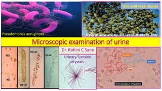
  • 1. Dr. Rohini C Sane Microscopic examination of urine Urinary Tyrosine crystals Uric acid renal stones Pseudomonas aeruginosa
  • 2.
  • 3. Standard Routine urine analysis ❖The Standard Routine urine analysis include : 1. Physical examination 2. Chemical examination 3. Microscopic examination of urine *
  • 4. Urine sample for the microscopic examination of urine Freshly voided ,midstream ,morning urine sample
  • 5. Precautions for the microscopic examination of the urine sample 1. Urine must be examined with the fresh sample . 24 hour specimen should be preserved in a refrigerator or a preservative should be added. 2. A thin preparation are made under the coverslip and should be without any air bubbles. 3. The condenser should be kept quite low and light cut down considerably while examining with the low power in particular.( If not, certain structures will be missed ,particularly hyaline casts). 4. Always examine using low power lens and then high power for finer details . Observe at least 10-15 different fields. 5. Before centrifugation see that the urine in the container is well mixed or else the cellular elements and other elements may have settled to the bottom leaving the supernatant urine clear.
  • 6. Procedure for the microscopic examination of urine Centrifuge≈10mlurineinacentrifugetubeatmoderatespeed(1500r.p.m*)for5minutes. Decant the supernatant. Tap the bottom of centrifuge tube to loosen the deposit. Place one small drop on a clean glass slide. Apply a coverslip ,avoiding formation of air bubbles . Examinethefieldwiththe⅔inobjective(lowpower)toobtainageneralimpressionofthe deposit,thenusethe¼inobjective(highpower)toidentifyalltheconstituents. Illumination of the field should not be too bright ,as casts and other structures may not be seen. *This speed is sufficient to bring to bottom of centrifuge tube ,tube cast , pus cells ,RBC , and crystals . However sedimentation of bacteria require centrifugation of urine at high speed (3000 R.P.M.) ↓ ↓ ↓ ↓ ↓ ↓
  • 7. Procedure for the microscopic examination of urine Centrifuge ≈ 10ml urine inacentrifugetubeatmoderatespeed(1500r.p.m*)for5minutes . Decant the supernatant. Tap the bottom of centrifuge tube to loosen the deposit. Place one small drop on a clean slide. Apply a coverslip ,avoiding formation of air bubbles . Examine the field with the ⅔ in objective (lowpower)to obtain a general impression of the deposit , then use the ¼ in objective (highpower)to identify all the constituents. Illuminationofthefieldshouldnotbetoobright,ascastsandotherstructuresmaynotbeseen.
  • 8. Clinical importance of Microscopic examination of urine:1 • Microscopic examination of the centrifuged urinary sediments is done to detect : 1. Cells : RBC , WBC/ Pus cells , bacteria, yeast cells ,parasites. 2. Crystals : e.g. Calcium oxalate ,Calcium phosphate, Uric acid , Amorphous phosphate , Cystine. 3. Cast : e.g. hyaline casts, granular casts , waxy cast, and blood cast. Examination of the urine for protein ,cells and casts give idea of an active lesions in the kidney. Presence of renal crystals /stones in urine are due to presence of specific type of renal calculi and renal disease. Microscopic examination of urinary sediment is completed by search for bacteria.
  • 9. Clinical Importance of microscopic examination of urine:2 ❖microscopic examination of urine provides information not given by chemical tests viz: 1. Evidence of kidney damage → shown by casts. 2. Differentiate between Hematuria and Hemoglobinuria by the presence of RBC. 3. Detection of bacteria, parasites and yeast. 4. Presence of crystals . 5. Detection of Pus cells → evidence of infection.
  • 10. Amorphous material present in normal urine Amorphous material present in normal urine : 1. Amorphous urates of Sodium ,Potassium or Calcium 2. Amorphous phosphates of Calcium and Magnesium
  • 11. Broad classification of urinary sediments :1 Unorganized sediment Organized sediment Crystalsfoundinacidurine Crystalsfoundinalkalineurine Renal Tube casts Uric acid and urates Triple phosphates(Ammonium Magnesium phosphate) Hyaline casts Calcium oxalate Disodium phosphate Granular casts Cystine Calcium carbonate Epithelial casts Leucine and Tyrosine Ammonium biurate Blood casts Sulfa Pus cell casts Cholesterol Fatty casts Hippuric acid Waxy casts Calcium sulphate Pus cells Red blood cells Bacteria , parasites and yeast
  • 12. Broad classification of urinary sediments :2
  • 13. Renal calculi and renal conditions • Renal stones are formed in urinary tract(kidney and bladder) due to an imbalance between conservation of water and excretion of insoluble substances by the kidneys. Crystal formation occurs when the urine is supersaturated with substances of low solubility . • They originate in renal papillae or in collecting tubules . • They then pass into the pelvis of kidneys and may increase in size so that they may not pass through ureters . Renal calculi may obstruct flow of urine(post - renal condition). • Other may pass to the bladder later may increase in size and obstruct the urethra.
  • 15. Unorganized deposits in urine • A knowledge of the reaction of the urine sample is of great assistance in identification of deposits. • Commonest crystalline deposits in acidic urine sample : Calcium oxalate, Sodium urates or Uric acid. • Commonest crystalline deposits in alkaline urine sample : Phosphates ,Calcium carbonate, Ammonium urate.
  • 16. Clinical significance of Urinary Crystals ❖Many crystals are found in urine have little clinical significance although they may be found in calculus formation ,metabolic disorders and in the regulation of medication .
  • 17. Characteristic of Urinary calculi Criteria Characteristic of Urinary calculi Composition of substances normally excreted in urine and found in part of the urinary tract . Size may vary from a pin head size to the size of an egg . Classification 1. Simple calculi : contain a single urinary constituent. 2. Mixed calculi : contain two or more substances present in urine. 3. Foreign body calculi : may be present due to introduction of some substances from outside. Substances found in calculi Uric acid, Urates, Triple phosphates ,Calcium carbonate, Cholesterol (Cystine extremely rare).
  • 18. Predisposing factors for renal stones formation ❖Predisposing factors for renal stones formation include : 1. High concentration of one of more normal constituents of the glomerular filtrate . 2. Alteration in urinary pH due to bacterial infection. 3. Obstruction to urinary flow. 4. Hyperparathyroidism and hypervitaminosis D. 5. Absence of inhibitors of crystal growth.
  • 19. Bacterial infection as a predisposing factor for renal calculi formation ❖Bacterial infection: • Imparts alkalinity to infected urine due to urease producing organism. • Formation of ammonium carbonate due to action of urease on urea. • Facilitate the formation of Triple phosphate and Calcium carbonate.
  • 20. Obstruction to urinary flow as a predisposing factor for renal calculi formation ❖Obstruction to urinary flow(at bladder neck): • May be due to prostatic hypertrophy. • Leads to stasis of urine. • Results in deposition of substances such as Calcium oxalate ,Uric acid which may be present in the urine in supersaturated state.
  • 21. Hyperthyroidism and hypervitaminosis D as a predisposing factor for renal calculi formation ❖Hyperthyroidism and hypervitaminosis D: ▪ leads to excessive mobilization and calcium excretion. ▪ results in saturation of calcium ions and hence renal stone formation.
  • 22. Formation of urinary calculi Formation of nucleus of stone can be obtained by presence of small lesion. Deposition of Crystals on the stone nucleus. Continuous growth of crystals of calculi. Adherence of calculi to the renal papillae.
  • 23. Consequences of renal calculi formation The presence of stone in renal pelvis ,ureter or bladder often interferes with normal flow of urine.
  • 24. Crystals found in acidic urine
  • 25. Substances present in Renal stones ❖The substances present in renal stones urine include : 1. Uric acid (5 % of total) 2. Calcium oxalate 3. Calcium phosphate (80% of total) 4. Struvite / MgNH4PO4 (15 % of total) 5. Amorphous urates 6. Sodium urates 7. Calcium sulphate 8. Cystine (1 % of total) 9. Tyrosine 10. Leucine 11. Cholesterol 12. Hippuric acid
  • 26. Urinary Uric acid ,Calcium oxalate , Hippuric acid and Ammonium Biurate crystals
  • 27. Cystine ,Calcium ,Uric acid and Struvite renal stones Presence of renal crystals /stones in urine are due to presence of specific type of renal calculi. Examination of urine for protein ,cells and casts give idea of an active lesions in the kidney.
  • 28. Uric acid renal crystals ❖Uric acid renal crystals : • are not found when urine is freshly passed but tend to develop when urine has been standing for sometime. . • occur in acidic urine (presence of uric acid can be a normal occurrence). • vary in shape(characteristic diamond rhombic or rosette form) and stained with urinary pigment as yellow or red brown. • not dissolved by heat ,acetic acid or by HCl. • soluble when heated with sodium hydroxide. ➢Excessive deposits of uric acid and urates in fresh urine are seen in concentrated urine(e.g. Fever/acute febrile conditions, diarrhea) , disturbances of uric acid metabolism(e.g. Gout), chronic nephritis, inflammatory bowel disease and myeloproliferative disorders. ➢Amorphous urates are small ,yellowish-brown ,radio- translucent granules and more common in men . They dissolve by heat and by sodium hydroxide not by acetic acid.
  • 29. Management of Uric acid renal stones ❖Management of Uric acid renal stones include: 1. Increasing fluid intake 2. Alkalization of urine 3. Administration of Allopurinol 4. Restriction of dietary purine intake 5. Lithotripsy Uric acid renal stones
  • 31. Calcium oxalate renal crystals ❖Calcium oxalate renal crystals are: 1. very hard, colorless. 2. have the typical envelope shape and may also appear like biconcave disk which have dump-bells shape(when viewed from outside) or octahedral or oval spheres . They have tendency to clump together to form stones(fusiform) . 3. occur frequently in neutral or slightly alkaline urine(occasionally in in alkaline urine). 4. soluble in strong hydrochloric acid and insoluble in acetic acid . 5. commonly found when diet consists of food like tomatoes , spinach, garlic, oranges asparagus and vitamin C. 6. found in the ureter. 7. Caused by pathological conditions( large number in freshly voided urine ) : a) Hypercalciuria with or without hypercalcemia (possibility of oxalate calculi). b) Hyperuricosuria due to stimulation of Calcium oxalate crystals by uric acid. c) Hyperoxaluria , which favors formation of Calcium oxalate crystals even when calcium excretion is normal. d) Diabetes mellites e) Liver diseases /chronic liver diseases
  • 32. Calcium oxalate renal crystals Envelope shape Dump-bells shape
  • 33. Management of Calcium oxalate renal stones ❖ Management of Calcium renal stones involves reducing urinary Calcium by: 1. Decreasing Calcium and oxalate intake. 2. Decreasing the intestinal absorption of calcium by administration of oral phosphates. 3. Management of hypercalcemia or urinary tract infection. 4. administration of thiazide diuretics which reduce urinary calcium excretion.
  • 34. Urinary Amorphous urates ❖Urinary Amorphous urates: • are urates of Sodium ,Potassium, Magnesium and Calcium. • occur as non-crystalline and amorphous. • appear yellow-red granular. • are soluble in alkali at 60ᴼC. • have no clinical significance. Thick sediment of amorphous urates
  • 35. Urinary Sodium urates ❖Urinary Sodium urates : ▪present as amorphous or as crystals(needles occurring in clusters). ▪are colorless or yellowish needles. ▪soluble in alkali at 60ᴼC. ▪have no clinical significance. Urinary Sodium urates
  • 36. Urinary Calcium sulphate crystals ❖Urinary Calcium sulphate crystals : ▪are long ,thin and colorless needles or prism. ▪soluble in acetic acid. ▪occur rarely in the urine. ▪no clinical significance.
  • 37. Urinary Hippuric acid crystals ❖Urinary Hippuric acid crystals : • occur in the form of elongated prisms or plates. • are yellow or brown or colorless. • soluble in water. • rarely seen in urine. • have no clinical significance. Urinary Hippuric acid crystals
  • 38. Urinary Cystine crystals ❖Urinary Cystine crystals: 1. are very rarely seen in urine . 2. highly refractile hexagonal plates with equal or unequal sides. 3. soluble in hydrochloric acid and ammonia but insoluble in acetic acid. 4. more soluble in alkaline urine. 5. can form calculi. 6. occur in patients with either congenital cystinosis or congenital cystinuria. 7. may form stones which are found in homocystinuria.
  • 39. Management of Cystine renal stones ❖Management of Cystine renal stones involves : 1. increasing fluid intake. 2. increasing urine volume. 3. alkalization of urine. 4. administration of penicillamine that binds cystine.
  • 40. Urinary Tyrosine and Leucine crystals 1. occur together. 2. are found in those conditions associated with severe liver damage. Consequently one would hesitate to identify crystals as Tyrosine or Leucine in absence of bile pigments. Urinary Tyrosine crystals Urinary Leucine crystals Fineneedlesarrangedassheavesorclusters withamarkedconstrictionatthemiddle. maybemergedtogetherascluster. Appearblackorrefractile. Slightlyyelloworbrown,oilylookingspheres.Manyof themwithradialandconstrictionatthemiddle. Solubleinammonia(ammoniumhydroxide), alcoholandhydrochloricacid.Insolubleinacetic acid. Solubleinalcohol.Insolubleinhydrochloricacidorether. Seen in severe liver disease , Tyrosinosis. Severehepatitis,Maplesyrupurine,acuteyellowatrophy
  • 41. Urinary Tyrosine and Leucine crystals Urinary Tyrosine crystals Urinary Leucine crystals
  • 42. Morner’s test for detection of Urinary Tyrosine crystals Morner’s reagent: Formalin : 1 ml Distilled water : 45 ml Concentrated H2 SO4 : 55ml Morner’s test for detection of tyrosine crystals: A Small quantity of crystals in a test tube + 1ml of Morner’s reagent → boil → green colour indicates presence of Tyrosine. Urinary tyrosine crystals
  • 43. Urinary Cholesterol crystals ❖Urinary Cholesterol crystals : ✓ Appearance : large , flat and in the form of transparent plates with notched corners. ✓Solubility : soluble in ether, chloroform and hot alcohol. ✓Pathological conditions: Nephritis , Nephrotic syndrome, Chyluria and excessive tissue breakdown.
  • 45. Urinary Uric acid ,Tyrosine , Leucine and cholesterol crystals Tyrosine crystals : soluble in acid /alcohol ,needle shaped. Seen in severe liver disease and Tyrosinosis . Leucine crystals : yellow spherical ,insoluble in acids ,soluble in alcohol. Seen in Severehepatitis,Maplesyrupurine andAcuteyellowatrophy .
  • 46. Urinary Sulpha crystal 1. found in urine when the patient is undergoing treatment with one of the sulphonamides group of drugs. 2. vary in shape. 3. Whenever unfamiliar crystals are seen ,particularly in acid urine. Sulpha crystals should be thought of. 4. Urine tested chemically for sulphonamides compounds/the patient questioned as to whether he/she has taken these drugs. ➢ Reference : Grathwohl's book of clinical laboratory methods and diagnosis. Urinary Sulpha crystal
  • 47. Biochemical Tests for detection of urinary Sulpha drugs Biochemical Test with Ehrlich’s reagent Observation Inference Dilute urine until it appears colorless +few drops of Ehrlich’s reagent. Picric acid yellow color Sulphonamides drug present. Lignin Test Observation Inference Wet the news paper /Whatman's paper with the urine + few drops of 5% diluted hydrochloric acid. Yellow color produced Sulpha drugs present. The arylamine group of sulphonamides drug reacts with crude cellulose(in the presence of acid).
  • 48. Interpretative microscopic urinalysis(crystals found in acidic urine ):1 Urinarymicroscopic Findings Findings in Normal urine Findings in Abnormal urine Pathology /etiology Cystine Absent Present Congenital cystinosis Congenital cystinuria Cholesterol Absent Present Lipoid nephrosis Nephritic conditions Leucine Absent Present Acute yellow atrophy Tyrosine Absent Present Destructive disease of liver Bilirubin Absent Present Bilirubinuria Sulphonamides Absent Present May cause kidney damage by blocking the tubules. Crystals found in acidic urine are suggestive of abnormal metabolism. (There are no non- pathogenic causes for these urinary crystals).
  • 49. Interpretative microscopic urinalysis(acidic crystals found in urine):2 Urinary microscopic Findings Findings in Normal urine Findings in Abnormal urine Pathology /etiology Non-pathologic Acidic crystals Uric acid Calcium sulphate (When) present in fresh urine in high proportion Presence of renal calculi -
  • 50. Types of renal stones
  • 51. Crystals found in alkaline urine
  • 52. Triple phosphates/ Struvite / Ammonium Magnesium phosphate (NH4MgPO4 ) renal crystals 1. Colorless. 2. appear prisms with three to six sides or coffin-lid crystals or as feathery or leaf like forms frequently with opaque ends. 3. dissolve /soluble in acetic acid. 4. Phosphates may occur as an amorphous deposit in alkaline urine. 5. frequently found in normal urine but can form crystals. 6. may indicate stones in kidney or bladder when they appear in freshly voided urine in large number. 7. are common in women and result from renal infection with a Proteus vulgaris (urease producing organism) . 8. enlarge ,assume a ‘staghorn’ appearance, fill the renal pelvis thereby causing obstruction. 9. can be managed by antibiotics that suppress infection and prevent recurrence .
  • 53. Triple phosphates/ Struvite / Ammonium Magnesium phosphate (NH4MgPO4 ) renal crystals Triple phosphates
  • 54. Urinary Amorphous phosphates ❖Urinary Amorphous phosphates: •are present in amorphous granular form. •spherical , dumb-bell shaped or granular type. •are soluble in acetic acid . Amorphous urates which appear similar to these phosphates are insoluble in acetic acid. •have no clinical significance.
  • 56. Disodium phosphate renal crystals 1. may appear in slightly alkaline or neutral urine. 2. colorless prisms arranged in stars and rosettes. Because of the shape of the crystals ,these are sometimes called “stellar phosphates ”. 3. The individual prisms are slender ,with are usually slender(thin) with one levelled wedge-like end , sometimes needle-like irregular or usually granular colorless plates. 4. soluble in acetic acid. 5. soft ,white ‘staghorn’ calculi in the renal pelvis that precipitate under alkaline conditions. 6. seen during chronic renal infection with a Proteus vulgaris(urease producing organism).
  • 57. Calcium carbonate renal crystal ❖Calcium carbonate renal crystal: ▪are sometimes mixed with the phosphate deposits. ▪occur usually as amorphous granules or more rarely as colorless spheres and dumb-bells. ▪are soluble in acetic acid with gas formation. ▪have no clinical significance.
  • 59. Calcium phosphate renal crystals ❖Calcium phosphate renal crystals : • are long, thin and colourless. • appear like prism with pointed end arranged as rosettes or stars or appearing as needles. • may also appear as irregular ,granular plates . • soluble in dilute acetic acid . • may be present in normal urine. • may also form renal calculi.
  • 61. Urinary Ammonium Biurate ❖Urinary Ammonium Biurate : • are generally found along with phosphates in decomposing urine when free ammonia is present. • form opaque ,yellow crystals usually in the form of spheres which are covered with or without long, fine or coarse(irregular) spicules. • soluble in acetic acid and rhombic plates of uric acid appear. • presence suggestive of pathological conditions, if found in a fresh urine.
  • 63. Interpretative microscopic urinalysis(acidic ,neutral or slightly alkaline crystals found in urine ) Urinary microscopic Findings Findings in Normal urine Findings in Abnormal urine Pathology /etiology Non-pathologic Acidic, neutral or slightly alkaline crystals Calcium oxalate Hippuric acid (When) present in fresh urine in high proportion Not significant May indicate presence of renal calculi. They are derived from various drugs a food items (spinach , vitamin C, berries, tomatoes).
  • 64. Interpretative microscopic urinalysis(alkaline, neutral or slightly acidic crystals found in urine) Urinary microscopic Findings Findings in Normal urine Findings in Abnormal urine Pathology /etiology Non-pathologic Alkaline, neutral or slightly acidic crystals Triple phosphates (Ammonium magnesium phosphate ) Present in high concentration May indicate presence of renal calculi. -
  • 65. Interpretative microscopic urinalysis(alkaline crystals found in urine) Urinary microscopic Findings Findings in Normal urine Findings in Abnormal urine Pathology /etiology Non-pathologic Alkaline crystals Calcium carbonate, Ammonium biurate, Calcium phosphate Not significant - -
  • 66. Chemical examination of urinary calculi for diagnostic purpose:1 Type Procedure Results Uric acid and Urates Make the powder of the stone by using mortar and pestle. Pinch of the powder in test tube . Add 1 drop of 20g/dL Na2CO3. Add 2 drops of phosphotungstic acid reagent . Formation of deep blue color.
  • 67. Chemical examination of urinary calculi for diagnostic purpose:2 Type Procedure Results Phosphates Make the powder of the stone by using mortar and pestle. Add 0.5 ml of Ammonium Molybdate reagent. Warm over a gas flame. Formation of yellow precipitate. Oxalates Makethepowderofthestonebyusing mortarandpestle. Add2-3dropsof10%HCl. CoolandaddpinchofMnO2.Donotmix. Formation of gas bubbles from bottom.
  • 68. Chemical examination of urinary calculi for diagnostic purpose:3 Type Procedure Results Sulphonamides Make the powder of the stone by using mortar and pestle. Add 2 drops of 10% HCl. After one minute ,add 2 drops of NaNO2. Wait for a minute. Add 2 drops of 0.5g/dl Ammonium sulfamate. Add 2 drops of reagent ‘S’(100mg/dl N(1- naphthylethylene diamine dihydrochloride in Distilled Water). Formation of Brownish pink to magenta color.
  • 69. Chemical examination of urinary calculi for diagnostic purpose:4 Type Procedure Results Cystine Make the powder of the stone by using mortar and pestle. Add 1 drop of Ammonium hydride reagent + one drop of NaCN reagent, wait for 5 minutes. Add 2drops of Sodium nitroprusside solution. Beet red color changes to orange on standing.
  • 70. Chemical examination of urinary calculi for diagnostic purpose:5 Type Procedure Results Carbonates Make the powder of the stone by using mortar and pestle. Take 0.5g in a test tube. Add 1ml of 10% HCl. Centrifugeandseparateacid extract.Usethisacidextract forfollowingtests. Formation of foaming effervescence. Calcium 0.2ml of acid extract + 2-3drops of 20g/dl NaOH. Fine white precipitate of Calcium oxalate . Dense whiteprecipitateofCalcium phosphate.
  • 71. Chemical examination of urinary calculi for diagnostic purpose:6 Type Procedure Results Magnesium 0.2ml of acid extract . Add 2 drops of 20 g/dl NaOH. Add 2 drops of reagent “M” (1 mg/dl p-nitrobenzene azo-resorcinol in 1N Sodium hydroxide). Formation of blue precipitate. Ammonium group 0.2ml of acid extract . Add 2 drops of 20 g/dl NaOH. Add 2-3 drops of Nessler’s reagent. Yellow –orange precipitate.
  • 72. Mucus in urine • Mucus in urine : • is derived from mucous glands the urinary tract. • appears as long translucent shreds . • presence of small amounts Mucous is considered normal .
  • 74. Broad classification of the urinary sediments
  • 75. Urinary Organized sediments • Organized sediments are more important sediments. • The principal organized structure in urinary sediments are : 1. Tube casts 2. Epithelial cells 3. White blood cells (pus cells) 4. Red cell cells 5. Spermatozoa 6. Bacteria 7. Yeast cells 8. Animal parasites
  • 76. Formation of Urinary casts ▪Site of formation of urinary casts: in the lumen of distal and collecting tubules of the kidney. This because the formation of casts require acidic conditions and high solute concentration. ▪Tamm- Horsfall protein :(a mucoprotein) secreted by renal tubular cells forms basic matrix of all casts. ▪Urinary casts can form as the result of : a. Precipitation of gelatin of Tamm- Horsfall mucoprotein. b. Clumping of cells on other material within protein matrix. c. The adherence of cells or cellular material to the matrix. d. Coagulation of material within the lumen.
  • 77. Properties of Casts of Renal tubules • Casts of Renal tubules : absent in a normal urine. • Renal dysfunction : casts present in urine(associated with albuminuria). • Appearance of casts : a. Sides: Parallel b. End : may be rounded or blunted c. May be convoluted ,straight or curved or broken off. • Solubility of renal casts: dissolve in alkaline urine.
  • 78. Renal Tube casts ❖Renal Tube casts : • Formation : are formed in renal tubules by the coagulation of albuminous material and are washed out by glomerular secretion into collecting tubules and finally in the bladder . • Shape : cylindrical with round or broken ends. • Length : varying • Diameter : not varying (as formed in tubules of kidney). • Diagnostic tests: to be performed in cases of renal albuminuria . • Clinical significance :associated with some pathological change in kidney (slight or transitory), temporary irritation and congestions (e.g. heart failure).
  • 79. Types of renal casts Hyaline Renal casts Cellular Renal casts Granular Renal casts ▪ Colorless/semi- transparent. ▪ Homogenous → consists of coagulated protein material ▪ Soluble in acetic acid. ▪ Few found in a normal urine. ▪ Occur in urine in large numbers in some diseases of kidneys(suggestive of mildest type of renal disease). ▪ Quantitative measurements by Addis count. ▪ To be examined by lowering the condenser and cutting light. ▪ Composedof epithelialorred bloodcellsorpuscellspartially orwholly. ▪ Epithelialcasts: coagulated proteininwhichare embeddedepithelialcells fromrenaltubulesand indicatekidneydisease. ▪ Red Bloodcasts:redblood cellsembeddedincoagulated proteinintubuleandindicate acuteglomerularnephritis. ▪ Puscellcasts:puscell embeddedincoagulated protein&indicaterenal infection. ▪ ContainlargeGranulesandare merelycoagulatedproteinsin whichnumerousgranulesare embeddedordueto aggregationordirect aggregationofserumproteinin Tamm-Horsefallmucoprotein. ▪ Granulesaredueto disintegration/degenerationof whitebloodcellsorepithelial cellsoftherenaltubules (Degeneratedcellularcasts). ▪ Notfoundinanormalurine. Indicateseverekidneydisease.
  • 80. Types of renal casts Hyaline Renal casts Cellular Renal casts Granular Renal casts ▪ Transparent , colorless ▪ Homogenous ▪ Composed of pus, epithelial or red blood cells partially or wholly. ▪ Granular in appearance. ▪ Degenerated cellular casts. Redbloodcellrenalcast White blood cell renal cast Granular Renal cast Hyaline renal cast
  • 82. Interpretative microscopic urinalysis(urinary casts ):1 Urinarymicroscopic finding Findings in Normal urine Findings in Abnormal urine Pathology /etiology Non- pathologic Hyaline casts Absent Present Glomerular damage , renal inflammation, renal infection , suggestive of mildest kind of renal disease Occasional– hyalinecastsmay bepresentdue tophysical exercise andor physiologic dehydration Redcellcasts (containonlyafew RBCinprotein matrixormanyRBC packedtogetherwithno visiblematrix) Absent Present Glomerular damage, Acute glomerulonephritis, Subacute bacterial endocarditis, renal infarction , severe pyelonephritis -
  • 83. Interpretative microscopic urinalysis(urinary casts ):2 Urinary microscopicfinding Findingsin Normal urine Findingsin Abnormalurine Pathology/etiology Non- pathologic White cell casts (WBC in cast are polymorphonucl ear neutrophils, packed together) Absent Present Renal infection and non- infectious inflammation, acute pyelonephritis, intestinal nephritis , glomerular disease Granular casts may be present after strenuous exercise. Granular casts Absent Present Significant renal disease - Epithelial cell casts Absent Present Tubular degeneration, necrosis, nephrotoxic agents or viruses, chronic renal disease -
  • 84. Interpretative microscopic urinalysis(urinary casts ):2 Urinary microscopic finding Findings in Normal urine Findingsin Abnormal urine Pathology/etiology Non- pathologic Waxycasts Absent Present Degeneratedwaxycasts, severechronicrenalfailure, malignanthypertension, tubularinflammation - Fattycasts Absent Present Fattydegenerationofthe tubularepithelium,nephrotic syndrome,chronic glomerulonephritis,toxicrenal poisoning -
  • 85. Urinary Fatty and waxy casts Fatty casts Waxy casts ▪ are formed by incorporated free oil droplets or oval fat bodies. ▪ Fatty casts can be stained by adding a few drops of Sudan III solution (take red stain). ▪ Not found in a normal urine. ▪ derived from degenerating tubular epithelial cells . ▪ suggestive of Nephrotic syndrome, Chronic glomerulonephritis and Toxic renal poisoning. • resemble hyaline casts but opaque with a fully waxy homogenous appearance. • have very high refractive index. • yellow or colorless. • result from the degeneration of granular cast. • found in terminal stages of Nephritis.
  • 86. Urinary Fatty and waxy casts Fatty casts Waxy casts Suggestive of nephrotic syndrome Found in terminal stages of nephritis
  • 87. Types of renal casts
  • 88. Urinary Mucous threads ❖Mucous threads : • are long ,thin waxy threads of ribbon like structures. • may be present in the normal urine. • Found in high proportion in the presence of inflammation or irritation of urinary tract.
  • 89. Renal Cylindroids ❖Renal Cylindroids: ▪resemble casts. ▪have one end which tapers out like a strand of mucous. ▪frequently hyaline. ➢Exact site and mechanism of their formation are under further study.
  • 90. Hyaline and granular Cylindroids
  • 92. Microscopic examination of urinary cells:1
  • 93. Microscopic examination of urinary cells:2 Urinary Epithelial cells Urinary Red blood cells(RBC) Leucocytes(white blood cells/pus cells) ▪ Originate from any site in genito-urinary tract from proximal convoluted tubule to urethra or from vagina. ▪ Normally (3-5 per h.p.f.) due to sloughing off old cells. ▪ Squamousarepresentinmany normalurinespecimens, especially innon-catheter samplesfromfemalepatients. ▪ Not present in the urine of normal males. ▪ In samples from female patients ,they may be of menstrual origin. ▪ Red cells may be normal, crenated or swollen , depending on whether the urine is isotonic, hypertonic or hypotonic. ▪ Enter urine anywhere from the glomerulus to urethra. ▪ Are found occasionally, generally not more than 2-3 per h.p.f./field using 4mm objective. ▪ Depending on the toxicity of the urine , they may normal, swollen or shrunken in size. ▪ whendegenerate,theyare sometimescalledpuscells.
  • 94. Microscopic examination of urinary cells:3 Urinary Epithelial cells Urinary Red blood cells(RBC) Urinary Leucocytes(WBC) . ↗ ↑ ← ↗
  • 95. Microscopic examination of urinary cells:4
  • 96. Interpretative microscopic urinalysis(urinary epithelial cells) Urinarymicroscopic finding Findingsin Normalurine Findingsin Abnormalurine Pathology/etiology Non-pathologic Epithelialcells Male: 2to3perHPF Female: 3to5perHPF >5epithelialcells perHPF 3Typesofepithelialcells: a) Tubular b) transitional c)squamous Increasednumberoftubular epithelialcellssuggesttubular damage.Itcanoccurin pyelonephritis,Acutetubular necrosis,Salicylatesintoxicationand kidneytransplantrejection. Transitionalepithelial cells:linethe urinarytractfrompelvisofthekidney toupperportionoftheurethra. Squamousepithelialcells:occur principallyinurethraandvaginamay bepresentinas urinarycontaminant fromvulvaorvaginainfemale. Few epithelial cellsfromthese sitescanbefound inurineasaresult of thenormal sloughingoff of oldcells. UrinaryEpithelial cellsoriginatefrom anysitein genitourinarytract fromproximal convolutedtubule tourethra orfrom vagina.
  • 97. Types of urinary epithelial cells RenalTubularepithelial cells Transitionalepithelial cells Squamousepithelial cells ▪ Slightly larger than leucocytes. ▪ Cuboidal ,flat or columnar. ▪ contain large round nucleus. ▪ Slightly larger than leucocytes. ▪ Pear or round shaped . ▪ contain large nucleus . Occasionally contain two nuclei. ▪ Large ,flat and irregularly shaped. ▪ containsmallcentral nucleusandabundant cytoplasm. Squamousepithelial cell RenalTubularepithelialcells
  • 98. Morphology of urinary epithelial cells
  • 99. Origin of Epithelial cells as the organized renal sediments ▪Normal urine : few epithelial cells. ▪Abnormal/pathological urine: a large number which indicate destruction of tissue in urinary tract. ▪Sources of urinary epithelial cells : RenalTubular epithelial cells Transitional epithelial cells Squamous epithelial cells Kidney tubules Kidney pelvis ,urethra or bladder Urethra or vagina
  • 100. Interpretative microscopic urinalysis(urinary pus cells) Urinary microscopic finding Findingsin Normal urine Findingsin Abnormalurine Pathology/etiology Non- pathologic Puscells (leucocytes) 2to3per HPF >5puscellsper HPF Pyuria(pusinurine):urinary tractinfection,inflammatory processinoradjacent in urinarytract. Non-infectiousconditions: acuteglomerulonephritis,renal tubularacidosis,dehydration, stress,fever,non-infectious irritationtotheureter,bladder orurethra. Few leucocytes cannormally befoundin secretionsfrom maleorfemale genitaltracts. Thereis possibilityof contamination andshouldbe considered.
  • 101. Pus cells as the organized renal sediments ▪Normal urine : few pus cells (2 to 3 WBC per high power field) ➢When pus cells are degenerating they tend to occur in clumps. ➢ When pus cells present in large number ,the urine will most certainly contain albumin . ➢Abnormal/pathological urine: • Pyuria(pusinurine):urinary tractinfection,inflammatoryprocessinor adjacent inurinary tract and in female, contamination with vaginal secretions. • Non-infectiousconditions: Acuteglomerulonephritis, Renaltubularacidosis, Dehydration, stress,fever,non-infectiousirritationtotheureter,bladderor urethra.
  • 102. Red blood cells as the organized renal sediments • Red blood cells in the fresh urine : ✓occasional presence(of erythrocytes). ✓ have a normal pale or yellow appearance. ✓ appear smooth ,biconcave disks about 7 μm diameter and 2 μm thick. ✓Do not contain nuclei. ✓Solubility of RBC : in acetic acid • Hypotonic urine : red blood cells swell up and lyse. • Hypertonic urine :red blood cells crenate. • Abnormal/pathological urine: a large number erythrocytes which may appear normal or crenated . • When red blood cells present in large number, the urine will most certainly contain albumin . ➢Reporting of RBC in urine : number of RBC per high power field .
  • 103. Microscopic features of urinary RBC Crenated RBC Abnormal/pathological urine: a large number erythrocyte(RBC)
  • 104. Yeast cells can be mistaken for RBCs .Yeast cells are ovoid and frequently contain buds. Microscopic view of Budding yeast cells in urine Budding yeast cells in urine
  • 105. Addis count for urinary casts /cells ❖Addis Count: quantitative measure of the urine sediment. • Specimen : 12 hours urine (mix thoroughly before centrifugation). • Preservative: formalin • Centrifugation : 10 ml urine in a graduated centrifuge tube at 1800 RPM for 5 minutes. • Procedure : remove 9ml of supernatant fluid and thoroughly mixed sediment. • Count of cast : using blood counting chamber and count same as a total WBC count under low power in all 9 large squares (0.0009 ml of urine). • V = accurate volume of 12hr urine in ml • S = volume of urine centrifuged • v = volume of in ml in which count is made • n= number of casts / cells counted • N = number of cell/casts in 12hours • Calculation: N= V X S X n v x 10 Normal range: Casts : 0-5000 RBC : 0-500000 WBC : 1,000,000 Spermatozoa may be sometimes found in urine from males.
  • 106. Quantitative evaluation of the urine sediments
  • 107. Addis count for urinary casts /cells
  • 108. Interpretative microscopic urinalysis of organisms or abnormal cells Urinarymicroscopic finding Findings in Normal urine Findings in Abnormal urine Pathology /etiology Non-pathologic Bacteria Absent Absent Renal infection Afterstorageat roomtemperature bacterialgrowth maytakeplacein urine. Yeast cells Absent Present May be present in acidic urine containing sugar. - Parasites Absent Present Trichomonas vaginalis from vagina and Trichomonas hominis from rectum. -
  • 109. Urinary Organisms • Organisms are of no significance when they are found in urine samples that have been standing overnight. • Presence of bacteria and pus cells in freshly voided urine : indicative of renal infection(finding should be reported).
  • 110. Urinary Bacteria • Normal urine : when fresh does not show any bacteria , when allowed to stand for some times may show bacteria. • Presence of large number of bacteria with many pus cells in the fresh urine : suggestive of bacterial infection of kidney(abnormal urine). Contamination may occur from bacteria present in urethra, vagina or external sources. • Precaution: collect urine aseptically(cleaned-up mid-stream specimens of urine in the sterile container). Catheter specimens should be avoided because of high incidence of bladder infections after catheterization. • Examination of bacteria : direct smear • Recommended stains for smears : a. Acid /alcohol fast stain : tubercule bacillus b. Gram’s stain : gonococcus ➢Culture of urine sample : e.g. Typhoid ➢Animal inoculation: diagnosis Weil’s syndrome
  • 111. Staining of bacteria Ziehl –Nielsen's staining of acid-fast bacteria e.g. Mycobacterium Tuberculosis Gram’s staining of Neisseria gonococcus
  • 113. Salmonella Typhi cause Typhoid fever Salmonella Typhi
  • 114. Common urinary pathogens Organisms Observationinwetpreparation Gramsmear E.Coli Motileornon-motile rods Gramnegative Staphylococcus saprophyticus Cocci(afterattachedtoepithelial cells) Grampositive staphylococci Proteusspecies Motilerods Gramnegativerods Pseudomonasaeruginosa Motilerods Gramnegativerods Streptococcusfaecalis (groupDstreptococci) Cocciinchains Klebsiellastrains Nonmotilerodsorcocobacilli Gramnegative capsulatedrods Enterococci cocciinshortchainsorpairs Grampositive streptococci
  • 115. Microscopic features of Common urinary pathogens Staphylococcus saprophyticus: ▪ Gram positive Cocci in groups. ▪ causative agent for urinary tract infection. Escherichia coli: ▪ Motile or non-motile gram negative rods. ▪ causative agent for urinary tract infection.
  • 116. MicroscopicfeaturesofurinaryProteusspeciesandPseudomonasaeruginosa Pseudomonas aeruginosa: Motilerods& causativeagentfor urinarytractinfection. Proteus species: Motile rods & cause urinary tract infection.
  • 117. Microscopic features of Streptococcus faecalis and Klebsiella pneumoniae Enterococcus faecalis:Grampositivecocciinchains andcausativeagentforurinarytractinfection. Klebsiella pneumoniae:non-motilecapsulatedgram negativerods&causativeagentofurinarytract infection.
  • 118. Concentration method for Acid fast bacteria ▪ Specimen : 24hr. Specimen is collected in sterile “ Winchester bottle ”. ▪ Preservative : to be added if culture is not to be carried out . ▪ Refrigeration of urine sample needed. No preservative ,if culture is to be carried out. ▪ Procedure : a. siphon off supernatant fluid. b. Acid treatment : treat deposit with equal volume of 6% sulphuric acid for 20 minutes. c. centrifugation : 3000 RPM for 30 minutes. d. Washing of deposit : 3 times in distilled water. e. Prepare a smears and examine after acid fast staining.
  • 119. Urinary yeast cells •smooth , colorless and usually avoid cells. •vary in size and have refractile walls. •often show budding. •insoluble in acid and alkali(unlike red blood cells). •will not stain with eosin. •may be found in urinary tract infections (mainly in diabetic patients). •may be present in urine as a result of skin or vaginal contamination.
  • 120. Microscopic examination of yeast cells(Candida Albicans and Clostridium perfringens) Candida Albicans
  • 121. Normal flora of Urethra and Vagina Body site of urinary tract Normal flora Urethra ▪ Candida species ▪ Mycobacterium smegmatis ▪ Mycoplasma species ▪ Acinetobacter species Vagina (between puberty and menopause) acid pH ▪ Lactobacillus species ▪ Streptococcus species ▪ Clostridium species ▪ Corynebacterium species ▪ Gardnerella vaginalis
  • 122. Parasites observed in microscopic examination of urine Trichomonas vaginalis Enterobius vermicularis ova Schistosoma haematobium ovum
  • 123. Trichomonas vaginalis in microscopic examination of urine
  • 124. Trichomonas vaginalis causes Trichomoniasis
  • 125. Enterobius vermicularis ova in microscopic examination of urine Enterobius vermicularis
  • 126. Schistosoma haematobium ovum microscopic examination of urine Schistosoma haematobium ovum
  • 127. Oval fat bodies and fat droplets ❖Oval fat bodies: • are renal tubular cells containing highly refractile fat droplets. • may be macrophages or polynuclear leucocytes which have ingested lipids or have undergone fatty degeneration. ❖Free fat droplets: ▪Present in urine in lipuria /chyluria(presence of lipids in urine). ▪Present in Nephrotic syndrome, Diabetes mellitus , Lipoid nephrosis in chronic glomerulonephritis and fat embolism.
  • 128. Chyluria ❖Chyluria : milky appearance of urine. • Etiology : rupture of wall of the bladder or kidney pelvis leading to escape of fat droplets in the urine. • Diagnosis of chyluria : a. Staining with Sudan III. b. Adding of little ether which dissolves the fat clearing the urine. c. Microscopic examinations : presence of Microfilaria. • Clinical condition associated with chyluria : Lymphatic Filariasis.
  • 130. Specimen collection for urogenital specimens • Wear rubber gloves. • Use sterile cotton swab moistened with normal saline to cleans around ulcer . • Collect serous exudate on a coverslip and invert it on a slide. • Examine immediately on dark-field microscopy. • Examine saline preparation for suspected Trichomoniasis.
  • 131. Examination of Urogenital specimens Perform gram’s staining and observe the following organisms : Suspected gonorrhea Pus cells containing Gram negative diplococci Suspected vaginitis Large gram positive yeast cells (Candida albicans or candida species) Suspected peripheral sepsis or septic abortion Large gram positive rod cells(Clostridium perfringens) Gram positive streptococci Gram negative rods (Bacteroides species)
  • 132. Gonorrhea • Gonorrhea : a sexually transmitted blood -borne infection. • Causative Organism: Neisseria gonorrhea • Diagnosis : in men , Gram stain of urethral discharge using swabs. In women, urethral ,cervical and or anal swabs. • Microscopic examination of urethral discharge : Gram Negative diplococci. • Biochemical test: purple color after pouring weak solution of tetra-methyl para phenylenediamine over the culture plate. • Clinical manifestation : inflammation of the urethra ,cervix , fallopian tubes, rectum, prostrate and /or pharynx spread to joints and skin. Congenitally transmitted infection in eye of newborn(neonatal conjunctivitis). Infection around the liver may result from peritoneal spread of the disease. • Management : Cephalosporins or fluoroquinolones (Tetracyclines used to treat coinfection with Chlamydia).
  • 133. Neisseria gonorrhea Intracellular in polymorphonuclear cells in the exudate from the gonorrheal sore. Oxidase-positive ,confirmed by typical sugar reactions.
  • 134. Spermatozoa in urine ▪Spermatozoa : have oval bodies and thin, long and delicate tails. ▪Presence of Spermatozoa in urine of man: after nocturnal emission, epileptic convulsions and diseases of genital organ. ▪After coitus, they may be present in the urine of both the sexes.
  • 135. Spermatozoa in urine Presence of when present in large number , they should be reported as it may suggest a lesion in the Genito-urinary tract.
  • 136. Artifacts in microscopic examination of urine Starch crystals : found occasionally in urine as round or oval highly refractile crystals. Fibers : come from clothing ,toilet paper or may be piece of lint from the air. Hair Air bubbles Talcum powder particles
  • 137. Artifacts in microscopic examination of the urine
  • 138. Alterations in urine test results Test Falsepositive Falsenegative Specificgravity Contaminationdingcollectionandstorage None pH Increasedwhilestandingduetoureaseproducing microorganisms Blood Hypochloritebacterialperoxidase Ascorbicacid,nitrites Protein Fever,cells,bacteria,concentratedurine Dilutionofurine Glucose Oxidizingagents Ascorbicacid Ketonebodies Captopril,M-dopa Prolongedstanding Bilirubin Rifampicin,chlorpromazine Sunlight,Ascorbicacid Urobilinogen Alkalineurine,Sulphonamide Broadspectrumantibiotics,sunlight Leukocyte esterase Oxidizingagents,Trichomonas Ascorbicacid,Tetracyclines, cephalosporin Nitrites Ascorbicacid,Mycobacterium
  • 139. Thank You Google Images