
Recomendados
Más contenido relacionado
La actualidad más candente
La actualidad más candente (20)
Destacado
Destacado (13)
Similar a General anatomy of muscles
Similar a General anatomy of muscles (20)
Último
Último (20)
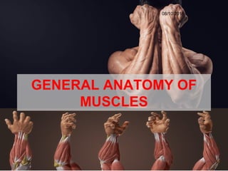
General anatomy of muscles
- 2. 1. Derivation of name Muscle (Latin Mus= mouse) are so named because many of them resemble a mouse, with their tendons representing the tail.
- 3. Definition • Muscle is a contractile tissue which brings about movements. • Muscles can be regarded as motors of the body.
- 4. • Other contractile cells: Myofibroblasts- seen in regenerating connective tissue. Myoepitheliocytes- associated with glands.
- 5. BASIC PROPERTIES • IRRITABILITY: – Sensitive to stimuli • CONTRACTILITY: – When stimulated, the contracts lengthwise leading to its shortening • EXTENSIBILITY: – Once stimuli removed, the muscle fibers return to their original length. • ELASTICITY: – Muscle assumes a desired shape.
- 6. Types of muscles There are three types of muscles : 1. Skeletal muscles 2. Smooth muscles 3. Cardiac muscles
- 7. Skeletal muscle tissue •Attached to skeleton •Cross-striated •Voluntarily controlled • Cells are cylindrical and multinucleated • They respond quickly but fatigue easily
- 8. Cardiac muscle tissue • Makes up myocardium of heart • Unconsciously (involuntarily) controlled • Microscopically appears striated • Cells are short, branching & have a single nucleus • Cells connect to each other at intercalated discs
- 9. Smooth (visceral) muscle tissue • Makes up walls of organs & blood vessels • Tissue is non-striated & involuntary • Cells are short, spindle-shaped & have a single nucleus • Tissue is extremely extensible, while still retaining ability to contract
- 10. Skeletal muscles SynonymsSynonyms 1. Striped muscles 2. Striated muscles 3. Somatic muscles 4. Voluntary muscles
- 11. Parts of a skeletal muscle A. Two ends 1. Origin is one end of the muscle which remains fixed during its contraction. 2. Insertion is the other end which moves during its contraction. In the limb muscles, origin is usually proximal to insertion.
- 12. 1. Fleshy part is contractile, and is called the ‘belly’. 2. Fibrous part is non- contractile and inelastic. When cord-like or rope-like, it is called ‘tendon’ ; when flattened it is called ‘aponeurosis’. B. Two parts
- 13. Skeletal muscle fiber (cell) Muscle Fascicle Surrounded b perimysium Surrounded by endomysium Skeletal muscle Surrounded by epimysium epimysium tendon A. CONTRACTILE TISSUE • Each muscle is composed of numerous muscle fascicles. • Each muscle fascicle has numerous muscle fibres. • Each muscle fibre is a multinucleated, cross- striated cylindrical cell (myocyte). Structure of striated muscle
- 14. MYOCYTE • Sarcolemma- membrane • Peripheral nuclei – Multinucleated • Sarcoplasm- cytoplasm • Longitudinal myofibrils
- 15. MYOFIBRIL • Each myofibril is composed of longitudinal protein filaments, called myofilaments: – Actin (thin) – Myosin (thick) • Each myofibril shows alternate dark and light bands – Dark bands : A bands (anisotropic) – Light bands: I bands (isotropic). • In the middle of the A band, there is a light H band with M line (dark) in the middle. • In the middle of the I band there is a dark Z disc or Krauses membrane.
- 16. A band I band H band Z LINEZ LINE
- 17. • The segment of myofibril between two Z discs is called sarcomere. • Sarcomere is the structural and functional unit of muscle.
- 18. B. SUPPORTING TISSUE • It helps in the organization of the muscle. • Epimysium • Perimysium • Endomysium • The connective tissue of the muscle becomes continuous with the tendon.
- 20. Slow and Fast Muscle Fibres 1. Type I (slow,red) fibres • show a slow ‘tonic’ contraction characteristic of postural muscles. • They are red in colour because of large amounts of myoglobin. • The fibres are rich in mitochondria and oxidative enzymes but poor in phosphorylases. • Because of well developed metabolism, slow fibers are highly resistant to fatigue.
- 21. 2. Type II (fast,white) fibres • show a fast ‘phasic’ contraction required for large-scale movements of body segments (non- postural muscles) • These are paler (white) in color because of small amounts of myoglobin. • These fibres are rich in glycogen and phosphorylases, but poor in mitochondria and oxidative enzymes. • Because of anaerobic glycolytic respiration, the fast fibres are quite easily fatigued.
- 22. FUNCTIONS • MOTION: – E.g. Walking, running • HEAT PRODUCTION: – Metabolism within muscle cell release heat as end product. – Rate of heat production increases when person performs strenuous exercise. • POSTURE AND BODY SUPPORT:
- 23. FASCICULAR ARCHITECTURE OF MUSCLES • The arrangement of muscle fibres varies according to –direction, –force –range of movement at joint.
- 24. CLASSIFICATION OF MUSCLE ACCORDING TO THE ARRANGEMENT OF THE FASCICULI A.Parallel Fasciculi B.Oblique Fasciculi C. Pennate Fasciculi D.Spiral or Twisted Fasciculi
- 26. When the fasciculi are parallel to the line of pull, the muscle may be: (1)quadrilateral e.g. thyrohyoid (2)straplike e.g. sternohyoid, sartorius A. Parallel Fasciculi
- 27. (3) strap-like with tendinous intersections e.g. rectus abdominis (4) fusiform e.g. biceps, digastric The range of movement in such muscle is maximum
- 28. 1. Triangular- e.g. adductor longus 2. Fan shaped e.g. temporalis B. Oblique Fasciculi
- 29. C. Pennate Fasciculi 1. Unipennate- e.g. flexor pollicis longus, extensor digitorum longus, peroneus tertius 2. Bipennate- e.g. rectus femoris, dorsal interossei, peroneus longus, flexor hallucis longus
- 30. 3. Multipennate- e.g. deltoid, subscapularis 4. Circumpennate- e.g. tibialis anterior
- 31. 1. Spiral fibres: found in trapezius, latissimus dorsi, pectoralis major, supinator etc. D. Twisted Fasciculi
- 32. 2. Cruciate fibres: In certain muscles the fasciculi are crossed. e.g. sternocleidomastoi d, masseter, adductor magnus.
- 33. NOMENCLATURE OF MUSCLES The muscles have been named in a number of ways SHAPE e.g. trapezius, rhomboideus, serratus anterior, latissimus dorsi, etc. NUMBER OF HEADS OF ORIGIN e.g., biceps, triceps, quadriceps, digastric, etc.
- 34. GROSS STRUCTURE e.g., semitendinosus, semimembranous, etc. LOCATION e.g temporalis, supraspinatus, intercostals, etc.
- 35. ATTACHMENTS e.g., stylohyoid, cricothyroid, etc. FUNCTION e.g., adductor longus, flexor carpi ulnaris, abductor pollicis longus, etc. DIRECTION OF FIBRES e.g., rectus abdominis, oblique abdominis, transversus, etc.
- 36. Lubricating Mechanisms 1. Synovial bursae: • To reduce friction between two mobile but tightly apposed surfaces. • Structurally it is a closed sac of synovial membrane with a capillary film of synovial fluid.
- 37. 2. Synovial sheaths: • Tendons, while passing under the fibrous bands, are surrounded by synovial sheaths. • Formed by invagination of the tendons into the sheath. • The two layers are separated by a capillary film of synovial fluid
- 38. Blood supply of skeletal muscle • Blood supply is derived from muscular branches from neighbouring arteries. • The arteries, veins and motor nerve pierce the muscle at a fairly constant point called neurovascular hilum. • The arteries divide repeatedly to form arterioles in the perimysium, and capillaries in the endomysium for nutritive circulation.
- 39. Nerve supply of skeletal muscle The nerve supplying a muscle is called a motor nerve. In fact it is a mixed nerve. 1. MOTOR FIBRES (60%) α efferents γ efferents Autonomic efferents Large Small Fine Myelinated Myelinated Non-myelinated Extrafusal Intrafusal Supply smooth muscles of blood vessels
- 40. 2. SENSORY FIBRES (40%) Myelinated Non-myelinated Proprioceptive Nociceptive Sense of position Sensation of pain Present in muscle, fascia, tendon, joint capsule etc Present in muscle
- 42. • MOTOR POINT: Site where motor nerve enters the muscle. It may be one or more than one. • MOTOR UNIT (MYONE): Single alpha motor neuron together with the muscle fibres supplied by it. The size of motor unit depends upon the precision the muscle control. – Small motor units (5-10 muscle fibres) are found in muscles of fine movements (extra-ocular muscles). – Large motor units (100-2000 muscle fibres) are found in muscles of gross movements (proximal limb muscles).
- 43. • NEUROMUSCULAR JUNCTIONS: • On approaching the muscle the axons of motor nerve loose their myelin sheath and break up into a number of branches to supply the individual muscle fibres. • These specialized motor nerve endings, rich in acetylcholine form junction with the muscle fibre called Neuromuscular junction.
- 44. • Nerve part: – Motor end plate – Pre synaptic membrane – Synaptic vesicle • Muscle part: – Sole plate – Synaptic Cleft – Granular sarcoplasm – Nuclei & mitochondria – Subneural clefts
- 45. Neuromuscular Spindle • Definition: – Spindle shaped sensory end organs within skeletal muscle provide sensory information to CNS to control tone of muscle. • Each spindle: – Intrafusal fibers – Extrafusal fibers
- 46. ACTION OF MUSCLES • Muscle Tone: The constant tension produced by muscles for long period of time. • Type of muscle contraction: • Isometric contraction – length of muscles do not change but tension does increases.. • Isotonic contraction – Tension produced by muscle is constant during contraction but length of muscle changes.
- 48. A. Prime movers (agonists) Bring about the desired movement. B. Antagonists (opponents) Produce movement opposite to prime mover. They help the prime movers by active controlled relaxation. C. Fixators They stabilize the origin of prime mover so it can act efficiently. D. Synergists: When the prime movers cross more than one joint, the undesired actions at the proximal joints are prevented by certain muscles known as synergists GROUP ACTION OF MUSCLES
- 49. APPLIED ANATOMY PARALYSIS Loss of motor power (power of movement) This is due to inability of muscles to contract either because of damage to motor pathways or by inherent disease of muscles (myopathy).
- 50. APPLIED ANATOMY MUSCULAR SPASM Localized muscle spasm is commonly caused by a ‘muscle pull’. Generalized muscle spasms occur in tetanus and epilepsy. DISUSE ATROPHY muscles which are not used for a long time become thin and weak. This is called disuse atrophy.
- 51. APPLIED ANATOMY OVERUSE HYPERTROPHY Excessive use of a particular muscle causes the better development of that muscle evident by increase in size and bulk. MUSCLE REGENERATION Muscles have a limited capability of regeneration.
- 52. Thank you…