SlideShare una empresa de Scribd logo
1 de 38
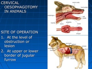
CERVICAL
 OESOPHAGOTOMY
 IN ANIMALS


SITE OF OPERATION
1. At the level of
  obstruction or
  lesion
2. At upper or lower
  border of jugular
  furrow
TOPOGRAPHIC
  ANATOMY
1. The oesophagus
  is three to three
  and or half feet
  long in medium
  sized animals and
  is comparatively
  small in dogs. It
  connects pharynx
  and the stomach.
2. The whole length of
  oesophagus is divided
  into cervical, thoracic
  and abdominal part in
  horses and dogs the
  abdominal part is absent
  in dogs.

3. The average diameter
  approximately one to
  two inches and is
  masculomembrane
  tube.
4. In cervical area it is
   almost in dorsal
   position at origin and
   passes gradually to
   left
side of the trachea at
   the level of about 4th
   cervical vertebrae.
   Thereafter it
occupies the left position
   of trachea upto 3rd
 thoracic vertebrae.
In the thoracic
region it is median in
  position and enters
  the abdominal
  cavity through
  hiatus
oesophagus and
  terminates at the
  cardia of the
  stomach.
5. As the oesophagus
  crosses to left side of
  the trachea it is
  accompanied by
  longus coli and longus
  capitis muscles
  dorsally, left carotid
  artery,
  vagosympathetic
 trunk, jugular vein and
  recurrent laryngeal
  nerve laterally.
Overlying the
oesophagus are skin,
  cervical fascia, cervical
  paniculus muscle and
  the

omohyoideus muscle,
  which crosses the
  jugular furrow obliquely
  from below
upward, forward and
  inward towards the
  median line.
6. Its wall is composed of
• fibrous sheath,

• the tunica adventitia,

• the muscular coat,

• the submucous and mucous

  coat. In cervical area the
  oesophageal wall is thicker.

7. The oesophagus is supplied
  by branches of
• carotid,

• brachio-oesophageal

• and gastric arteries.
8. The nerve supply to oesophagus is
  by
• vagus,

• glosso-pharyngeal

• and sympathetic nerves.
INDICATIONS
1. Oesophageal
  obstruction and
  wounds of
  oesophagus.
2. Stricture of
  oesophagus or
  oesophageal
  stenosis.
3. Neoplastic
  growths inside the
  oesophagus
4. Oesophageal
  diverticulum.
CONTROL AND ANAESTHESIA
1. The position of animal is right
  lateral recumbency after proper
  sedation.
2. Anaesthesia is by general
  anaesthesia in small animals or by
  local in filtration
 analgesia at the site of operation.
SURGICAL TECHNIQUE
1. At the marked site, a long
   incision is made on skin
   and subcutaneous tissue,
sufficient enough to extract
   the obstruction, if present.
2. The omohyoideus muscle
   is separated from upper
   and lower structure. The
   areolar
tissue is bluntly dissected with
   the help of fingers.
3. The trachea is recognized
   to locate the oesophagus
   on its lateral surface.
4. The oesophagus is
  drawn out and fixed in
  position by placing
  blunt instrument
under it.
5. Make an incision on
  dorsal wall of
  oesophagus either
  anterior or posterior to
obstruction. The incision
  should be large
  enough to extract the
  obstruction/foreign
body.
6. The repair of oesophageal incision can be
  done in two layers. The mucous
membrane can be sutured with mattress sutures
  or continuous sutures.
The
muscularis layer is to be sutured with connell
pattern or continuous lock stitch
pattern. Chromic catgut or silk suture is used for
suturing.
7. The oesophagus is replaced in its
  original position.
8. The skin wound is closed in routine
  manner or it is left as open wound.
POST OPERATIVE CARE
1. Do not allow solid food for few days and
  intravenous fading is done twice
daily.
2. A course of antibiotics is to be completed
  (4-5 days)
3. Antiseptic dressing of the wound should
  be carried one till healing is complete
or when sutures are removed after 8-12
  days.
IMPORTANT CONSIDERATION/ REMARKS
1. Check hemorrhage during surgery
2. If oesophagus is empty it is recognized by passing
  a stomach tube.
3. During dissection,
  prevent damage to
  recurrent laryngeal
  nerve.
4. Suturing only
  oesophagus and
  leaving the skin
  wound open is the
  procedure of
choice because
a) It favours early closure of
  oesophageal wound
 b) It prevents escape of alimentary
  matter during swallowing.
 c) It permits drainage of any
  material, if present.
TRACHETOMY AND
  TRACHEOSTOMY IN ANIMALS
 Mostly indicated in
   buffalo and cattle.
SITE OF OPERATION
At the junction of upper
   and middle third portion
   of the neck on mid
   ventral
line.
TOPOGRAPHIC ANATOMY
1. Trachea is a musculo-
   membrano-
   cartilagenous tube
   extending from the
larynx to
the hilus of
  the lungs. It
  occupies a
  median
  position in
  the ventral
  aspect of the
  neck.
2. It is composed of
  incomplete cartilaginous
  rings, which helps to
  keep the trachea
permanently open. In
  ruminants these rings
  are 45 to 60 in number.
  These rings
are enclosed and
  connected by
  fibroelastic membrane
  and constitute the
  tracheal
annular ligament.
3. The cervical part
  of the trachea is
  related dorsally to
  the longus coli
  muscle and
oesophagus and
  laterally to the
  thyroid gland, the
  carotid artery, the
  jugular vein,
the vagus, sympathetic and recurrent
  laryngeal nerves, the tracheal lymph
  duct
and cervical lymph gland.
4. The sternocephalicus muscle
  converges from below to above and
  crosses the
trachea obliquely, passing from the
  ventral surface, forward its sides and
diverging to reach the angle of jaws.
  The left-over area of trachea is
  covered only
with skin, subcutaneous tissue and areolar
  tissue between the two halves of
sternothyroideus muscles which lie on the
  ventral surface.

•   5.
•   The branches of common carotid artery
    supply the trachea

•   nerve supply is
    by vagus and sympathetic nerves.
INDICATIONS
1. Obstruction in the upper
  respiratory passage.(rattle snake
  bite, regional lymph node
  abscessation due to streptococcus,
  nasopharyngeal neoplasia, excessive
  distension of gutteral pouches etc. )
2. Paralysis of intrinsic muscles of the
  larynx
3. Fracture of tracheal ring causing
  obstruction of trachea.
CONTROL AND ANAESTHESIA
1. The animal is positioned in lateral
  recumbency with neck extended.
2. Head is kept in lower position to prevent
  aspiration of fluids.
3. The anaesthesia is local linear infiltration
  analgesia at the site of incision.
SURGICAL TECHNIQUE
1. A mid line 7-10 cm long incision is
  made through the skin and subcutaneous
tissues.

2. Separate two portions of
  sternothyroideus muscle and exposed the
  trachea after
bluntly dissecting the areolar tissue.

3. Two tracheal rings are selected,
  exposed at wound edges and fixed with
  the help
of two sharp hooks through the inter-
  annular ligament.
4. If temporary
  tracheotomy is
  desired, an incision
  is made on the inter-
  annular
ligament. just enough
  to permit the
  passage of tracheal
  tube.
5. If permanent
  tracheotomy
  (tracheostomy) is
  desired, the incision is
  made in the
tracheal ring using
  either of following
  techniques.
  a) Incise the inter-
  annular ligament and
  the tracheal ring in its
  transverse plane
going semi circularly
  leaving half portion of
  the tracheal ring
  intact.
Repeat the
  sameprocedure on
  opposite tracheal ring.
  The incised portion of
  the cartilage along
  with
inter-annular ligament
  is removed. An oval
  opening in the trachea
  will be created.
Instead of oval, square
  opening can also be
  made.
OR
 b) Make a
  longitudinal incision
  on two or three
  tracheal rings and
  with the
help of traction
  sutures, applied
  through
  cartilaginous rings
  on either side of
  incision,
the tracheal lumen is
  exposed.
6. Insert the tracheostomy
 tube through these
 openings into the tracheal
 lumen and
then keep in position by
 suturing it to trachea
7. The remainder
  tracheostomy
  incision is sutured,
  applying
  continuous suture
pattern using silk
  thread.
8. The skin wound is
  closed in routine
  manner.
POST OPERATIVE CARE
1. The tube is cleaned daily for first few days.
2. The opening of tracheostomy tube should be
  covered with gauze to prevent
entrance of any foreign material.
3. The course of antibiotics for 5 days must be
  completed.
4. Daily/alternate day antiseptic dressing of wound
  till complete healing when
sutures are removed (normally 8-12 days after
  operation).

Más contenido relacionado

La actualidad más candente

Caesarean section in cow
Caesarean section in cowCaesarean section in cow
Caesarean section in cowRekha Pathak
 
Ovariohysterectomy in bitch
Ovariohysterectomy in bitchOvariohysterectomy in bitch
Ovariohysterectomy in bitchDr.Jigdrel Dorji
 
Affection of horn
Affection of hornAffection of horn
Affection of hornBikas Puri
 
Hernias and its classification in small animals
Hernias  and its classification in small animalsHernias  and its classification in small animals
Hernias and its classification in small animalsskuast jammu
 
Vaginal & uterine prolapse in cattle
Vaginal & uterine prolapse in cattleVaginal & uterine prolapse in cattle
Vaginal & uterine prolapse in cattleIVRI
 
Perineal hernia in dogs
Perineal hernia in dogs Perineal hernia in dogs
Perineal hernia in dogs girjesh upmanyu
 
Surgical procedures in the bovine
Surgical procedures in the bovineSurgical procedures in the bovine
Surgical procedures in the bovinelizzette mudindo
 
Fetotomy in bovines by Dr Dushyant Yadav BASU, Patna INDIA
Fetotomy  in bovines by  Dr Dushyant Yadav BASU, Patna INDIAFetotomy  in bovines by  Dr Dushyant Yadav BASU, Patna INDIA
Fetotomy in bovines by Dr Dushyant Yadav BASU, Patna INDIADrDushyant Yadav
 
Caessarean section in bovines
Caessarean section in bovinesCaessarean section in bovines
Caessarean section in bovinesIVRI
 
Internal fixation techniques
Internal fixation techniquesInternal fixation techniques
Internal fixation techniquesIVRI
 
Affection of guttral pouch
Affection of guttral pouchAffection of guttral pouch
Affection of guttral pouchBikas Puri
 
Mammary glands power point by Dr. Rekha Pathak senior scientist IVRI
Mammary glands power point by Dr. Rekha Pathak senior scientist IVRIMammary glands power point by Dr. Rekha Pathak senior scientist IVRI
Mammary glands power point by Dr. Rekha Pathak senior scientist IVRIRekha Pathak
 
Sub-clinical endometritis and its effect on the fertility of dairy cattle
Sub-clinical endometritis and its effect on the fertility of dairy cattleSub-clinical endometritis and its effect on the fertility of dairy cattle
Sub-clinical endometritis and its effect on the fertility of dairy cattlehabtamu kenide
 
auricular Hematoma in Dogs
auricular Hematoma in Dogsauricular Hematoma in Dogs
auricular Hematoma in Dogsghulam abbas
 

La actualidad más candente (20)

Caesarean section in cow
Caesarean section in cowCaesarean section in cow
Caesarean section in cow
 
Ovariohysterectomy in bitch
Ovariohysterectomy in bitchOvariohysterectomy in bitch
Ovariohysterectomy in bitch
 
Affection of horn
Affection of hornAffection of horn
Affection of horn
 
Hernia
HerniaHernia
Hernia
 
FRACTURE PPT.pptx
FRACTURE PPT.pptxFRACTURE PPT.pptx
FRACTURE PPT.pptx
 
Hernias and its classification in small animals
Hernias  and its classification in small animalsHernias  and its classification in small animals
Hernias and its classification in small animals
 
Vaginal & uterine prolapse in cattle
Vaginal & uterine prolapse in cattleVaginal & uterine prolapse in cattle
Vaginal & uterine prolapse in cattle
 
Uterine prolapse
Uterine prolapseUterine prolapse
Uterine prolapse
 
Perineal hernia in dogs
Perineal hernia in dogs Perineal hernia in dogs
Perineal hernia in dogs
 
Surgical procedures in the bovine
Surgical procedures in the bovineSurgical procedures in the bovine
Surgical procedures in the bovine
 
C section
C sectionC section
C section
 
Fetotomy in bovines by Dr Dushyant Yadav BASU, Patna INDIA
Fetotomy  in bovines by  Dr Dushyant Yadav BASU, Patna INDIAFetotomy  in bovines by  Dr Dushyant Yadav BASU, Patna INDIA
Fetotomy in bovines by Dr Dushyant Yadav BASU, Patna INDIA
 
Horse Castration
Horse CastrationHorse Castration
Horse Castration
 
Caessarean section in bovines
Caessarean section in bovinesCaessarean section in bovines
Caessarean section in bovines
 
Internal fixation techniques
Internal fixation techniquesInternal fixation techniques
Internal fixation techniques
 
Affection of guttral pouch
Affection of guttral pouchAffection of guttral pouch
Affection of guttral pouch
 
Minor surgical affections
Minor surgical affectionsMinor surgical affections
Minor surgical affections
 
Mammary glands power point by Dr. Rekha Pathak senior scientist IVRI
Mammary glands power point by Dr. Rekha Pathak senior scientist IVRIMammary glands power point by Dr. Rekha Pathak senior scientist IVRI
Mammary glands power point by Dr. Rekha Pathak senior scientist IVRI
 
Sub-clinical endometritis and its effect on the fertility of dairy cattle
Sub-clinical endometritis and its effect on the fertility of dairy cattleSub-clinical endometritis and its effect on the fertility of dairy cattle
Sub-clinical endometritis and its effect on the fertility of dairy cattle
 
auricular Hematoma in Dogs
auricular Hematoma in Dogsauricular Hematoma in Dogs
auricular Hematoma in Dogs
 

Destacado

Scientix 9th SPNE Brussels 6 November 2015: D3Mobile
Scientix 9th SPNE Brussels 6 November 2015: D3MobileScientix 9th SPNE Brussels 6 November 2015: D3Mobile
Scientix 9th SPNE Brussels 6 November 2015: D3MobileBrussels, Belgium
 
Tetrology of fallot
Tetrology of fallotTetrology of fallot
Tetrology of fallotRekha Pathak
 
Dr. B Ch 24_lecture_presentation
Dr. B Ch 24_lecture_presentationDr. B Ch 24_lecture_presentation
Dr. B Ch 24_lecture_presentationTheSlaps
 
Tetralogy of Fallot (TOF) - Symptoms, Tests and Treatment
Tetralogy of Fallot (TOF) - Symptoms, Tests and TreatmentTetralogy of Fallot (TOF) - Symptoms, Tests and Treatment
Tetralogy of Fallot (TOF) - Symptoms, Tests and Treatmentheartdoctor123456
 
Hematology basics pt 1 jpg
Hematology basics pt 1 jpgHematology basics pt 1 jpg
Hematology basics pt 1 jpgeyedogtor
 

Destacado (8)

Scientix 9th SPNE Brussels 6 November 2015: D3Mobile
Scientix 9th SPNE Brussels 6 November 2015: D3MobileScientix 9th SPNE Brussels 6 November 2015: D3Mobile
Scientix 9th SPNE Brussels 6 November 2015: D3Mobile
 
Part 1 oral surgery
Part 1 oral surgeryPart 1 oral surgery
Part 1 oral surgery
 
Tetrology of fallot
Tetrology of fallotTetrology of fallot
Tetrology of fallot
 
Dr. B Ch 24_lecture_presentation
Dr. B Ch 24_lecture_presentationDr. B Ch 24_lecture_presentation
Dr. B Ch 24_lecture_presentation
 
Foregion Body Esophagus
Foregion Body EsophagusForegion Body Esophagus
Foregion Body Esophagus
 
Tetralogy of Fallot (TOF) - Symptoms, Tests and Treatment
Tetralogy of Fallot (TOF) - Symptoms, Tests and TreatmentTetralogy of Fallot (TOF) - Symptoms, Tests and Treatment
Tetralogy of Fallot (TOF) - Symptoms, Tests and Treatment
 
Tetralogy of fallot
Tetralogy of fallotTetralogy of fallot
Tetralogy of fallot
 
Hematology basics pt 1 jpg
Hematology basics pt 1 jpgHematology basics pt 1 jpg
Hematology basics pt 1 jpg
 

Similar a Tracheotomy, By Dr. Rekha Pathak, Senior scientist IVRI

Similar a Tracheotomy, By Dr. Rekha Pathak, Senior scientist IVRI (20)

Perineal lacerations
Perineal lacerationsPerineal lacerations
Perineal lacerations
 
Mid Penile Hypospadias
Mid Penile HypospadiasMid Penile Hypospadias
Mid Penile Hypospadias
 
Affection of guttral pouch
Affection of guttral pouchAffection of guttral pouch
Affection of guttral pouch
 
Vaginal approach for Stress Urinary Incontinence surgery
Vaginal approach for Stress Urinary Incontinence surgeryVaginal approach for Stress Urinary Incontinence surgery
Vaginal approach for Stress Urinary Incontinence surgery
 
1- Laparotomy.pdf
1- Laparotomy.pdf1- Laparotomy.pdf
1- Laparotomy.pdf
 
ANATOMY OF ESOPHAGUS-Dr.Neeraj Kumar Banoria
ANATOMY OF ESOPHAGUS-Dr.Neeraj Kumar BanoriaANATOMY OF ESOPHAGUS-Dr.Neeraj Kumar Banoria
ANATOMY OF ESOPHAGUS-Dr.Neeraj Kumar Banoria
 
Total laryngectomy
Total laryngectomyTotal laryngectomy
Total laryngectomy
 
Atresi ani and Recto-vaginal fistula.pptx
Atresi ani and Recto-vaginal fistula.pptxAtresi ani and Recto-vaginal fistula.pptx
Atresi ani and Recto-vaginal fistula.pptx
 
8 abdominal cavity surgery helpexams.ppt
8 abdominal cavity surgery helpexams.ppt8 abdominal cavity surgery helpexams.ppt
8 abdominal cavity surgery helpexams.ppt
 
MATERNAL ANATOMY.pptx
MATERNAL ANATOMY.pptxMATERNAL ANATOMY.pptx
MATERNAL ANATOMY.pptx
 
Prepared by: Abdulmalik Omar Qahtan
Prepared by: Abdulmalik Omar QahtanPrepared by: Abdulmalik Omar Qahtan
Prepared by: Abdulmalik Omar Qahtan
 
Hindgut
HindgutHindgut
Hindgut
 
Hindgut
HindgutHindgut
Hindgut
 
Fistulainanosiap 170820115528
Fistulainanosiap 170820115528Fistulainanosiap 170820115528
Fistulainanosiap 170820115528
 
Fistula in ANO
Fistula in ANOFistula in ANO
Fistula in ANO
 
BHARAT GANDER
BHARAT GANDERBHARAT GANDER
BHARAT GANDER
 
Acetabulum ant approaches
Acetabulum ant approachesAcetabulum ant approaches
Acetabulum ant approaches
 
Journal club presentation
Journal club presentationJournal club presentation
Journal club presentation
 
DOC-20221014-WA0013.pptx
DOC-20221014-WA0013.pptxDOC-20221014-WA0013.pptx
DOC-20221014-WA0013.pptx
 
Anatomy of larynx
Anatomy of larynxAnatomy of larynx
Anatomy of larynx
 

Más de Rekha Pathak

spinal instability...pptx
spinal instability...pptxspinal instability...pptx
spinal instability...pptxRekha Pathak
 
Ligament Injuries.pptx
Ligament Injuries.pptxLigament Injuries.pptx
Ligament Injuries.pptxRekha Pathak
 
Fracture Healing.pptx
Fracture Healing.pptxFracture Healing.pptx
Fracture Healing.pptxRekha Pathak
 
Fracture Fixation Techniques.pptx
Fracture Fixation Techniques.pptxFracture Fixation Techniques.pptx
Fracture Fixation Techniques.pptxRekha Pathak
 
Current trends in treatment of fracture.pptx
Current trends in treatment of fracture.pptxCurrent trends in treatment of fracture.pptx
Current trends in treatment of fracture.pptxRekha Pathak
 
Bone disease 1.pptx
Bone disease 1.pptxBone disease 1.pptx
Bone disease 1.pptxRekha Pathak
 
Traumatic reticuloperitonitis , traumatic pericarditis
Traumatic reticuloperitonitis , traumatic pericarditisTraumatic reticuloperitonitis , traumatic pericarditis
Traumatic reticuloperitonitis , traumatic pericarditisRekha Pathak
 
Diaphragmatic hernia
Diaphragmatic hernia Diaphragmatic hernia
Diaphragmatic hernia Rekha Pathak
 
Neuroanatomy and physiology
Neuroanatomy and physiologyNeuroanatomy and physiology
Neuroanatomy and physiologyRekha Pathak
 
X rays discovered on nov
X rays discovered on novX rays discovered on nov
X rays discovered on novRekha Pathak
 
External fixation techniques
External fixation techniquesExternal fixation techniques
External fixation techniquesRekha Pathak
 
The concept of peripheral nerve repair
The concept of peripheral nerve repairThe concept of peripheral nerve repair
The concept of peripheral nerve repairRekha Pathak
 
Surgical instruments
Surgical instrumentsSurgical instruments
Surgical instrumentsRekha Pathak
 
Management of fractures
Management of fracturesManagement of fractures
Management of fracturesRekha Pathak
 
Gutteral pouches, By Dr. Rekha Pathak, senior scientist IVRI
Gutteral pouches, By Dr. Rekha Pathak, senior scientist IVRIGutteral pouches, By Dr. Rekha Pathak, senior scientist IVRI
Gutteral pouches, By Dr. Rekha Pathak, senior scientist IVRIRekha Pathak
 

Más de Rekha Pathak (20)

spinal instability...pptx
spinal instability...pptxspinal instability...pptx
spinal instability...pptx
 
Ligament Injuries.pptx
Ligament Injuries.pptxLigament Injuries.pptx
Ligament Injuries.pptx
 
Fracture Healing.pptx
Fracture Healing.pptxFracture Healing.pptx
Fracture Healing.pptx
 
Fracture Fixation Techniques.pptx
Fracture Fixation Techniques.pptxFracture Fixation Techniques.pptx
Fracture Fixation Techniques.pptx
 
Current trends in treatment of fracture.pptx
Current trends in treatment of fracture.pptxCurrent trends in treatment of fracture.pptx
Current trends in treatment of fracture.pptx
 
Bone disease 1.pptx
Bone disease 1.pptxBone disease 1.pptx
Bone disease 1.pptx
 
Traumatic reticuloperitonitis , traumatic pericarditis
Traumatic reticuloperitonitis , traumatic pericarditisTraumatic reticuloperitonitis , traumatic pericarditis
Traumatic reticuloperitonitis , traumatic pericarditis
 
Diaphragmatic hernia
Diaphragmatic hernia Diaphragmatic hernia
Diaphragmatic hernia
 
Thoracic Surgery
Thoracic SurgeryThoracic Surgery
Thoracic Surgery
 
Neuroanatomy and physiology
Neuroanatomy and physiologyNeuroanatomy and physiology
Neuroanatomy and physiology
 
Brain structure
Brain structureBrain structure
Brain structure
 
X rays discovered on nov
X rays discovered on novX rays discovered on nov
X rays discovered on nov
 
External fixation techniques
External fixation techniquesExternal fixation techniques
External fixation techniques
 
The concept of peripheral nerve repair
The concept of peripheral nerve repairThe concept of peripheral nerve repair
The concept of peripheral nerve repair
 
First aid hindi-2
First aid hindi-2First aid hindi-2
First aid hindi-2
 
Surgical instruments
Surgical instrumentsSurgical instruments
Surgical instruments
 
Management of fractures
Management of fracturesManagement of fractures
Management of fractures
 
Cataract
CataractCataract
Cataract
 
Gutteral pouches, By Dr. Rekha Pathak, senior scientist IVRI
Gutteral pouches, By Dr. Rekha Pathak, senior scientist IVRIGutteral pouches, By Dr. Rekha Pathak, senior scientist IVRI
Gutteral pouches, By Dr. Rekha Pathak, senior scientist IVRI
 
Radiation therapy
Radiation therapyRadiation therapy
Radiation therapy
 

Último

PNEUMOTHORAX AND ITS MANAGEMENTS.pdf
PNEUMOTHORAX   AND  ITS  MANAGEMENTS.pdfPNEUMOTHORAX   AND  ITS  MANAGEMENTS.pdf
PNEUMOTHORAX AND ITS MANAGEMENTS.pdfDolisha Warbi
 
Basic principles involved in the traditional systems of medicine PDF.pdf
Basic principles involved in the traditional systems of medicine PDF.pdfBasic principles involved in the traditional systems of medicine PDF.pdf
Basic principles involved in the traditional systems of medicine PDF.pdfDivya Kanojiya
 
SYNDESMOTIC INJURY- ANATOMICAL REPAIR.pptx
SYNDESMOTIC INJURY- ANATOMICAL REPAIR.pptxSYNDESMOTIC INJURY- ANATOMICAL REPAIR.pptx
SYNDESMOTIC INJURY- ANATOMICAL REPAIR.pptxdrashraf369
 
Presentation on General Anesthetics pdf.
Presentation on General Anesthetics pdf.Presentation on General Anesthetics pdf.
Presentation on General Anesthetics pdf.Prerana Jadhav
 
Wessex Health Partners Wessex Integrated Care, Population Health, Research & ...
Wessex Health Partners Wessex Integrated Care, Population Health, Research & ...Wessex Health Partners Wessex Integrated Care, Population Health, Research & ...
Wessex Health Partners Wessex Integrated Care, Population Health, Research & ...Wessex Health Partners
 
LUNG TUMORS AND ITS CLASSIFICATIONS.pdf
LUNG TUMORS AND ITS  CLASSIFICATIONS.pdfLUNG TUMORS AND ITS  CLASSIFICATIONS.pdf
LUNG TUMORS AND ITS CLASSIFICATIONS.pdfDolisha Warbi
 
Glomerular Filtration rate and its determinants.pptx
Glomerular Filtration rate and its determinants.pptxGlomerular Filtration rate and its determinants.pptx
Glomerular Filtration rate and its determinants.pptxDr.Nusrat Tariq
 
PULMONARY EMBOLISM AND ITS MANAGEMENTS.pdf
PULMONARY EMBOLISM AND ITS MANAGEMENTS.pdfPULMONARY EMBOLISM AND ITS MANAGEMENTS.pdf
PULMONARY EMBOLISM AND ITS MANAGEMENTS.pdfDolisha Warbi
 
call girls in green park DELHI 🔝 >༒9540349809 🔝 genuine Escort Service 🔝✔️✔️
call girls in green park  DELHI 🔝 >༒9540349809 🔝 genuine Escort Service 🔝✔️✔️call girls in green park  DELHI 🔝 >༒9540349809 🔝 genuine Escort Service 🔝✔️✔️
call girls in green park DELHI 🔝 >༒9540349809 🔝 genuine Escort Service 🔝✔️✔️saminamagar
 
call girls in paharganj DELHI 🔝 >༒9540349809 🔝 genuine Escort Service 🔝✔️✔️
call girls in paharganj DELHI 🔝 >༒9540349809 🔝 genuine Escort Service 🔝✔️✔️call girls in paharganj DELHI 🔝 >༒9540349809 🔝 genuine Escort Service 🔝✔️✔️
call girls in paharganj DELHI 🔝 >༒9540349809 🔝 genuine Escort Service 🔝✔️✔️saminamagar
 
Report Back from SGO: What’s New in Uterine Cancer?.pptx
Report Back from SGO: What’s New in Uterine Cancer?.pptxReport Back from SGO: What’s New in Uterine Cancer?.pptx
Report Back from SGO: What’s New in Uterine Cancer?.pptxbkling
 
Music Therapy's Impact in Palliative Care| IAPCON2024| Dr. Tara Rajendran
Music Therapy's Impact in Palliative Care| IAPCON2024| Dr. Tara RajendranMusic Therapy's Impact in Palliative Care| IAPCON2024| Dr. Tara Rajendran
Music Therapy's Impact in Palliative Care| IAPCON2024| Dr. Tara RajendranTara Rajendran
 
COVID-19 (NOVEL CORONA VIRUS DISEASE PANDEMIC ).pptx
COVID-19  (NOVEL CORONA  VIRUS DISEASE PANDEMIC ).pptxCOVID-19  (NOVEL CORONA  VIRUS DISEASE PANDEMIC ).pptx
COVID-19 (NOVEL CORONA VIRUS DISEASE PANDEMIC ).pptxBibekananda shah
 
Big Data Analysis Suggests COVID Vaccination Increases Excess Mortality Of ...
Big Data Analysis Suggests COVID  Vaccination Increases Excess Mortality Of  ...Big Data Analysis Suggests COVID  Vaccination Increases Excess Mortality Of  ...
Big Data Analysis Suggests COVID Vaccination Increases Excess Mortality Of ...sdateam0
 
Culture and Health Disorders Social change.pptx
Culture and Health Disorders Social change.pptxCulture and Health Disorders Social change.pptx
Culture and Health Disorders Social change.pptxDr. Dheeraj Kumar
 
call girls in aerocity DELHI 🔝 >༒9540349809 🔝 genuine Escort Service 🔝✔️✔️
call girls in aerocity DELHI 🔝 >༒9540349809 🔝 genuine Escort Service 🔝✔️✔️call girls in aerocity DELHI 🔝 >༒9540349809 🔝 genuine Escort Service 🔝✔️✔️
call girls in aerocity DELHI 🔝 >༒9540349809 🔝 genuine Escort Service 🔝✔️✔️saminamagar
 
Informed Consent Empowering Healthcare Decision-Making.pptx
Informed Consent Empowering Healthcare Decision-Making.pptxInformed Consent Empowering Healthcare Decision-Making.pptx
Informed Consent Empowering Healthcare Decision-Making.pptxSasikiranMarri
 
Apiculture Chapter 1. Introduction 2.ppt
Apiculture Chapter 1. Introduction 2.pptApiculture Chapter 1. Introduction 2.ppt
Apiculture Chapter 1. Introduction 2.pptkedirjemalharun
 
SWD (Short wave diathermy)- Physiotherapy.ppt
SWD (Short wave diathermy)- Physiotherapy.pptSWD (Short wave diathermy)- Physiotherapy.ppt
SWD (Short wave diathermy)- Physiotherapy.pptMumux Mirani
 
Primary headache and facial pain. (2024)
Primary headache and facial pain. (2024)Primary headache and facial pain. (2024)
Primary headache and facial pain. (2024)Mohamed Rizk Khodair
 

Último (20)

PNEUMOTHORAX AND ITS MANAGEMENTS.pdf
PNEUMOTHORAX   AND  ITS  MANAGEMENTS.pdfPNEUMOTHORAX   AND  ITS  MANAGEMENTS.pdf
PNEUMOTHORAX AND ITS MANAGEMENTS.pdf
 
Basic principles involved in the traditional systems of medicine PDF.pdf
Basic principles involved in the traditional systems of medicine PDF.pdfBasic principles involved in the traditional systems of medicine PDF.pdf
Basic principles involved in the traditional systems of medicine PDF.pdf
 
SYNDESMOTIC INJURY- ANATOMICAL REPAIR.pptx
SYNDESMOTIC INJURY- ANATOMICAL REPAIR.pptxSYNDESMOTIC INJURY- ANATOMICAL REPAIR.pptx
SYNDESMOTIC INJURY- ANATOMICAL REPAIR.pptx
 
Presentation on General Anesthetics pdf.
Presentation on General Anesthetics pdf.Presentation on General Anesthetics pdf.
Presentation on General Anesthetics pdf.
 
Wessex Health Partners Wessex Integrated Care, Population Health, Research & ...
Wessex Health Partners Wessex Integrated Care, Population Health, Research & ...Wessex Health Partners Wessex Integrated Care, Population Health, Research & ...
Wessex Health Partners Wessex Integrated Care, Population Health, Research & ...
 
LUNG TUMORS AND ITS CLASSIFICATIONS.pdf
LUNG TUMORS AND ITS  CLASSIFICATIONS.pdfLUNG TUMORS AND ITS  CLASSIFICATIONS.pdf
LUNG TUMORS AND ITS CLASSIFICATIONS.pdf
 
Glomerular Filtration rate and its determinants.pptx
Glomerular Filtration rate and its determinants.pptxGlomerular Filtration rate and its determinants.pptx
Glomerular Filtration rate and its determinants.pptx
 
PULMONARY EMBOLISM AND ITS MANAGEMENTS.pdf
PULMONARY EMBOLISM AND ITS MANAGEMENTS.pdfPULMONARY EMBOLISM AND ITS MANAGEMENTS.pdf
PULMONARY EMBOLISM AND ITS MANAGEMENTS.pdf
 
call girls in green park DELHI 🔝 >༒9540349809 🔝 genuine Escort Service 🔝✔️✔️
call girls in green park  DELHI 🔝 >༒9540349809 🔝 genuine Escort Service 🔝✔️✔️call girls in green park  DELHI 🔝 >༒9540349809 🔝 genuine Escort Service 🔝✔️✔️
call girls in green park DELHI 🔝 >༒9540349809 🔝 genuine Escort Service 🔝✔️✔️
 
call girls in paharganj DELHI 🔝 >༒9540349809 🔝 genuine Escort Service 🔝✔️✔️
call girls in paharganj DELHI 🔝 >༒9540349809 🔝 genuine Escort Service 🔝✔️✔️call girls in paharganj DELHI 🔝 >༒9540349809 🔝 genuine Escort Service 🔝✔️✔️
call girls in paharganj DELHI 🔝 >༒9540349809 🔝 genuine Escort Service 🔝✔️✔️
 
Report Back from SGO: What’s New in Uterine Cancer?.pptx
Report Back from SGO: What’s New in Uterine Cancer?.pptxReport Back from SGO: What’s New in Uterine Cancer?.pptx
Report Back from SGO: What’s New in Uterine Cancer?.pptx
 
Music Therapy's Impact in Palliative Care| IAPCON2024| Dr. Tara Rajendran
Music Therapy's Impact in Palliative Care| IAPCON2024| Dr. Tara RajendranMusic Therapy's Impact in Palliative Care| IAPCON2024| Dr. Tara Rajendran
Music Therapy's Impact in Palliative Care| IAPCON2024| Dr. Tara Rajendran
 
COVID-19 (NOVEL CORONA VIRUS DISEASE PANDEMIC ).pptx
COVID-19  (NOVEL CORONA  VIRUS DISEASE PANDEMIC ).pptxCOVID-19  (NOVEL CORONA  VIRUS DISEASE PANDEMIC ).pptx
COVID-19 (NOVEL CORONA VIRUS DISEASE PANDEMIC ).pptx
 
Big Data Analysis Suggests COVID Vaccination Increases Excess Mortality Of ...
Big Data Analysis Suggests COVID  Vaccination Increases Excess Mortality Of  ...Big Data Analysis Suggests COVID  Vaccination Increases Excess Mortality Of  ...
Big Data Analysis Suggests COVID Vaccination Increases Excess Mortality Of ...
 
Culture and Health Disorders Social change.pptx
Culture and Health Disorders Social change.pptxCulture and Health Disorders Social change.pptx
Culture and Health Disorders Social change.pptx
 
call girls in aerocity DELHI 🔝 >༒9540349809 🔝 genuine Escort Service 🔝✔️✔️
call girls in aerocity DELHI 🔝 >༒9540349809 🔝 genuine Escort Service 🔝✔️✔️call girls in aerocity DELHI 🔝 >༒9540349809 🔝 genuine Escort Service 🔝✔️✔️
call girls in aerocity DELHI 🔝 >༒9540349809 🔝 genuine Escort Service 🔝✔️✔️
 
Informed Consent Empowering Healthcare Decision-Making.pptx
Informed Consent Empowering Healthcare Decision-Making.pptxInformed Consent Empowering Healthcare Decision-Making.pptx
Informed Consent Empowering Healthcare Decision-Making.pptx
 
Apiculture Chapter 1. Introduction 2.ppt
Apiculture Chapter 1. Introduction 2.pptApiculture Chapter 1. Introduction 2.ppt
Apiculture Chapter 1. Introduction 2.ppt
 
SWD (Short wave diathermy)- Physiotherapy.ppt
SWD (Short wave diathermy)- Physiotherapy.pptSWD (Short wave diathermy)- Physiotherapy.ppt
SWD (Short wave diathermy)- Physiotherapy.ppt
 
Primary headache and facial pain. (2024)
Primary headache and facial pain. (2024)Primary headache and facial pain. (2024)
Primary headache and facial pain. (2024)
 

Tracheotomy, By Dr. Rekha Pathak, Senior scientist IVRI

  • 1. CERVICAL OESOPHAGOTOMY IN ANIMALS SITE OF OPERATION 1. At the level of obstruction or lesion 2. At upper or lower border of jugular furrow
  • 2.
  • 3. TOPOGRAPHIC ANATOMY 1. The oesophagus is three to three and or half feet long in medium sized animals and is comparatively small in dogs. It connects pharynx and the stomach.
  • 4. 2. The whole length of oesophagus is divided into cervical, thoracic and abdominal part in horses and dogs the abdominal part is absent in dogs. 3. The average diameter approximately one to two inches and is masculomembrane tube.
  • 5. 4. In cervical area it is almost in dorsal position at origin and passes gradually to left side of the trachea at the level of about 4th cervical vertebrae. Thereafter it occupies the left position of trachea upto 3rd thoracic vertebrae.
  • 6. In the thoracic region it is median in position and enters the abdominal cavity through hiatus oesophagus and terminates at the cardia of the stomach.
  • 7. 5. As the oesophagus crosses to left side of the trachea it is accompanied by longus coli and longus capitis muscles dorsally, left carotid artery, vagosympathetic trunk, jugular vein and recurrent laryngeal nerve laterally.
  • 8. Overlying the oesophagus are skin, cervical fascia, cervical paniculus muscle and the omohyoideus muscle, which crosses the jugular furrow obliquely from below upward, forward and inward towards the median line.
  • 9. 6. Its wall is composed of • fibrous sheath, • the tunica adventitia, • the muscular coat, • the submucous and mucous coat. In cervical area the oesophageal wall is thicker. 7. The oesophagus is supplied by branches of • carotid, • brachio-oesophageal • and gastric arteries.
  • 10. 8. The nerve supply to oesophagus is by • vagus, • glosso-pharyngeal • and sympathetic nerves.
  • 11. INDICATIONS 1. Oesophageal obstruction and wounds of oesophagus. 2. Stricture of oesophagus or oesophageal stenosis.
  • 12. 3. Neoplastic growths inside the oesophagus 4. Oesophageal diverticulum.
  • 13. CONTROL AND ANAESTHESIA 1. The position of animal is right lateral recumbency after proper sedation. 2. Anaesthesia is by general anaesthesia in small animals or by local in filtration analgesia at the site of operation.
  • 14. SURGICAL TECHNIQUE 1. At the marked site, a long incision is made on skin and subcutaneous tissue, sufficient enough to extract the obstruction, if present. 2. The omohyoideus muscle is separated from upper and lower structure. The areolar tissue is bluntly dissected with the help of fingers. 3. The trachea is recognized to locate the oesophagus on its lateral surface.
  • 15. 4. The oesophagus is drawn out and fixed in position by placing blunt instrument under it. 5. Make an incision on dorsal wall of oesophagus either anterior or posterior to obstruction. The incision should be large enough to extract the obstruction/foreign body.
  • 16. 6. The repair of oesophageal incision can be done in two layers. The mucous membrane can be sutured with mattress sutures or continuous sutures. The muscularis layer is to be sutured with connell pattern or continuous lock stitch pattern. Chromic catgut or silk suture is used for suturing.
  • 17. 7. The oesophagus is replaced in its original position. 8. The skin wound is closed in routine manner or it is left as open wound.
  • 18. POST OPERATIVE CARE 1. Do not allow solid food for few days and intravenous fading is done twice daily. 2. A course of antibiotics is to be completed (4-5 days) 3. Antiseptic dressing of the wound should be carried one till healing is complete or when sutures are removed after 8-12 days.
  • 19. IMPORTANT CONSIDERATION/ REMARKS 1. Check hemorrhage during surgery 2. If oesophagus is empty it is recognized by passing a stomach tube.
  • 20. 3. During dissection, prevent damage to recurrent laryngeal nerve. 4. Suturing only oesophagus and leaving the skin wound open is the procedure of choice because
  • 21. a) It favours early closure of oesophageal wound b) It prevents escape of alimentary matter during swallowing. c) It permits drainage of any material, if present.
  • 22. TRACHETOMY AND TRACHEOSTOMY IN ANIMALS Mostly indicated in buffalo and cattle. SITE OF OPERATION At the junction of upper and middle third portion of the neck on mid ventral line. TOPOGRAPHIC ANATOMY 1. Trachea is a musculo- membrano- cartilagenous tube extending from the
  • 23. larynx to the hilus of the lungs. It occupies a median position in the ventral aspect of the neck.
  • 24. 2. It is composed of incomplete cartilaginous rings, which helps to keep the trachea permanently open. In ruminants these rings are 45 to 60 in number. These rings are enclosed and connected by fibroelastic membrane and constitute the tracheal annular ligament.
  • 25. 3. The cervical part of the trachea is related dorsally to the longus coli muscle and oesophagus and laterally to the thyroid gland, the carotid artery, the jugular vein,
  • 26. the vagus, sympathetic and recurrent laryngeal nerves, the tracheal lymph duct and cervical lymph gland.
  • 27. 4. The sternocephalicus muscle converges from below to above and crosses the trachea obliquely, passing from the ventral surface, forward its sides and diverging to reach the angle of jaws. The left-over area of trachea is covered only
  • 28. with skin, subcutaneous tissue and areolar tissue between the two halves of sternothyroideus muscles which lie on the ventral surface. • 5. • The branches of common carotid artery supply the trachea • nerve supply is by vagus and sympathetic nerves.
  • 29. INDICATIONS 1. Obstruction in the upper respiratory passage.(rattle snake bite, regional lymph node abscessation due to streptococcus, nasopharyngeal neoplasia, excessive distension of gutteral pouches etc. ) 2. Paralysis of intrinsic muscles of the larynx
  • 30. 3. Fracture of tracheal ring causing obstruction of trachea. CONTROL AND ANAESTHESIA 1. The animal is positioned in lateral recumbency with neck extended. 2. Head is kept in lower position to prevent aspiration of fluids. 3. The anaesthesia is local linear infiltration analgesia at the site of incision.
  • 31. SURGICAL TECHNIQUE 1. A mid line 7-10 cm long incision is made through the skin and subcutaneous tissues. 2. Separate two portions of sternothyroideus muscle and exposed the trachea after bluntly dissecting the areolar tissue. 3. Two tracheal rings are selected, exposed at wound edges and fixed with the help of two sharp hooks through the inter- annular ligament.
  • 32. 4. If temporary tracheotomy is desired, an incision is made on the inter- annular ligament. just enough to permit the passage of tracheal tube.
  • 33. 5. If permanent tracheotomy (tracheostomy) is desired, the incision is made in the tracheal ring using either of following techniques. a) Incise the inter- annular ligament and the tracheal ring in its transverse plane going semi circularly leaving half portion of the tracheal ring intact.
  • 34. Repeat the sameprocedure on opposite tracheal ring. The incised portion of the cartilage along with inter-annular ligament is removed. An oval opening in the trachea will be created. Instead of oval, square opening can also be made.
  • 35. OR b) Make a longitudinal incision on two or three tracheal rings and with the help of traction sutures, applied through cartilaginous rings on either side of incision, the tracheal lumen is exposed.
  • 36. 6. Insert the tracheostomy tube through these openings into the tracheal lumen and then keep in position by suturing it to trachea
  • 37. 7. The remainder tracheostomy incision is sutured, applying continuous suture pattern using silk thread. 8. The skin wound is closed in routine manner.
  • 38. POST OPERATIVE CARE 1. The tube is cleaned daily for first few days. 2. The opening of tracheostomy tube should be covered with gauze to prevent entrance of any foreign material. 3. The course of antibiotics for 5 days must be completed. 4. Daily/alternate day antiseptic dressing of wound till complete healing when sutures are removed (normally 8-12 days after operation).