

Recomendados
Más contenido relacionado
La actualidad más candente
La actualidad más candente (20)
Similar a Nose and paranasal sinuses
Similar a Nose and paranasal sinuses (20)
Último
Último (20)
Nose and paranasal sinuses
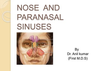
- 1. NOSE AND PARANASAL SINUSES By Dr. Anil kumar (First M.D.S)
- 2. CONTENTS INTRODUCTION DEVELOPMENT FUNCTIONS ANATOMY BLOOD SUPPLY NERVE SUPPLY LYMPHATIC DRAINAGE APPLIED ASPECTS RADIOGRAPHIC VIEWS SURGICAL APPROACHES
- 3. INTRODUCTION
- 4. INTRODUCTION
- 5. DEVELOPMENT OF NOSE 4 weeks
- 6. DEVELOPMENT OF NOSE 5 weeks
- 7. DEVELOPMENT OF NOSE 6 weeks
- 8. DEVELOPMENT OF NOSE 7 weeks
- 9. DEVELOPMENT OF PARANASAL SINUSES At about 25 – 28 weeks of gestation, three medially directed projections arise from the lateral wall of the nose. Between these projections small lateral diverticula invaginate into the primitive choana to eventually form the meati of the nose. This serves as the beginning of the development of paranasal sinuses. Sinuses begin developing as small sacculations of the mucosa of the nasal meati and recesses. As the pouches or sacs develop and grow they will invade the respective bones to form air sinuses and cells. Development is brougt about by resorption of inner surface and apposition on the outer surface by remodelling to accomidate the stresses.
- 10. Maxillary sinus - first to be developed and aerated at birth. shows biphasic growth. The first growth phase during the first three years of life, and the next growth phase occur between 7 – 18 years. Initially located medial to the orbit, later sinus extends laterally & Inferiorly. Floor of sinus does not extend below the level of nasal cavity until the eruption of permenant teeth. 1=newborn, 2=12 yrs 3=adult
- 11. Sphenoidal sinus is undevoleped and non-aereated at birth. Aeration begins at age 3years and then progresses posteriorly. sphenoid: 1=newborn, 2=3yo, 3=5yo, 4=7yo, 5=12yo, 6=adult,
- 12. Ethmoid air cells-develop during puberty and develop slowly until approximately 17-18 years of age. Pneumatization of this sinus begins during the 4th year of childhood and gets completed by the 17th year of life
- 13. Frontal sinus is last sinus to develop ,as a direct continuation or by upward migration of anterior ethmoidal air cells. Remains as a small blind sac within the frontal bone till 2 years of age,from 2 to 9 years secondary pneumatization of frontal bone proceeds. frontal: 4=newborn, 5=1yo, 6=4yo, 7=7yo,9=adult
- 14. CONGENITAL ANOMALIES OF THE NOSE HALF NOSE due to unilateral absence of nasal placode ARHINIA due to bilateral absence of nasal placodes
- 15. CONGENITAL ANOMALIES OF THE NOSE PROBOSCIS LATERALIS due to imperfect fusion between the maxillary process and the lateral nasal process. POLYRRHINIA duplication of the medial nasal processes.
- 16. CONGENITAL ANOMALIES OF THE NOSE NASAL CLEFTS : failure of the frontal nasal process to develop appropriately results into two separated halves of the nose. SUPERNUMERARY NOSTRIL MIDLINE NASAL SINUS: incomplete fusion of the right and left medial nasal prominence
- 17. FUNCTIONS OF NOSE & PARANASAL SINUSES To breath Olfaction To filter the air To taste Humidifying and warming inspired air Increasing surface area for olfaction Lightening the skull Resonance Absorbing shock Contribute to facial growth
- 18. ANATOMY OF NOSE & PARANASAL SINUSES
- 19. EXTERNAL NOSE Radix/root of nose Bridge Nasofacial angle Dorsum Alar nasal angle Pronasale (tip) Ala nasi (nasal wings) columella Anterior nares /nostrils
- 20. EXTERNAL NOSE Upper 1/3 rd (nasion region) Middle 1/3 rd Lower 1/3 rd
- 21. BONY PARTS Frontal process of the maxillae Nasal bones Nasal processes of the frontal bones
- 22. CARTILAGINOUS PARTS Greater alar cartilage Lesser alar cartilage Dense connective tissue Lateral nasal cartilage
- 23. BASAL VEIW Middle crus Lateral crus Medial crus
- 24. NASAL CAVITY Extends from nostrils to posterior nasal
- 25. WALLS OF NASAL CAVITY Roof Floor Medial/septal wall Lateral wall
- 26. ROOF OF NASAL CAVITY Frontal & nasal bones Cribriform plate of ethmoid Body of sphenoid
- 27. The Floor of Nasal Cavity Palatine process of maxilla Horizontal plate of palatine bone
- 28. MEDIAL WALL Divides nasal cavity into two halves. Seldom lies in midlineVestibule of nose
- 29. NASAL SEPTUM Vertical plate of ethmoid Septal cartilage vomer
- 30. LATERAL WALL
- 31. LATERAL WALL OF NOSE Marked by 3 projections: Superior concha Middle concha Inferior concha The space below each concha is called a meatus.
- 32. SUPERIOR MEATUS Space below the superior concha. Superior concha is a process of ethmoid bone. Smallest of all meatus. Posterior ethmoidal sinuses opens into it. Sphenoethmoidal recess is space above superior concha. Sphenoidal sinus opens into it.
- 33. MIDDLE MEATUS Space below middle concha. Middle concha is the medial process of ethmoidal labrynth. Hiatus semilunaris (curved opening) -frontal & maxillary sinuses Bulla ethmoidalis (rounded opening) -middle ethmoidal air cells.
- 34. INFERIOR MEATUS Largest of the meatuses. Space below the inferior concha. Inferior concha is thin, curved ,independent bone. Naso lacrimal duct opens in the anterior part.
- 35. SKIN OVER THE NOSE Skin is mobile over upper thirds but firmly adherent in the lower part to cartilages.
- 36. MUCOUS MEMBRANE Upper 1/3 rd –olfactory region, mucous membrane- more delicate and yellowish. Lower 2/3 rd – Respiratory region, Lined by pseudo stratified ciliated columnar epithelium, mucoperiosteum-Thick ,spongy ,highly vascular with numerous mucous glands. Mucous membrane covering vestibule of nose carries stiff hairs / vibrissae. Contains Arteriovenous anastamosis –warms the air passing through it.
- 38. MUSCLES OF THE NOSE
- 39. MUSCLES OF THE NOSE PROCERUS: most cephalic muscle of the nose, pyramidal shaped. Origin –facial aponeurosis. Insertion –from glabellar area. Assists in dilatation of the nares.
- 40. NASALIS: It has 2 components: (1)transverse nasalis /compressor nasi: the muscle spans the dorsum of the nose, covering the upper lateral cartilages. ORIGIN :maxilla above and lateral to incisive fossa. INSERTION: with its counterpart and procerus & levator labi superioris aeque nasi muscle. (2) the pars alaris (alar nasalis). ORIGIN: above lateral and canine.(more lateral & slightly caudal to the bony origin of the depressor septi nasi muscles). INSERTION : into ala above lateral crus of the
- 41. Levator labii superioris alaequae nasi Origin: upper part of frontal process of maxilla. It extends lateral to the nose in a cephalocaudal direction and has fibers that are attached to the nostril, contributing to the dilatation of the nares. Insetion:lateral crus of major alar cartilage and lateral part of upper lip. Release of the muscles will dilate the nostrils,
- 42. Dilator naris anterior Small muscle Origin:ULC and alar part of nasalis. Encircles nares Primary dilator of nose Depressor alae or myrtiforme. originates from the border of the pyriform crest and then rises vertically, like a fan, up to the ala, acting as a depressor and constrictor of the nostrils. Depressor septi Arises from the maxilla (just below the nasal spine), sometimes fuses with some fibers of the orbicularis oris muscle. Inserted along the columella, medial crus of alar cartilage.
- 43. NERVE SUPPLY Olfactory neves Anterior ethmoidal nerve Nasal branches of pterigo palatine ganglion Nasopalatine nerve External nose –Infra orbital nerve, Infra trochlear, External nasal nerve.
- 45. BLOOD SUPPLY EXTERNAL NOSE: Dorsal nasal artery Angular artery Superior labial artery INTERNAL NOSE: Anterior & posterior ethmoidal arteries Sphenopalatine artery Superior labial artery Infraorbital and superior dental arteries Pharyngeal branch of maxillary artery Greater palatine artery
- 46. KESSELBACHS PLEXUS These arteries are (mnemonic –LEGS) Superior Labial Anterior Ethmoidal Greater palatine Sphenopalatine Little's area, is a region in the anteroinferior part of the nasal septum, where there is confluence of 4 arteries forming this plexus.
- 47. LYMPHATIC DRAINAGE Submandibular lymphnodes: from the external nose and anterior part of the nasal cavity. Upper deep cervical nodes: drain the rest of the nasal cavity, either directly or through the retropharyngeal nodes.
- 49. MAXILLARY SINUS Antrum of Highmore. largest of all paranasal sinuses Pyramidal shaped , Lying just under the cheek. Capacity of 30ml.
- 50. Anterior wall (anterolateral wall)–lateral wall of the maxilla (canine fossa). Posterior wall – temporal surface of maxilla. Roof – floor of the orbit(infraorbital vessels and nerve). Floor – alveolar process of maxilla & hard palate. Medial wall (base of maxillary sinus)- lateral wall of the nasal cavity. Laterally (apex of sinus) – zygomatic bone
- 51. OSTIUM OF MAXILLARY SINUS Opens in the Posteroinferior end of hiatus semilunaris. Close to roof of sinus. Unfavorable for drainage of sinus. In children the floor lies at or above the level of the floor of the nasal fossa. In adults it lies about 1.25cm below the floor of the nasal fossa
- 52. SPHENOIDAL SINUS Lie within the body of the sphenoid bone Below sella turcica (extends between dorsum sellae and post clinoid processes) The average capacity is 7ml.
- 53. Superiorly – Pituitary gland (hypophysial fossa) Lateral wall – Optic nerve and internal carotid artery Floor – Nerve of pterygoid canal RELATIONS OF SPHENOIDAL SIN
- 54. OPENING OF SPHENOID SINUS Opens into the sphenoethmoidal recess above the superior concha. Ostium -Size (0.5-4mm)
- 55. ETHMOIDAL SINUS They are anterior, middle, and posterior. They are contained within the ethmoid bone, between the nose and the orbit. Anterior & middle drains into middle nasal meatus Posterior drain into superior nasal meatus
- 56. FRONTAL SINUS Second largest sinuses ◦ 2 – 2.5 cm Rarely symmetrical Contained within the frontal bone . Separated from each other by a bony septum. Each sinus is roughly triangular Extending upward above the medial end of the eyebrow and backward into the medial part of the roof of the orbit.
- 57. FRONTONASAL DUCT Opens into the middle meatus The average capacity is about 7 ml in the adult. True frontonasal duct only in 15% of people.
- 59. OSTEOMEATAL COMPLEX This is the area bounded by the middle turbiante medially, the lamina papyracea laterally, and the basal lamella superiorly and posteriorly. The inferior and anterior borders of the osteomeatal complex are open. This is in fact a narrow anatomical region consisting of : 1. Multiple bony structures (Middle turbinate, uncinate process, Bulla ethmoidalis) 2. Air spaces (Frontal recess, ethmoidal infundibulum, middle meatus) 3. Ostia of anterior ethmoidal, maxillary and frontal sinuses.
- 60. APPLIED ASPECTS
- 61. Sinus infections refer to the inflammation of the para- nasal cavities caused by irritation of the sinus membranes. Sinus cavities get irritated / infected Overproduction of mucus Sinus cavity openings may swell and block Blocked/congested Any accumulated mucus can become a haven for bacteria propagation Unbearable pain SINUSITIS
- 62. Acute, which last for 3 weeks or less Chronic, which usually last for 3 to 8 weeks but can continue for months or even years Recurrent, which are several acute attacks within a year Sinusitis can be classified based on which sinus cavities it affects: Antritis/ maxillary sinusitis Ethmoiditis / ethmoid sinusitis. Sphenoiditis, sphenoid Frontal sinusitis
- 63. Symptoms of Sinusitis location of pain depends on which sinus is affected. Headache when you wake up in the morning is typical of a sinus problem. Infection in the maxillary sinuses can cause your upper jaw and teeth to ache and your cheeks to become tender to the touch. Fever ,Weakness ,Tiredness etc. A cough that may be more severe at night. Runny nose (rhinitis) or nasal congestion . postnasal drip.
- 64. General measures: Drinking plenty of fluids to thin the secretions and keep them flowing. Hot showers to loosen the mucus. Alternate hot and cold compresses- place the hot compress across your sinuses for 3 minutes, then the cold compress for 30 second. Nasal irrigation
- 68. NOSEBLEED (EPISTAXIS) Traumatic Iatrogenic Inflammatory Foreign bodies Trauma (including nose picking) allergy Benign & malignant neoplasms Anterior nasal bleed Posterior nasal bleed
- 69. General measures: The nostrils are compressed against the nasal septum. The patient is told not to swallow blood running down the pharynx. The patient is kept in an upright posture An ice bag can be placed on the back of the neck to induce reflex vasoconstriction. Treatement Anterior nasal bleed is treated by packing the area with gauze soaked in L.A, by using electrocautery, or with silver nitrate If bleeding persists anterior nasal packing is performed.
- 70. Anterior nasal packing If it is posterior nasal bleed, posterior nasal packing has to be done. Reliable method is by using Foleys catheter. posterior nasal packin
- 71. CYSTIC FIBROSIS Cystic fibrosis is a systemic disease of unknown etiology affecting the mucus producing exocrine glands of upper respiratory tract, liver, pancreas, intestine and the non-mucus producing salivary and sweat glands. The abnormal secretions produced may lead to disease in any of the involved organ systems. The paranasal sinuses are ultimately involved with the viscous secretions generally result in chronic pansinusitis.
- 72. RHINITIS Seasonal/Acute: Itching, Sneezing, Rhinorrhea. Perennial/Chronic: The blocked nose Main causative organisms – Rhinoviruses coronaviruses
- 73. ALLERGIC RHINITIS local manifestation of an allergic reaction.
- 74. SEPTAL DEVIATION
- 75. NASAL FRACTURES Type I – inferior one half of nasal bones. Type II –entire nasal bone separated at nasofrontal suture. Type III –nasal bones and frontal process of maxilla. Type IV – nasal bones frontal process of maxilla. nasal spine of frontal bone and ethmoid bone. ASCH FORCEPS.
- 76. RADIOGAPHIC VIEWS Lateral view PA (Caldwell) view Waters view Open mouth Waters view Sub Mento Vertex view
- 77. Image is done in vertical position. air fluid level is clearly demonstrated Image is done vertically, but CR is angled 45 degrees. gradual fading of the fluid line. Image is done horizontally and the CR is vertical no evidence of an air-fluid level. Exudate in the sinuses is not a fluid but is commonly a heavy semi gelatinous material that clings to the walls of cavity and takes several minutes to shift the position .so you must position the patient for several minutes to allow the exudate to gravitate to the desired location before the exposure is made.
- 79. CALDWELL / PA AXIAL FRONTAL SINUS ANT. ETHMOIDAL SINUS
- 81. OPEN MOUTH WATERS SPHENOID SINUS MAXILLARY SINUS
- 82. SUB MENTO VERTEX ETHMOIDAL SINUSES SPHENOIDAL SINUSES NASAL SEPTUM ZYGOMATIC ARCHS
Notas del editor
- Part of MID FACE. Entrance of the respiratory tract. Plays role in warming ,humidifying and filtering the air. Important role in facial esthetics. Nostrils opens into nasal cavities. Nasal cavities are housed in a frame work of bones and fibroelastic cartilages. The bones surrounding the nasal cavities contains air filled cavities called paranasal sinuses.
- Brain occupies most of head region. Eyes are laterally located. Stomodeum represents future mouth. At the upper margin of Stomodeum – fronto nasal process formed from mesoderm. The frontonasal process inferiorly differentiates into two projections known as “Nasal Placodes”.
- Nasal pits are continuous with stomodeum, Sink to form the nostril. Nasal pits –partly surrounded by unevenly grown median and lateral nasal processes. Around the gut tube pharyngeal arches are formed. The oronasal membrane is fully formed by the end of 5th week of development. It gives rise to the floor of the nose (palate develops from this membrane).
- Later medial process joins the maxillary process forming closed maxillary arch. Lateral nasal swelling also join maxillary process and gives nasolacrimal duct at their junction. frontonasal prominence gives rise to inferior mesodermic projection-form the nasal septum dividing the nose into two cavities.
- Medial swellings on both the sides fused forming middle part of nose,philtrum & premaxilla. Lateral swellings forms the alae of nose.
- Anterior most ethmoidal air cell is known as agger nasi. Large agger nasi air cell can impede frontal sinus drainage due to its close proximity to the frontal sinus drainage pathway. Haller cells belong to the anterior ethmoidal group of air cells. These cells are also known as infra orbital cells. Enlargement of this cell may block drainage of the maxillary sinus. Extension of posterior ethmoidal cells supero lateral to the sphenoid sinus is known as onodi cell. This cell lies in close proximity to optic nerve. Inflammation of this cell may cause blindness. This anatomy is also crucial in endoscopic sinus surgical procedures Separated from the orbit by a thin plate of bone so that infection can readily spread from the sinuses into the orbit .