
Recomendados
Más contenido relacionado
La actualidad más candente
La actualidad más candente (20)
Destacado
Destacado (20)
Similar a Fundoscopy Exam Guide
Similar a Fundoscopy Exam Guide (20)
Más de meducationdotnet
Más de meducationdotnet (20)
Fundoscopy Exam Guide
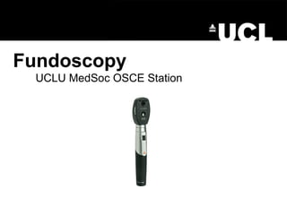
- 1. Fundoscopy UCLU MedSoc OSCE Station
- 2. First of all… • Don’t panic! The actual examination itself is easy! Secondly • Know the slides and know the basic pathologies
- 3. Learn how it works- they might ask you to put it together before you begin the station! The ophthalmoscope
- 4. The examination •WIPER •Inspection •Fundoscopy •To finish… Wash hands Introduce yourself Permission ‘Is it alright if look into your eyes today? This will involve shining a bright light into your eye. You can let me know if it gets too uncomfortable. I will also have to stand quite close in order to see properly. Is this OK? Expose Reposition Facing forward in a chair, ask to look at a point in the corner of the room.
- 5. The examination •WIPER •Inspection •Fundoscopy •To finish… Around the bed Patient General (Briefly!)
- 6. The examination •WIPER •Inspection •Fundoscopy •To finish… Ready the ophthalmoscope Put it together, turn the light setting so it’s on a white circle. Add the patient’s prescription to your own before turning the number dial. Comment on pupil dilation and then red reflex What would you use? Differential for abnormal red reflex? Examine the retina Comment on the disc- approach the patient (right eye to right eye), aim your view towards the nasal retina to find the optic disc. Colour, contours and cupping. Follow the arcades around, comment on vasculature and 4 quadrants then finally examine the macula by asking the patient to look into the light.
- 8. What might you see? The ophthalmology slides, obviously. If you haven’t got a copy of them already, get one! But it’s a good idea to know about the basic pathology of each of the main conditions: •Diabetic retinopathy •Hypertensive retinopathy •Glaucoma •Retinal vein/artery occlusion •Others signs e.g. papilloedema, optic atrophy
- 9. Background
- 11. Proliferative
- 12. Diabetic retinopathy- making the diagnosis • Background -Microaneurysms, blot/flame haemorrhages, hard exudate • Pre-proliferative -As above (but in all 4 quadrants), cotton wool spots (soft exudate), venous loops • Proliferative -New vessels, pre-retinal/vitreous haemorrhage, pre-retinal fibrosis, tractional retinal detachment • Maculopathy -Can occur at any stage
- 14. Hypertensive retinopathy- making the diagnosis Grades: 1. To torture someone you: 2. Nip ‘em 3. Flame ‘em 4. Pap ‘em
- 16. Retinal vein/artery occlusion- making the diagnosis • RVO -’Stormy sunset’, widespread haemorrhage without other features, may be branch vein occlusion • RAO -Retinal pallor, cherry red spot, may even see cholesterol embolus
- 20. Now watch us…
- 21. Now try it for yourselves…
- 25. Questions?