SlideShare a Scribd company logo
1 of 42
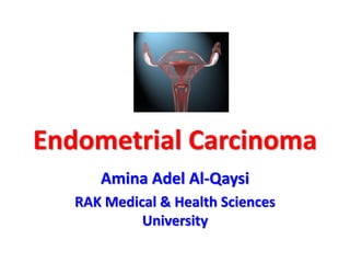
Endometrial Carcinoma
Amina Adel Al-Qaysi
RAK Medical & Health Sciences
University
Objectives
• Introduction
• Epidemiology
• Risk Factors
• Protective Factors
• Classification
• Spread
• Clinical Presentation
• Investigations
• Staging & Grading
• Treatment
• Follow Up
• Recurrence
• Prognosis
• Prevention
• Screening
Endometrial Carcinoma
• Carcinoma of the endometrial
lining of the uterus.
• Most common gynaecological
malignancy in postmenopausal women.
• 4th most common malignancy in women
(following breast, bowel, & lungs).
• Majority are adenocarcinoma.
Endometrial Carcinoma - Gross
Uterine Corpus
Cancers
Glands:
Endometrial
Carcinoma
Stroma:
Sarcoma
Epidemiology
• Most common gynaecological malignancy.
• 8th leading site of cancer-related mortality.
• 2-3% of women develop it in lifetime.
• Disease of postmenopausal women.
• 15%-25% of postmenopausal women with
bleeding have endometrial cancer.
• Mean age is 60 years.
• Uncommon before age of 40 years.
Risk Factors
• Older age.
• Early menarche.
• Late menopause.
• Nulliparity.
• Unopposed estrogen (Obesity, PCOS, HRT).
• Chronic Tamoxifen use.
• Previous pelvic irradiation.
• Hypertension, Diabetes mellitus.
Any agent/factor that rises the level
or time of exposure to estrogen is a
risk factor for endometrial
carcinoma
Risk Factors Cont’d
• Hx of other estrogen-
dependent neoplasm (breast,
ovary).
• Family Hx of endometrial
carcinoma.
• Estrogen-secreting ovarian
cancer (e.g. granulosa cell
tumor).
• Genetic: Lynch II $ (HNPCC).
Protective Factors
• Multiparity.
• Smoking.
• COCP.
• Physical activity.
Any agent/factor that lowers the
level or time of exposure to
estrogen is a protective factor
against endometrial carcinoma
Classification
TYPE 1
• Associated hyperestrogenism.
• Associated with hyperplasia.
• Patients usually peri-
memopausal.
• Estrogen & progesterone
receptors common.
• Usually endometrioid &
mucinous subtypes.
• Favourable prognosis.
TYPE 2
• Not related to
hyperestrogenism.
• Usually atrophic
endometrium.
• Postmenopausal patients.
• Estrogen & progesterone
receptors uncommon.
• Usually serous or clear cell
subtypes.
• Aggressive, poor prognosis.
Classification Cont’d
• Endometrioid - Adenocarcinoma (most
common 80%).
• Adenosquamous carcinoma (15%).
• Papillary serous carcinoma, Clear cell
carcinoma (3-4% overall).
Spread
• Direct extension .. MC
• Lymphatics
• Transtubally
• Haematogenous
(Lungs)
Clinical Presentation
Patient Profile
• Postmenopausal
• Nullipara
• Hx of early menarche & delayed menopause
• Obese
• Hypertension
• Diabetes mellitus
Clinical Presentation Cont’d
• Asymptomatic (˂ 5% of cases).
• Abnormal bleeding:
Postmenopausal bleeding *
Menorrhagia
Post-coital spotting
Intermenstrual bleeding
• Blood-stained vaginal discharge.
• If + cervical stenosis: Hematometra, Pyometra,
purulent vaginal discharge.
• Colicky abdominal pain.
Clinical Presentation Cont’d
Signs:
• Patient’s profile.
• Pallor (varying degree).
• Pelvic examination:
Speculum Exam: Normal looking cervix, blood or
purulent discharge through external os.
Bimanual exam: Uterus either atrophic, normal,
or enlarged. Uterus is mobile unless in late stage.
Per-rectal examination.
Regional lymph nodes & Breast examination.
Diagnosis
• Majority are diagnosed early, when surgery
alone may be adequate for cure.
• History + Physical examination.
• CBC
• Transvaginal Ultrasound (endometrial thickness).
• Endometrial biopsy.
• Hysteroscopy & endometrial biopsy (Gold
standard).
Transvaginal Ultrasound
Findings suggestive of endometrial
carcinoma:
Endometrial thickness ˃5 mm.
Hyper-echogenic endometrium with irregular
outline.
Increased vascularity with low vascular resistance.
Intrauterine fluid.
Diagnosis Cont’d
• Pap smear is not diagnostic, but a finding of
abnormal glandular cells of unknown
significance (AGCUS) leads to further
investigations.
• Abnormal Pap smears is the presentation
Of 1-5% of endometrial carcinoma cases.
• Pap smear/endocervical
curettage is required to evaluate
cervical involvement.
Diagnosis Cont’d
Pre-operative Evaluation:
• Physical examination
• Blood: CBC, postprandial sugar, urea & creatinine, S.E,
LFTs, CA-125.
• Urine: protein, sugar, pus cells.
• ECG
• Chest x-ray
• Pelvic USG
• Abdomeno-pelvic CT scan
• MRI
• PET
FIGO Staging
• Based on surgical & pathological evaluation.
Stage 0: Atypical hyperplasia.
Stage I: Tumor limited to the uterus
I A: Limited to the endometrium
I B: Invasion ˂ 1/2 of myometrium
I C: Invasion ˃ 1/2 of myometrium
Stage II: Extension to cervix
II A: Involves endocervical glands only
II B: Invasion of cervical stroma
FIGO Staging Cont’d
Stage III: Spread adjacent to uterus
III A: Invades serosa or adnexa, or positive cytology
III B: Vaginal invasion
III C: Invasion of pelvic or para-aortic lymph nodes
Stage IV: Spread further from uterus
IV A: Involves bladder or rectum
IV B: Distant metastasis
FIGO Staging
Grades
Treatment
• Surgery
• Chemotherapy
• Radiotherapy
• Hormonal therapy
Treatment Cont’d
• Based on tumour grade and depth of
myometrial invasion.
• Surgical: TAH+BSO and pelvic washings ±
pelvic and periaortic node dissection
Stage 1: TAH+BSO and washings.
Stages 2&3: TAH+BSO and washings and node
dissection.
Stage 4: No surgical option.
Treatment Cont’d
• Hormonal therapy: Progestins for recurrent
disease.
• Chemotherapy: In advanced, recurrent, or
metastatic disease.
Treatment Cont’d
Radiotherapy:
• Indications:
Patient medically unfit for surgery.
Surgically inoperable disease.
Those with high risk of recurrence
Stage III or IV disease
• Contraindications: Pelvic kidney, pyometra,
pelvic abscess, prior pelvic radiation, previous
laparotomy/adhesions.
Follow Up
• Thorough physical examination, CXR.
• Regular serum CA-125 estimation.
• Mammography, CT, MRI: When indicated.
• Every 4 months for the first 2 years.
• Every 6 months for the next 2 years.
• Thereafter annually.
Recurrent Disease
• Most commonly in the vagina & pelvis.
• Majority (60%) present within 6 years of initial
therapy.
• Management:
Radiation therapy (for isolated recurrence)
Hormonal therapy.
Chemotherapy.
Surgery: Of limited value.
Prognostic Factors
• Histologic grade (single most important).
• Depth of myometrial invasion (Second).
• Histologic type.
• Original tumor volume.
• Pelvic lymph nodes involvement.
• Extension to the cervix, adnexal metastasis,
positive peritoneal washings.
Prognosis Cont’d
5-years survival rate:
Stage 5-year survival (%)
Stage I 83
Stage II 71
Stage III 39
Stage IV 27
Screening
• There is no effective screening test.
• Occasionally, cervical smears contain
endometrial cancer cells, or endometrial
ultrasonic thickness of more than 5 mm
indicates the need for endometrial sampling.
Prevention
• Controlling obesity, blood pressure, and
diabetes help reduce risk.
• Restrict the use of estrogen after menopause
in non-hysterectomised women.
• Estrogen + cyclical progesterone.
• Women report any abnormal vaginal
bleeding or discharge to the doctor.
• Screening of high risk women in
postmenopausal period.
References
• Obstetrics & Gynaecology, Beckmann.
• Hacker & Moore’s Essentials of Obstetrics &
Gynaecology.
• Textbook of Gynaecology, Dutta.
• Current diagnosis & treatment, Obstetrics &
Gynaecology.
• Gynaecology By Ten Teachers, 18th edition.
Endometrial Carcinoma

More Related Content

What's hot

What's hot (20)

Endometrial Carcinoma
Endometrial Carcinoma Endometrial Carcinoma
Endometrial Carcinoma
 
Postmenopausal bleeding for undergraduate
Postmenopausal bleeding for undergraduatePostmenopausal bleeding for undergraduate
Postmenopausal bleeding for undergraduate
 
Ovarian teratoma
Ovarian teratomaOvarian teratoma
Ovarian teratoma
 
Abnormal uterine bleeding
Abnormal  uterine bleedingAbnormal  uterine bleeding
Abnormal uterine bleeding
 
Uterine fibroids
Uterine fibroidsUterine fibroids
Uterine fibroids
 
Abnormal Uterine Bleeding by Dr Kemi Dele
Abnormal Uterine Bleeding by Dr Kemi DeleAbnormal Uterine Bleeding by Dr Kemi Dele
Abnormal Uterine Bleeding by Dr Kemi Dele
 
Molar pregnancy
Molar pregnancyMolar pregnancy
Molar pregnancy
 
PUBERTY MENORRHAGIA & BLEEDING DISORDERS Made Easy Dr Sharda Jain
PUBERTY MENORRHAGIA & BLEEDING DISORDERS  Made Easy Dr Sharda Jain PUBERTY MENORRHAGIA & BLEEDING DISORDERS  Made Easy Dr Sharda Jain
PUBERTY MENORRHAGIA & BLEEDING DISORDERS Made Easy Dr Sharda Jain
 
Endometrial hyperplasia
Endometrial hyperplasiaEndometrial hyperplasia
Endometrial hyperplasia
 
Benign breast disease
Benign breast diseaseBenign breast disease
Benign breast disease
 
Pre cancerous lesions of cervix.pptx
Pre cancerous lesions of cervix.pptxPre cancerous lesions of cervix.pptx
Pre cancerous lesions of cervix.pptx
 
Ovarian cysts
Ovarian cystsOvarian cysts
Ovarian cysts
 
Cervical intra epithelial neoplasia
Cervical intra epithelial neoplasiaCervical intra epithelial neoplasia
Cervical intra epithelial neoplasia
 
Post menopausal bleeding
Post menopausal bleedingPost menopausal bleeding
Post menopausal bleeding
 
Endometriosis
EndometriosisEndometriosis
Endometriosis
 
Fibroadenoma
FibroadenomaFibroadenoma
Fibroadenoma
 
ENDOMETRIAL CANCER
ENDOMETRIAL CANCERENDOMETRIAL CANCER
ENDOMETRIAL CANCER
 
Benign ovarian tumours
Benign ovarian tumoursBenign ovarian tumours
Benign ovarian tumours
 
Pelvic mass
Pelvic massPelvic mass
Pelvic mass
 
Classification of ovarian tumors
Classification of ovarian tumorsClassification of ovarian tumors
Classification of ovarian tumors
 

Similar to Endometrial Carcinoma

ENDOMETRIAL CANCER (1).pptx
ENDOMETRIAL CANCER (1).pptxENDOMETRIAL CANCER (1).pptx
ENDOMETRIAL CANCER (1).pptxBhavesh SOBHANI
 
endometrial carcinoma.pptx
endometrial carcinoma.pptxendometrial carcinoma.pptx
endometrial carcinoma.pptxPoonamJhamb3
 
caendometrium-1-220817052735-f5d0c990.pdf
caendometrium-1-220817052735-f5d0c990.pdfcaendometrium-1-220817052735-f5d0c990.pdf
caendometrium-1-220817052735-f5d0c990.pdfharishgurawaliya1
 
Overview of Gynaecological Malignancies & Management
Overview of  Gynaecological Malignancies  &  ManagementOverview of  Gynaecological Malignancies  &  Management
Overview of Gynaecological Malignancies & ManagementKavya Liyanage
 
gynecologic cancers
gynecologic cancersgynecologic cancers
gynecologic cancersHiba Ahmed
 
Srishti Gupta, 503 A.M., Uterine Cancer.pptx
Srishti Gupta, 503 A.M., Uterine Cancer.pptxSrishti Gupta, 503 A.M., Uterine Cancer.pptx
Srishti Gupta, 503 A.M., Uterine Cancer.pptxSrishtiGupta304
 
14.tumor of reproductive organs
14.tumor of reproductive organs14.tumor of reproductive organs
14.tumor of reproductive organsHishgeeubuns
 
invasise Cervical carcinoma
invasise Cervical carcinomainvasise Cervical carcinoma
invasise Cervical carcinomaAisha Nazeer
 
Malignant ovarian tumours Dr H.K.Cheema
Malignant ovarian tumours  Dr H.K.CheemaMalignant ovarian tumours  Dr H.K.Cheema
Malignant ovarian tumours Dr H.K.CheemaDr H.K. Cheema
 
updated overview in management of ovarian cancer
updated overview in management of ovarian cancerupdated overview in management of ovarian cancer
updated overview in management of ovarian cancerSajan Thapa
 
Cervical neoplasia 2021
Cervical neoplasia 2021Cervical neoplasia 2021
Cervical neoplasia 2021Musa Abusabha
 

Similar to Endometrial Carcinoma (20)

ENDOMETRIAL CANCER (1).pptx
ENDOMETRIAL CANCER (1).pptxENDOMETRIAL CANCER (1).pptx
ENDOMETRIAL CANCER (1).pptx
 
endometrial carcinoma.pptx
endometrial carcinoma.pptxendometrial carcinoma.pptx
endometrial carcinoma.pptx
 
Cervical Malignancy.pptx
Cervical Malignancy.pptxCervical Malignancy.pptx
Cervical Malignancy.pptx
 
Ca endometrium-1.pptx
Ca endometrium-1.pptxCa endometrium-1.pptx
Ca endometrium-1.pptx
 
caendometrium-1-220817052735-f5d0c990.pdf
caendometrium-1-220817052735-f5d0c990.pdfcaendometrium-1-220817052735-f5d0c990.pdf
caendometrium-1-220817052735-f5d0c990.pdf
 
Endometrial carcinoma
Endometrial carcinomaEndometrial carcinoma
Endometrial carcinoma
 
Overview of Gynaecological Malignancies & Management
Overview of  Gynaecological Malignancies  &  ManagementOverview of  Gynaecological Malignancies  &  Management
Overview of Gynaecological Malignancies & Management
 
Ca endometrium
Ca endometriumCa endometrium
Ca endometrium
 
gynecologic cancers
gynecologic cancersgynecologic cancers
gynecologic cancers
 
Ovarian cancer
Ovarian cancer Ovarian cancer
Ovarian cancer
 
Srishti Gupta, 503 A.M., Uterine Cancer.pptx
Srishti Gupta, 503 A.M., Uterine Cancer.pptxSrishti Gupta, 503 A.M., Uterine Cancer.pptx
Srishti Gupta, 503 A.M., Uterine Cancer.pptx
 
ca cervix.pdf
ca cervix.pdfca cervix.pdf
ca cervix.pdf
 
Athira
Athira Athira
Athira
 
14.tumor of reproductive organs
14.tumor of reproductive organs14.tumor of reproductive organs
14.tumor of reproductive organs
 
Ca endometrium
Ca endometriumCa endometrium
Ca endometrium
 
invasise Cervical carcinoma
invasise Cervical carcinomainvasise Cervical carcinoma
invasise Cervical carcinoma
 
Ovarian Cancer
Ovarian CancerOvarian Cancer
Ovarian Cancer
 
Malignant ovarian tumours Dr H.K.Cheema
Malignant ovarian tumours  Dr H.K.CheemaMalignant ovarian tumours  Dr H.K.Cheema
Malignant ovarian tumours Dr H.K.Cheema
 
updated overview in management of ovarian cancer
updated overview in management of ovarian cancerupdated overview in management of ovarian cancer
updated overview in management of ovarian cancer
 
Cervical neoplasia 2021
Cervical neoplasia 2021Cervical neoplasia 2021
Cervical neoplasia 2021
 

More from meducationdotnet

More from meducationdotnet (20)

No Title
No TitleNo Title
No Title
 
Spondylarthropathy
SpondylarthropathySpondylarthropathy
Spondylarthropathy
 
Diagnosing Lung cancer
Diagnosing Lung cancerDiagnosing Lung cancer
Diagnosing Lung cancer
 
Eczema Herpeticum
Eczema HerpeticumEczema Herpeticum
Eczema Herpeticum
 
The Vagus Nerve
The Vagus NerveThe Vagus Nerve
The Vagus Nerve
 
Water and sanitation and their impact on health
Water and sanitation and their impact on healthWater and sanitation and their impact on health
Water and sanitation and their impact on health
 
The ethics of electives
The ethics of electivesThe ethics of electives
The ethics of electives
 
Intro to Global Health
Intro to Global HealthIntro to Global Health
Intro to Global Health
 
WTO and Health
WTO and HealthWTO and Health
WTO and Health
 
Globalisation and Health
Globalisation and HealthGlobalisation and Health
Globalisation and Health
 
Health Care Worker Migration
Health Care Worker MigrationHealth Care Worker Migration
Health Care Worker Migration
 
International Institutions
International InstitutionsInternational Institutions
International Institutions
 
Haemochromotosis brief overview
Haemochromotosis brief overviewHaemochromotosis brief overview
Haemochromotosis brief overview
 
Ascities overview
Ascities overviewAscities overview
Ascities overview
 
Overview of the Liver
Overview of the LiverOverview of the Liver
Overview of the Liver
 
Overview of Antidepressants
Overview of AntidepressantsOverview of Antidepressants
Overview of Antidepressants
 
Gout Presentation
Gout PresentationGout Presentation
Gout Presentation
 
Review of orthopaedic services: Prepared for the Auditor General for Scotland...
Review of orthopaedic services: Prepared for the Auditor General for Scotland...Review of orthopaedic services: Prepared for the Auditor General for Scotland...
Review of orthopaedic services: Prepared for the Auditor General for Scotland...
 
Sugammadex - a revolution in anaesthesia?
Sugammadex - a revolution in anaesthesia?Sugammadex - a revolution in anaesthesia?
Sugammadex - a revolution in anaesthesia?
 
Ophthamology Revision
Ophthamology RevisionOphthamology Revision
Ophthamology Revision
 

Endometrial Carcinoma

  • 1. Endometrial Carcinoma Amina Adel Al-Qaysi RAK Medical & Health Sciences University
  • 2. Objectives • Introduction • Epidemiology • Risk Factors • Protective Factors • Classification • Spread • Clinical Presentation • Investigations • Staging & Grading • Treatment • Follow Up • Recurrence • Prognosis • Prevention • Screening
  • 3. Endometrial Carcinoma • Carcinoma of the endometrial lining of the uterus. • Most common gynaecological malignancy in postmenopausal women. • 4th most common malignancy in women (following breast, bowel, & lungs). • Majority are adenocarcinoma.
  • 6. Epidemiology • Most common gynaecological malignancy. • 8th leading site of cancer-related mortality. • 2-3% of women develop it in lifetime. • Disease of postmenopausal women. • 15%-25% of postmenopausal women with bleeding have endometrial cancer. • Mean age is 60 years. • Uncommon before age of 40 years.
  • 7. Risk Factors • Older age. • Early menarche. • Late menopause. • Nulliparity. • Unopposed estrogen (Obesity, PCOS, HRT). • Chronic Tamoxifen use. • Previous pelvic irradiation. • Hypertension, Diabetes mellitus. Any agent/factor that rises the level or time of exposure to estrogen is a risk factor for endometrial carcinoma
  • 8.
  • 9.
  • 10. Risk Factors Cont’d • Hx of other estrogen- dependent neoplasm (breast, ovary). • Family Hx of endometrial carcinoma. • Estrogen-secreting ovarian cancer (e.g. granulosa cell tumor). • Genetic: Lynch II $ (HNPCC).
  • 11. Protective Factors • Multiparity. • Smoking. • COCP. • Physical activity. Any agent/factor that lowers the level or time of exposure to estrogen is a protective factor against endometrial carcinoma
  • 12.
  • 13. Classification TYPE 1 • Associated hyperestrogenism. • Associated with hyperplasia. • Patients usually peri- memopausal. • Estrogen & progesterone receptors common. • Usually endometrioid & mucinous subtypes. • Favourable prognosis. TYPE 2 • Not related to hyperestrogenism. • Usually atrophic endometrium. • Postmenopausal patients. • Estrogen & progesterone receptors uncommon. • Usually serous or clear cell subtypes. • Aggressive, poor prognosis.
  • 14. Classification Cont’d • Endometrioid - Adenocarcinoma (most common 80%). • Adenosquamous carcinoma (15%). • Papillary serous carcinoma, Clear cell carcinoma (3-4% overall).
  • 15. Spread • Direct extension .. MC • Lymphatics • Transtubally • Haematogenous (Lungs)
  • 16.
  • 17. Clinical Presentation Patient Profile • Postmenopausal • Nullipara • Hx of early menarche & delayed menopause • Obese • Hypertension • Diabetes mellitus
  • 18. Clinical Presentation Cont’d • Asymptomatic (˂ 5% of cases). • Abnormal bleeding: Postmenopausal bleeding * Menorrhagia Post-coital spotting Intermenstrual bleeding • Blood-stained vaginal discharge. • If + cervical stenosis: Hematometra, Pyometra, purulent vaginal discharge. • Colicky abdominal pain.
  • 19. Clinical Presentation Cont’d Signs: • Patient’s profile. • Pallor (varying degree). • Pelvic examination: Speculum Exam: Normal looking cervix, blood or purulent discharge through external os. Bimanual exam: Uterus either atrophic, normal, or enlarged. Uterus is mobile unless in late stage. Per-rectal examination. Regional lymph nodes & Breast examination.
  • 20. Diagnosis • Majority are diagnosed early, when surgery alone may be adequate for cure. • History + Physical examination. • CBC • Transvaginal Ultrasound (endometrial thickness). • Endometrial biopsy. • Hysteroscopy & endometrial biopsy (Gold standard).
  • 21. Transvaginal Ultrasound Findings suggestive of endometrial carcinoma: Endometrial thickness ˃5 mm. Hyper-echogenic endometrium with irregular outline. Increased vascularity with low vascular resistance. Intrauterine fluid.
  • 22.
  • 23.
  • 24. Diagnosis Cont’d • Pap smear is not diagnostic, but a finding of abnormal glandular cells of unknown significance (AGCUS) leads to further investigations. • Abnormal Pap smears is the presentation Of 1-5% of endometrial carcinoma cases. • Pap smear/endocervical curettage is required to evaluate cervical involvement.
  • 25. Diagnosis Cont’d Pre-operative Evaluation: • Physical examination • Blood: CBC, postprandial sugar, urea & creatinine, S.E, LFTs, CA-125. • Urine: protein, sugar, pus cells. • ECG • Chest x-ray • Pelvic USG • Abdomeno-pelvic CT scan • MRI • PET
  • 26. FIGO Staging • Based on surgical & pathological evaluation. Stage 0: Atypical hyperplasia. Stage I: Tumor limited to the uterus I A: Limited to the endometrium I B: Invasion ˂ 1/2 of myometrium I C: Invasion ˃ 1/2 of myometrium Stage II: Extension to cervix II A: Involves endocervical glands only II B: Invasion of cervical stroma
  • 27. FIGO Staging Cont’d Stage III: Spread adjacent to uterus III A: Invades serosa or adnexa, or positive cytology III B: Vaginal invasion III C: Invasion of pelvic or para-aortic lymph nodes Stage IV: Spread further from uterus IV A: Involves bladder or rectum IV B: Distant metastasis
  • 30. Treatment • Surgery • Chemotherapy • Radiotherapy • Hormonal therapy
  • 31. Treatment Cont’d • Based on tumour grade and depth of myometrial invasion. • Surgical: TAH+BSO and pelvic washings ± pelvic and periaortic node dissection Stage 1: TAH+BSO and washings. Stages 2&3: TAH+BSO and washings and node dissection. Stage 4: No surgical option.
  • 32.
  • 33. Treatment Cont’d • Hormonal therapy: Progestins for recurrent disease. • Chemotherapy: In advanced, recurrent, or metastatic disease.
  • 34. Treatment Cont’d Radiotherapy: • Indications: Patient medically unfit for surgery. Surgically inoperable disease. Those with high risk of recurrence Stage III or IV disease • Contraindications: Pelvic kidney, pyometra, pelvic abscess, prior pelvic radiation, previous laparotomy/adhesions.
  • 35. Follow Up • Thorough physical examination, CXR. • Regular serum CA-125 estimation. • Mammography, CT, MRI: When indicated. • Every 4 months for the first 2 years. • Every 6 months for the next 2 years. • Thereafter annually.
  • 36. Recurrent Disease • Most commonly in the vagina & pelvis. • Majority (60%) present within 6 years of initial therapy. • Management: Radiation therapy (for isolated recurrence) Hormonal therapy. Chemotherapy. Surgery: Of limited value.
  • 37. Prognostic Factors • Histologic grade (single most important). • Depth of myometrial invasion (Second). • Histologic type. • Original tumor volume. • Pelvic lymph nodes involvement. • Extension to the cervix, adnexal metastasis, positive peritoneal washings.
  • 38. Prognosis Cont’d 5-years survival rate: Stage 5-year survival (%) Stage I 83 Stage II 71 Stage III 39 Stage IV 27
  • 39. Screening • There is no effective screening test. • Occasionally, cervical smears contain endometrial cancer cells, or endometrial ultrasonic thickness of more than 5 mm indicates the need for endometrial sampling.
  • 40. Prevention • Controlling obesity, blood pressure, and diabetes help reduce risk. • Restrict the use of estrogen after menopause in non-hysterectomised women. • Estrogen + cyclical progesterone. • Women report any abnormal vaginal bleeding or discharge to the doctor. • Screening of high risk women in postmenopausal period.
  • 41. References • Obstetrics & Gynaecology, Beckmann. • Hacker & Moore’s Essentials of Obstetrics & Gynaecology. • Textbook of Gynaecology, Dutta. • Current diagnosis & treatment, Obstetrics & Gynaecology. • Gynaecology By Ten Teachers, 18th edition.

Editor's Notes

  1. Uterine sarcoma: progressive uterine enlargement in postmenopausal pt Postmenopausal bleeding + pelvic pain + uterine enlargement
  2. Tamoxifen: agonistic on endometrium, antagonistic on breast
  3. Prophylactic hysterectomy with bilateral salpingo-oophorectomy is an effective strategy for preventing endometrial and ovarian cancer in women with the Lynch syndrome.
  4. Adenoacanthoma: adenocarcinoma + benign squamous element less than 10% of histologic picture
  5. Usually spreads throughout endometrial cavity first Ca endometrium spreads hematogenously more readily than ca cervix or ovary Adnexal invasion: through lymphatics, or transtubal invasion
  6. Postmenopausal bleeding: after 6 months of amenorrhea in pt diagnosed as menopause Postmenopausal bleeding: atrophic endometritis, hormonal therapy, endometrial ca, endometrial poly, endometrial hyperplasia Pressure symptoms
  7. Normal uterus on pelvic exam doesn’t R/O endometrial ca
  8. Fractional biopsy: 1- endocervical sampling/pap smear 2-dilate & take endometrial biopsy
  9. Endometrial thickness on TVUS in postmenopausal pt should be ˂ or equal 4 mm, if pt on HRT then ˂ 10 mm In menstruating women ˂ or equal to 14 mm Normal endometrial thickness on TVUS doesn't R/O carcinoma especially in presence of risk factors
  10. CA-125 usually elevated in pt of advanced disease
  11. International Federation of Gynecology and Obstetrics
  12. Well differentiated, moderately differentiated, poorly differentiated Worse the differentiation: more the solid growth
  13. Complete surgical staging = pelvic washings, bilateral pelvic & para-aortic Lymphadenectomy, complete resection of all the tumor Sampling of common iliac LNs may also be done LNs palpation is anaccurate, shouldn’t substitute resection Complete surgical staging & retroperitoneal LNs assessment is not only therapeutic but also improves survival Surgical staging not required if: young or perimenopausal pt with grade 1 endometrioid adenocarcinoma associated with atypical hyperplasia, pt at increased risk of mortality secondary to morbidities
  14. In hysterectomy u try to dissect & remove the ligaments as near as possible to uterus, except in radical hysterectomy in which u dissect their attachement to pelvic walls
  15. In stage 1 disease: radiotherapy may reduce recurrence risk but doesn’t improve survival Stage 3C disease (LNs positive): radiotherapy is critical in improving survival rate.
  16. Pt tt with radiotherapy have decreased risk of vaginal recurrence, but fewer therapeutic options to tt recurrence Vaginal recurrence pt have better prognosis than pelvic recurrence & distant mets Chemootherapy has occassional favourable short term results, but good long-term results Major advantage of hormonal therapy is the fewer complications
  17. Estrogen therapy is CI in pt previously treated for endometrial Ca