BASICS of CT Head
•Descargar como PPTX, PDF•
1,582 recomendaciones•514,172 vistas

this was prepared for a class presentation during my MD course

Recomendados
Más contenido relacionado
La actualidad más candente
La actualidad más candente (20)
Destacado
Destacado (20)
Similar a BASICS of CT Head
Similar a BASICS of CT Head (20)
Más de Kunal Mahajan
Más de Kunal Mahajan (19)
Último
Último (20)
BASICS of CT Head
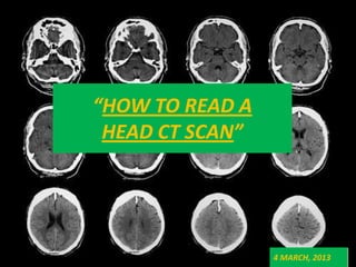
- 1. “HOW TO READ A HEAD CT SCAN” 4 MARCH, 2013
- 2. MUST FOR EVERY PHYSICIAN • CT HEAD is an extremely useful diagnostic tool used routinely in the care of A&E patients. • The treating physician needs to be able to accurately interpret and act upon certain CT findings without specialist (e.g., radiologist) assistance, because many disease processes are time dependent and require immediate action. • It has been shown that even a brief educational intervention can significantly improve the physician’s ability to interpret cranial CT scans.
- 3. SCHEME OF THE LECTURE • BASIC PRINCIPLES OF CT SCAN • NORMAL NEUROANATOMY AS SEEN ON HEAD CT SCANS • ILLUSTRATIONS
- 4. BASIC PRINCIPLES OF CT SCAN
- 5. HISTORY • Sir Godfrey hounsfield-1972 • Nobel prize in 1979 • Original scanners took approximately 6 minutes to perform a rotation (one slice) and 20 minutes to reconstruct. Despite many technological advances since then, the principles remain the same.
- 6. PARTS 1) Gantry- which houses X ray apparatus 2) X ray tube-akin to that in a X ray machine. 3) Detectors 4) Patient couch 5) Viewing console
- 8. PRINCIPLE • Uses X rays applied in sequence of slices across the organ • Images reconstructed from X ray absorption data • X ray beam moves around the patient in a circular path
- 10. PRINCIPLE….. • CT scan provides a 3D display of the intracranial anatomy built up from a vertical series of transverse axial tomograms. • Each tomogram represents a horizontal slice through the patient’s head.
- 11. TECHNIQUE….. Slice thickness may vary, but in general, it is between 5 and 10 mm for a routine Head CT
- 13. BASICS…. • X-RAYS ARE ABSORBED TO DIFFERENT DEGREES BY DIFFERENT TISSUES • Always describe CT findings as densities- isodense/hypodense/hyperdense. • Higher the density = whiter is the appearance • Lower the density = darker the appearance • Brain is the reference density • Anything of the density as brain= isodense • Higher density than brain= hyperdense ( skull is the best example) • Anything darker (lower density) than brain= hypodense( CSF and air are classical examples)
- 14. HOUNSFIELD UNITS • Related to composition & nature of tissue • Represent the density of tissue • Also called as CT NUMBER
- 15. air --- 1000 fat ---70 Pure water 0 Csf +8 White matter +30 Gray matter +45 blood +70 Bone/calcification +1000
- 16. Densities on ct scan…….
- 17. NORMAL NEUROANATOMY AS SEEN ON HEAD CT SCANS
- 18. AXIAL SECTIONS OF CT HEAD POSTERIOR FOSSA CUTS -ABOVE THE FORAMEN MAGNUM LEVEL -LEVEL OF THE FOURTH VENTRICLE -ABOVE THE FOURTH VENTRICULAR LEVEL -TENTORIAL SUPRATENTORIAL CUTS -THIRD VENTRICULAR LEVEL -LATERAL VENTRICULAR LEVEL -ABOVE THE VENTRICULAR LEVEL
- 19. Lateral View of Brain
- 20. NORMAL ANATOMY……. A= ORBIT , B= SPHENOID SINUS , C= TEMPORAL LOBE, D=EXTERNAL AUDITORY CANAL E= MASTOID AIR CELLS F= CEREBELLAR HEMISPHERES
- 21. NORMAL ANATOMY……. A=Frontal Lobe, B= Frontal Bone (Superior Surface of Orbital Part), C= Dorsum Sellae, D=Basilar Artery E= Temporal Lobe F= Mastoid Air Cells G=Cerebellar Hemisphere
- 22. NORMAL ANATOMY……. A=FRONTAL LOBE B= SYLVIAN FISSURE C=TEMPORAL LOBE D=SUPRASELLAR CISTERN E=MIDBRAIN F=FOURTH VENTRICLE G= CEREBELLAR HEMISPHERE
- 23. NORMAL ANATOMY…….. A=FALX CEREBRI B=FRONTAL LOBE C=ANTERIOR HORN LAT VENTRICLE D=THIRD VENTRICLE E=QUADRIGEMINAL PLATE CISTERN F=CEREBELLUM
- 24. NORMAL ANATOMY…….. A=ANTERIOR HORN LAT VENTRICLE B=CAUDATE NUCLEUS C=ANT LIMB INT CAPSULE D=GLOBUS PALLIDUS AND PUTAMEN E=POST LIMB INT CAPSULE F=THIRD VENTRICLE G=QUADRIGEMINAL PLATE CISTERN H=CEREBELLAR VERMIS I=OCCIPITAL LOBE
- 25. NORMAL ANATOMY…….. A=GENU OF CORPUS CALLOSUM B=ANT HORN OF LATERAL VENTRICLE C=INT CAPSULE D=THALAMUS E=PINEAL GLAND F=CHOROID PLEXUS G=STARAIGHT SINUS
- 26. NORMAL ANATOMY……. A=FALX CEREBRI B=FRONTAL LOBE C=BODY OF LATERAL VENTRICLE D=SPLENIUM OF CORPUS CALLOSUM E=PARIETAL LOBE F=OCCIPITAL LOBE G=SUPERIOR SAGITTAL SINUS
- 27. NORMAL ANATOMY…….. A=FALX CEREBRI B=SULCUS C=GYRUS D=SUPERIOR SAGGITAL SINUS
- 32. 1. Frontal bone 2. Superior frontal gyrus 3. Coronal suture 4. Precentral sulcus 5. Falx cerebri 6. Precentral gyrus 7. Parietal bone 8. Paracentral lobule 9. Central sulcus 10. Postcentral gyrus 11. Superior parietal lobule 12. Precuneus 13. Sagittal suture 14. Superior saggital sinus
- 33. Frontal bone Falx cerebri Central sulcus Parietal bone Superior saggital sinus
- 34. WHENEVER THE BRAIN SWELLS , THE GYRI BECOME LARGER AND THE SULCI SHRINK
- 36. BASICS….
- 38. 1. Frontal bone 2. Superior saggital sinus 3. Superior frontal gyrus 4. Coronal suture 5. Falx cerebri 6. Middle frontal gyrus 7. Longitudinal cerebral fissure 8. Precentral sulcus 9. Precentral gyrus 10. Central sulcus 11. Cerebral white matter (centrum semiovale) 12. Postcentral gyrus 13. Paracentral lobule 14. Supramarginal gyrus 15. Parietal bone 16. Inferior parietal lobule 17. Precuneus 18. Parieto-occipital sulcus 19. Occipital bone
- 39. CORONA RADIATA CORPUS CALLOSUM
- 41. 2 Frontal sinus 5 Falx cerebri 6 Caudate nucleus (head) 9 Corpus callosum (genu) 11 Lateral ventricle 12 Third ventricle 13 Central sulcus 14 Precentral gyrus 15 Fornix 16 Postcentral gyrus 17 Interventricular foramen (foramen of Monro) 18 Lateral sulcus 19 Claustrum 20 insular Cistern 22 Insula 23 Thalamus 25 Pineal gland 31 Vermis of cerebellum 32 Lateral ventricle (trigone with choroid plexus) 33 Straight sinus 34 Middle temporal gyrus 37 Superior sagittal sinus 38 Occipital gyri
- 44. 2 Frontal sinus 3 Falx cerebri 7 Corpus callosum (genu) 13 External capsule 14 Putamen 15 Septum verum (precommissural septum) 16 Cistern of lateral cerebral fossa (insular cistern) 17 Hypothalamus 19 Third ventricle 20 Claustrum 21 Superior temporal gyrus 22 Extreme capsule 27 Hippocampus 28 Thalamus 30 Pineal gland (calcified) 31 Tentorium cerebelli 32 Quadrigeminal plate 33 Vermis of cerebellum 34 Quadrigeminal and ambient cisterns 35 Straight sinus 37 Superior sagittal sinus 38 Lateral ventricle (trigone)
- 46. 1 Frontal sinus 2 Frontal bone 3 Falx cerebri 4 Orbital gyri 5 Straight gyrus 6 Anterior cerebral artery 7 Anterior communicating artery 8 Internal carotid artery 9 Superior temporal gyrus 10Mi ddle temporal gyrus 11 Middle cerebral artery 12 Posterior communicating artery 13 Optic chiasm 14 Amygdaloid body 15 Pituitary stalk 16 Lateral ventricle (temporal horn) 17 Dorsum sellae 18 Hippocampus 19 Pentagon of basal cisterns 20Infe rior temporal gyrus 21 Posterior cerebral artery 22 Parahippocampal gyrus 23 Tentorium cerebelli 24 Basilar artery and basal sulcus 25 Pons 26 Sigmoid sinus 27 Cerebellar peduncle (middle) 28 Fourth ventricle 29 Dentate nucleus 30V ermis of cerebellum (superior part) 31 Temporal bone 32 Confluence of the sinuses 33 Cerebellar hemisphere 34 Transverse sinus 35 Occipital bone
- 48. 1 Frontal bone 2 Frontal sinus 3 Straight gyrus 4 Temporal muscle 5 Orbital gyri 6 Roof of orbit 7 Superior temporal gyrus 8 Optic nerve 9 Internal carotid artery 10Pi tuitary gland 11 Middle temporal gyrus 12 Dorsum sellae 13 Parahippocampal gyrus 14 Basilar artery 15 Lateral ventricle (temporal horn) 16 Inferior temporal gyrus 17 Trigeminal nerve (V) 18 Trochlear nerve 19 Pontine cistern 20Mas toid antrum 21 Tentorium cerebelli 22 Fourth ventricle 23 Pons 24 Temporal bone 25 Cerebellar peduncle 26 Vermis of cerebellum 27 Sigmoid sinus 28 Cerebellar hemisphere 29 Dentate nucleus 30Occip ital sinus 31 Occipital bone 32 Semispinalis capitis muscle
- 51. 1 Nasal bone 2 Eyeball 3 Medial rectus muscle 4 Nasal septum 5 Ethmoidal cells 6 Zygomatic bone 7 Pterygopalatine fossa 8 Inferior rectus muscle 9 Occipital bone (basilar part) 10T emporal muscle 11 Foramen ovale with mandibular nerve 12 Sphenoidal sinus 13 Temporal bone (apex of the petrous pyramid) 14 Zygomatic arch 15 Internal carotid artery 16 Masseter muscle 17 Jugular vein (bulb) 18 Lateral pterygoid muscle (superior head) 19 External auditory meatus 20Auditor y tube 21 Medulla oblongata 22 Head of mandible 23 Mastoid process 24 Foramen lacerum 25 Sigmoid sinus 26 Vertebral arteries 27 Petro-occipital fissure 28 Flocculus 29 Cerebellar tonsil 30Dig astric muscle 31 Splenius capitis muscle 32 Cerebellar hemisphere (caudal lobe) 33 Rectus capitis posterior minor muscle 34 Cisterna magna (posterior cerebellomedullary cistern) 35 Rectus capitis posterior major muscle 36 Occipital bone 37 Semispinalis capitis muscle 38 Trapezius muscle
- 52. medulla Cerebellar hemisphere Cisterna magna
- 53. THANKS
- 54. 1. Frontal bone 2. Superior frontal gyrus 3. Falx cerebri 4. Middle frontal gyrus 5. Cingulate sulcus 6. Coronal suture 7. Pericallosal artery 8. Precentral gyrus 9. Corona radiata 10. Central sulcus 11. Corpus callosum 12. Postcentral gyrus 13. Lateral ventricle(choroid plexus) 14. Postcentral sulcus 15. Parietal bone 16. Supramarginal gyrus 17. Precuneus 18. Angular gyrus 19. Parieto-occipital sulcus 20. Occipital gyri 21. Cuneus 22. Occipital bone 23. Superior saggital sinus