SlideShare una empresa de Scribd logo
1 de 119
Functions Of Kidney
I. Elimination Of Metabolic Waste Products.
II. Fluid , Electrolytes & Acid-base Status
Regulation.
III. Hormonal Regulation With Erythropoietin &
Renin Production ,Vit.D Activation.
Routine Urine Examination
PHYSICAL
EXAMINATION
CHEMICAL
EXAMINATION
MICROSCOPIC
EXAMINATION
I. Physical Examination
1) Total Volume
Normal Volume:600-2000ml/Day
a) Polyuria:urine Volume >2000ml/Day
 Causes :-Osmotic Diuresis
:-Polydipsia(diabetes Insipidus)
:-Diuretics
:-Chronic Renal Failure
• Oliguria
b) EXCRETION <400ML/DAY
 CAUSES :-DEHYDRATION
:-CHF
:-SHOCK
:-RENAL ARTERY EMBOLISM
:-ACUTE GLOMERULONEPHRITIS
:-ATN
:-RENAL FAILURE
:-GU TRACK OBSTRUCTION
C) ANURIA :- EXCRETION <100ML/DAY
Causes :-Bilateral Complete Urinary
Tract Obstruction
:-Acute Cortical Necrosis
:-Necrotizing
Glomerulonephritis
2) Specific Gravity: It Reflects The Degree Of
Concentration Of Urine.
Methods:
1)reagent Strips
2)refractometer
3)urinometer
4)falling Drop Method
1.Reagent Strips
 Indirect Method
 Reagent Area Contains 3 Main
Ingredients:1)polyelectrolytes
2)indicator Substance
3)buffer
 PRINCIPLE:IT IS BASED ON THE Pka CHANGE OF THE
PRE-TREATED POLYELECTROLYTES IN RELATION TO THE
IONIC CONC.Of Urine.When The Ionic Conc.IS HIGH
,Pka IS DECREASED AS IS THE Ph.The Indicator
Substance Then Change Color Relative To The Ionic
Conc & This Is Translated To The Specific Gravity
Values.
 Advantage:not Affected By High Amounts Of
Glucose,protein Or Radiographic Contrast Media.
2)refractometer
 Principle:refractive Index Of A Solution Is
Related To The Content Of Dissolved Solids
Present.
 It Is Temperature Compensated Hand
Model(60 To 100f)
 Requires Only Few Drops Of Urine.
3)URINOMETER
IT IS A HYDROMETER ADOPTED
TO DIRECTLY MEASURE THE
SPECIFIC GRAVITY OF URINE AT
ROOM TEMP.
4)falling Drop Method
 Direct Method
 More Accurate Than Refractometer
 More Precise Than Urinometer
Interpretation:
Normal-1.016 To 1.022/24 Hr
Increased In:-proteinuria
:-Diabetes Melitus
:-Diuretics
:-Antibiotics
:-Acute Glomerulonephritis
Decreased In:-renal Insufficiency
Diurnal Variation:higher In The Morning
&Lower In Afternoon
Fixed Specific Gravity:sp.GRAVITY OF ANY
SAMPLE OF URINE COLLECTED REMAINS FIXED
E.G.1.010
Seen In Chronic Glomerulonephritis
3.Color
COLOR CONDITIONS
PALE YELLOW NORMAL URINE
RED Hb
ANILNE DYES
PHENOPHTHELIN
SMOKY RED OR BROWN BLOOD
DRUGS LIKE LEVODOPA
YELLOWISH ACRIFLAVIN
YELLOWISH GREEN BILE PIGMENTS
CAROTENE
ACRIFLAVIN
YELLOW BROWN BILIRUBIN
BROWN BLACK HOMOGENTISIC ACID
MELANIN
METHEMOGLOBIN
PORTWINE PHORPHYRINS
Physical Examination
4) Appearnce
• Normal:clear
• Milky:lipiduria
:Chyluria
:PRESENCE OF Pmns
• Cloudy:phosphates
:Carbonates
:Uric Acid
:Bacteria
:Sperm
Physical Examination
5)odour
• Normal:urinoid
• Maple Syrup-maple Syrup Urine Disease
• Mousy-phenylketonuria
• Sweet-ketosis
• Fishy-tyrosinemia
• Sulfurous-cystinuria
• Lack Of Odour- ARF(due To ATN)
Chemical Examination
1)proteins
Commonly Perfomed Methods Are:
I) REAGENT STRIPS:
MECHANISM:THIS METHOD TAKES ADVANTAGE
OF THE PROTEIN ERROR OF Ph INDICATORS.
PROTEINS CARRY A CHARGE AT
PHYSIOLOGICAL Ph , THEIR PRESENCE WILL
ELICIT A Ph CHANGE.
Chemical Examination
-In The Absence Of Protein The Strip Is Yellow.
-30-60 Sec. Following Urine Application, Variable
Shades Of Green Depending On The Type And
Conc. Of Protein Present.
-Results Read In A “Plus” System As:
Negative
Trace
1+ ( 30 Mg% )
2+ ( 100 Mg%)
3+ (300mg%)
4+ (>2gm%)
Chemical Examination
Ii) HEAT & ACETIC ACID TEST
• Principle: Proteins Are Precipitated When
Boiled In Acid Solution.
• Method:
• -Fill A Test Tube With Urine Up To 2/3.
• -Boil The Upper Portion By Holding The Tube
Up At The Bottom
Chemical Examination
-Appearance Of Cloudiness Or Precipitates On
Top;may Be Due To Proteins Or Phosphates.
-Add 1 T0 3 Drops Of 10%acetic Acid & Boil The
Top Portion Again.
-Cloudiness Due To Phosphates Will Disappear
But Not Due To Proteins.
Chemical Examination
Iii) SULPOSALICYLIC ACID TEST
-Mix Equal Amounts Of Urine With 5% Salphosalicylic Acid In A Test
Tube.
-Grading
Negative: No Turbidity(5mg/Dl Or Less)
TRACE: PERCEPTIBLE Turbidity(20mg/Dl)
1+ :Distinct Turbidity,no Discrete
Granulation(50mg/Dl)
2+ :Turbidity With Granulation,no
Flocculation(200mg/Dl)
3+ :Turbidity With Granulation And
Flocculation(500mg/Dl)
4+ :Clumps Of Precipitated Protein Or Solid Precipitate
(1gm/Dl OR MORE)
 Causes Of Proteinuria
• It Is Divided In To:
1)pre-renal:-chf
-Cerebral Injury
-Severe Infections
-Fever
2)renal:-glomerulonephritis
-Nephrosclerosis
-Diabetes Glomerulosclerosis
-Nephrotic Syndrome
-Pyelonephritis
3)post-renal:-inflammation Of The
Pelvis Of Kidney, Ureter, Bladder
Quantification Of Proteinuria
• HEAVY Proteinuria(>4gm/Day)
- Nephrotic Syndrome Associated With
1 Primary Renal Disease:
Idiopathic
ARPGN
Ch. Gn
2 Systemic Disease With Renal Involvement
Dm
Sickle Cell Disease
Sle
3 Others
Severe CHF
Renal Vein Thrombosis
Malignant HT
Quantification Of Proteinuria
• MODERATE PROTEINURIA (1 To 4 Gm/Day)
- Nephrosclerosis
- Multiple Myeloma
- Toxic Nephropathy
- Presence Of Calculi
• MINIMAL Proteinuria(<1gm/Day)
- Chronic Pyelonephritis
- Chronic Interstitial Nephritis
- Congenital- Polycystic Kidney Disease
- Medullary Cystic Disease
Qualitative Proteinuria
• It Requires Electrophoretic Separation Of Urine
Proteins.
1. Glomerular Pattern:
Less Selective Proteinuria Suggest Severe Glomerular
Damage.
2. Tubular Pattern:
Loss Of LMW Proteins E.G. B2microglobulin
Light Chain Ig
Tubular Proteinuria Seen In:
Fanconi’s Syndrome
Cystinosis
Wilson’s Disease
Pyelonephritis
Qualitative Proteinuria
3. Overflow Proteinuria
- Due To The Overflow Of Excess Levels Of A
Protein In The Circulation.
- Initially Not Associated With Glomerular Or
Tubular Damage But Themselves Cause Renal
Damage E.G. Myoglobin Causing ATN
Qualitative Proteinuria
4.Bence- Jones Proteinuria
- It Represents Either Kappa Or Lambda Ig Light
Chain.
• Causes
-Multiple Myeloma
-Monoclonal Gammopathy
-Cryoglobulinemia
-Primary Amyloidosis
-Adult Fanconi’s Syndrome
1) Heat Method
-To 10 Ml Of Fresh Clear Urine ,Add 2ml Of
Saturated Sod. Chloride Soln To Prevent
Precipitation Of Mucin.
-Add Acetic Acid Soln Drop By Drop To Make Ph
Slightly Acidic.
-Heat The Urine In A Beaker Or Waterbath
Controlling The Temp.With A Thermometer.
-If B-J Proeins Are Present ,The Urine Becomes
Cloudy Between 40 & 50C And A Flocculant
Precipitate Appears To 50c.
-On Raising The Temp.To 100c The Precipitate
Dissolves Completely Or Partially & Reappears
On Cooling.
Qualitative Proteinuria
2) Electrophoresis
-Presence Of B-J Globoulin Is Indicated By Single
Sharp Peak In The Globulin Region On
Electrophoresis.
-More Specific Than Heat Method.
-Positive Test For B-j Proteins By Heat Method
Should Always Be Confirmed By
Electrophoresis
Qualitative Proteinuria
5. Micro Albuminuria
- Presence Of Albumin In Urine Above The Normal
Level But Below The Detectable Range Of
Conventional Urine Dip Stick Method.
- Measured By
1 Immunological Method
2 Nephelometric Method
3 Radioimmuno Assay
*Causes:
- Indicator Of Early & Possibly Reversible
Glomerular Damage
- Dm
- Ht
Chemical Examination
 Othe Types Of Proteinuria
1)postural Proteinuria:first Morning Urine Before Arising
Shows High Specific Gravity But No Protein.Protein
Only Appears After Person Is Upright.
-Usually <1.5gm/Day
-Benign Condition
-Slowly Disappears With Time
-Occurs In 15% Of Apparently Healthy Persons
-SEEN IN SOME Pts. With Resolving Acute
Pyelonephritis Or Gn.
Chemical Examination
2)transient Proteinuria:commonly Found In Routine Urinalysis
Of Asymptomatic Healthy Children & Young Adults Initially
Progressive Renal Disease Is Not Present.
Usually < 2 Gm/Day
Disappear With Recovery From Precipitating Cause.
-Associated With High Fever
-Chf
-Hypertension
-Stress
-Exposure To Cold
-Strenous Exercise
-Seizures
Chemical Examination
3)persistent Proteinuria
A)glomerular :
IDIOPATHIC
o Membrenoproliferative Gn
o Membrenous Glomerulopathy
o Minimal Change Disease
o Focal Segmental Glomerulosclerosis
o Amyloidosis
Chemical Examination
 Secondary
o Infections
o Vascular
o Drugs(nsaids,heroin,gold,penicillamine
o Autoimmune
o Neoplasia
o Hereditary & Metabolic Ds.
B)decreased Tubular Reabsorption
 Acquired
o Drugs
o Heavy Metals
o Sarcoidosis
o Acute Tubular Necrosis
Chemical Examination
o Interstitial Nephritis
o Acute & Chronic Pyelonephritis
o Renal Graft Rejection
 Congenital
o Fanconi Syndrome
o Oculo-cerebral-renal Syndrome
 Hereditary
o Wilson Ds.
o Sickle Cell Ds.
o Medullary Cystic Ds.
Chemical Examination
2)sugar & Other Reducing Substances
Methods
I.Reagent Strips
-Based On A Specific Glucose Oxidase & Peroxidase Method
-Specific For Glucose
-Doesn’t React With Other Reducing Substances
• Interpretation
-Negative
-Trace ( 100mg%)
-1+ (250 Mg%)
-2+ (500mg%)
-3+ (1 Gm% )
-4+ (> 2 Gm%)
 False Positive Results
-Exposed To Air
-Peroxidase Contamination
-Oxidising Agents
 False Negative Results
-ASCORBIC ACID >25mg%
-Drugs(aspirin)
-Specific Gravity >1.020
-HIGH Ph
Chemical Examination
Ii. Benedict’s Qualitative Test
Principle:cupric Ions Are Reduced To Cuprous
Form By Sugar & Other Reducing Sustances When
Heated.
Method :
1] To 5ml Of Qualitative Benedict’s Reagent,8
Drops Of Urine Are Added & Mixture Is Boiled For
2 Minutes
2] Examine After Cooling
Chemical Examination
 Interpretation :Test Is Positive If The Originally Blue
Reagent Is Discoloured With The Formation Of
Precipitate; Olive Green,yellowish,orange Or Brick
Red(depending Upon The Amount Of The Reducing
Substances Present)
 Sugars Reducing The Benedict’s Reagent:
-Glucose
-Fructose
-Galactose
-Pentoses
Chemical Examination
Other Reducing Substances :
-Homogentisic Acid
-Gluconates
-Creatinine
-Uric Acid
-Certain Drugs
Chemical Examination
 Causes
1)hereditary
I. Galactosemia
II. Galactokinase Deficiency
III. Sever Liver Disease With Galactose Intolerance
IV. Hereditary Fructose Intolerance
V. Phenylketonuria
2) Due To Hyperglycemia
I. Diabetes Melitus
II. Cushing’s Syndrome
III. Administration Of Hormones
IV. Drugs(morphine,anesthetic Drugs)
Chemical Examination
3)renal
I. Tubular Origin(s.Glucose<180mg%,oral & INTRAVENOUS
GLUCOSE TOLERANCE TESTS ARE NORMAL,KETOSIS IS
ABSENT)
II. Fanconi Syndrome
III. Toxic Renal Tubular Acidosis
IV. Inflammatory Renal Disease
V. Glomerular Due To Increased Glomerular Filtration Rate
Without Tubular Damage)
4)lactosuria
-During Lactation & Late In Normal Pregnancy
-Sepsis
Chemical Examination
3)ketone Bodies
Acetone,aceto-acetic Acid & 3-hydroxybutyrate Are 3
Ketonbodies Seen In Urine.
Methods
I.Reagent Strips
-Based On A Nitroprusside Reaction For Ketones
1)chemistrip
-Detects About 10mg /Dl Of Acetoacetic Acid & 70mg/Dl
Of Acetone
2)multistix
-Detects 5-10mg/Dl Of Aceto-acetic Acid
-Doesn’t React With Acetone
Chemical Examination
Ii.Rothera’s Test For Acetone & Aceto-acetic Acid
Method
-Take 5 Ml Of Urine In A Test Tube
-Add Approx.1gm OF AMMONIUM SULPHATE & 3
DROPS OF FRESHLY PREPARED SODIUM
NITROPRUSSIDE SOLN.
-Now Run Along The Side Of The Tube,liquor
Ammonia So As To Form A Layer On The Top.
-If Permangenate Colour Ring Appears At The
Junction Of The Two Fluid,test Is Positive.
Chemical Examination
3)gerhardt’s Test For Acetoacetic Acid
-To 10 Ml Of Fresh Urine ,Add A Few Drops Of
10%ferric Chloride Drop By Drop Until A
Precipitate Is Formed.
-Filter The Precipitate & To Filtrate Add More
Ferric Chloride Drop By Drop .
-If Test Is Positive,a Violet Red Colour Develops.
Causes Of Ketonuria
-Metabolic Conditions
E.G.Dm
Renal Glycosuria
-Dietary Conditions
E.G.Starvation
High Fat Diets
-Increased Metabolic Requirements
E.G.Hyperthyroidism
Fever
Chemical Examination
4)bile Salts & Bile Pigments
Methods:
A)gmelin’s Test For Bile Pigments
-Take 3 Ml Of Conc. Nitric Acid In A Test Tube.
-Place Equal Amount Of Urine On It.
-Shake The Tube Gently From Side To Side.
-Note The Color Change.
-Interpretation: If Bile Pigments Are Present ,
There Is Play Of Colors: Yellow,red,violet,blue &
Green.
B) Harrison Fouchet’s Test For Bile Pigments
- 10 Ml Urine + 5 Ml Bacl2 Soln.
-It Gives Precipitates.
-Filter It Through Whatman No.2 Filter Paper.
-Gently Transfer Ppt. On Other Filter Paper.
-Now Add 1 To 2 Drops Of Fouchet’s Reagent
On It.
* Interpretation : It Gives Green Or Blue Color
If Bilirubin Is Present.
c) Hay’s Sulphur Flower Test For Bile Salts
-Take 5 Ml Of Urine In A Large Test Tube Or
Beaker.
-Sprinkle Sulphur Powder On The Surface.
* Interpretation : If Bile Salts Are Present,
Sulphur Particles Sink To Bottom Of The Tube.
o Inreased In Jaundice.
Microscopic Examination
• 15ml Of Mixed Sample Of Urine Is Spun At
2000rpm For 10mins.
• The Sediment Is Suspended In 1 To 2 Drops Of
Urine & Coverslip Preparation Is Made For
Microscopy.
• It Is Performed To Detect Cells,casts &
Crystals.
Microscopic Examination
1}cells
A)red Blood Cells
-Pale Biconcave Discs.
Normal: 0 To 2 Cells/Hpf
Abnormal: >3 Cells/Hpf
Causes: Trauma
Gn
Intestitial Nephritis
Calculus
Tumors
Acute Infection
Tb
Atn
• DYSMORPHIC Rbcs
-Cells With Protusions Or Fragmentation
-Their Presence Strongly Suggests Renal
Glomerular Bleeding.
B)leucocytes
I)neutrophils:granular Sphere About 12um With
Multilobulated Nuclei.
Normal:<5 Leucocytes /Hpf
Females May Show Higher Number
• Pyuria-increased No.Of Leucocytes In Urine
Causes:pyelonephritis
Cystitis
Prostatitis
Urethritis
Acute Urethral Syndrome
Fever
Physiological-following Exercise
C)eosinophils
-Not Normally Seen
-Seen In -Tubuloiterstitial Ds Associated With
Hypersensitivity To Drugse.G.Penicillin
-Disorder Of GUT
-Uti
-Renal Transplant Rejection
D)lymphocytes & Mononuclear Leucocytes
-Small Lymphocytes Are Normally +Nt In Urine
-Mononuclear Cells>30% Suggests Chronic
Inflammation
E)epithelial Cells
I)squamous Epi.Cells
-Seen In Normal Urine
Ii)transitional Epi.Cells
Iii)renal Tubular Epi.Cells
-Small No.+Nt Normally
-Increased In-atn
-Drugs
-Heavy Metals
Iv)collecting Duct Cells
-Increased In :
-Renal Transplant
Rejection
-Atn
-Ischemic Injuries
-Malignant Nephrosclerosis
-Acute GN With Tubular Damage
-Drugs & Chemicals
• 2}casts
-These Are Cylindrical Structures With Parallel Edges,
Basically These Are Precipitates Of Proteins Formed In
DCT And Collecting Tubules.
-Tamm-horsfall Protein,constituting About 1/3 Of Total
Urinary Protein, Forms Matrix Of All Casts.
-Classification Of Casts
I.Matrix
Ii.Cellular
Iii.Inclusions
Iv.Pigments
I.Matrix Casts
1)hyaline Casts
-0 To 2 Casts/Lpf Is Normal
-Increased In-renal Ds.
-Transiently Increased In-exercise
-Heat Exposure
-Dehydration
-Fever
-Chf
-Diuretic Therapy
2)waxy Casts
Seen In
-Tubular Inflammation
& Damage
-Crf
-Acute & Chronic Renal Allograft Rejection
Ii.Cellular Casts
1)rbcs CASTS
Seen In:agn
Ig Nephropathy
Lupus Nephritis
Sabe,infarction
2)leucocyte Casts
Seen In-tubulointerstitial Ds
-Glomerular Ds.
-Interstitial Nephritis.
-Lupus Nephritis
3)renal Tubular Epithelial Cell Cast
Seen In
-ATN
-Viral Ds E.G.Cmv
-Exposure To Drugs
-Heavy Metal Poisoning
-Allograft Rejection
4)mixed Cells
-Erythrocytes,neutrophils & Renal Tubular Cells
Iii.Inclusions
1)granular Cast
Seen In
-Glomerular & Tubular Ds
-Tubulointerstitial Ds.
-Renal Allograft Rejection
-Pyelonephritis
-Viral Infections
-Chronic Lead Poisoning
2)fatty Casts
Seen In Nephrotic Syndrome
3)crystal Cast
-Cast Containing Urate,ca Oxlate &
Sulfonamides.
4)haemosiderin Granules
5)melanin Granules
Iv.Pigmented Casts
1.Hb Cast
Seen With -Erythrocyte Cast
-Glomerular Ds.
2.Myoglobin Cast
Seen In Myoglobinuria
Arf
3.Bilirubin & Other Drug Casts
Seen In Obstructive Jaundice.
V.Broad Casts
Seen In CRF
Indicates Tubular Dilatation
3}crystals
I.Crystals In Acidic Urine
A) Ca.Oxlate Crystals
-Oxaluria
-Crohn’s Ds.
-Oxalate Stones
-Methoxyfurane Toxicity
B) Amorphous Urate
- Precipitate On Standing In Conc. Urine
C) Crystalline Uric Acid
Seen In Pt. On Chemotherapy
Lesch-nyhan Syndrome
Uric Acid Stones
Ii.Crystals Found In Alkaline Urine
A)triple Phosphate
B)amorphous
Phosphate
C)ca Carbonate
D)ammonium Biuret
- All Are Seen In UTI
Iii. Other Crystals In Urine
A)cysteine Crystals
-Cystinuria
-Cystine Calculi
B)tyrosine Crystals
-Severe Liver Disease
C)leucine Crystals
-Severe Liver Disease
D)sulfadiazine Crystals
-Pt On Sulfonamide Therapy
E)ampicillin (High Dose)
F)radiographic Media
-After I.V. Radiographic Studies
4) Abnormal Cells & Other Formed Elements
I)tumor Cells
-Exfoliated From Renal Pelvis,ureter,bladder
Wall & Urethra
Ii)viral Inclusion Cells
Herpes –Synctial Giant Cells Containing
Eosinophilic ,Intranuclear Inclusions
CMV -Enlarged Cells With Basophilic
Intranuclear Inclusions
Polyoma –Dense,basophilic ,Homogenous
Intanuclear Inclusions
Iii) Platelets
Up To 30,000/Ul Seen In HUS
Iv)bacteria
Significant When >100000 Organisms/Ml With
Gram’s Stain
V)fungi
Candidia
Yeast
Vi) Parasites
Commonly Found Parasites Are
-Trichomonas Vaginalis
-Schistosoma Haematobium
-Entamoeba Histolytica
Reference Values For The Urinary
Sediment Considered As Abnormal
• Erythrocytes>5 Cells At Hpf
• Leukocytes>5 Cells At Hpf
• Renal Tubular Cells>2 Cells At Hpf
• Transitional Cells>5 Cells At Hpf
• Squamous Cells Rarely Significant
• Hyaline Casts>3 Cast At Lpf
• Granular Casts>1 Cast At Lpf
• Pathological Casts Any
• Crystals Any
Special Examination
1) Quantitative Sugar Estimation
-By Quantitative Benedict’s Test
-Having Prognostic Value In DM
-But Now A Days Not Useful Because
Of Availability Of Glycosylated Hb.
2) Quantitative Protein Estimation
-For This 24 Hrs Urine Collection Sample Is
Required
A)esbach’s ALBUMINOMETER
-Urine Is Filled Up To “U” Mark.
-Esbach’s Reagent Is Filled Up To “R” Mark.
-Keep It Overnight.
-Next Day Precipitated Protein Form Whitish Clot
Like Coagulant Will Be Read As Per Marking On
Instrument.
-Result Is Recorded In Gm/Lit/24 Hrs.
• B) Biuret Method
– Colorimetric Method
– Less Time Consuming
– More Specific
– Routinely Used And More Reliable
 PRINCIPLE :PEPTIDES OF PROTEIN REACT WITH ALKALINE
COPPER TARTARATE SOLN.TO GIVE A VOILET COLOURED
COMPLEX WHICH IS MEASURED COLORIMRTRICALLY AT
540nm WHICH IS DIRECTLY PROPORTIONAL TO THE
CONC.Of Protein In Specimen.
C)sulphosalicylic Acid /Tca Method
– Turbidimetric & Semi-quantitative Methods
3)urobilinogen
-Normally Present In Urine.
-Unstable In Acid Urine
-When Exposed To Light,reduced To Form A Pigment
Urobilin,hence Urine Sample Is Collected In Amber
Coloured Bottle.
Method
Principle:formation Of Red Coloured Azo Dye With Diazonium
Compound
-To 10ml Of Urine,add 1ml Of Ehrlich’s Reagent
-If Intense Red Colour Develops,further Testing Is Done By
Using Serial Dilution Of Urine .
-Reading Is Recorded Up To The Test Tube Showing Faint Pink
Colour.
NORMAL: 1:10 To 1:30 Dilution
Significance:
*Hemolytic Jaundice-increased
*Hepatocellular Jaundice-early Stage->increased
Late Stage->decreased
*Obstructive Jaundice-absent Or Decreased
Biochemical Tests Perfomed In Blood
 Blood Urea
 Serum Creatinine
 Blood Urea
• End Product Of Proteins & Amino Acid
Metabolism
• Produced By Liver
• Excreted Mainly By Kidneys
• Measured By Following Methods
I.Berthelot Method
Principle:urea Is Converted To Ammonium By
The Use Of Urease.Ammonium Ion Then
Reacts With A Mixture Of Salicylate,sodium
Nitroprusside & Hypochlorite To Yield A Blue –
Green Chromophore.The Intensity Of The
Color Formed Is Proportional To The Urea
Concentration In The Sample.
It Is Colorimetric End Point Test.
Reagents
Reagent I : Urea Enzyme Reagent
Reagent II : Urea Color Developer
Urea Standard : 50mg/Dl
Sample
Serum From Plain Bulb
Plasma From Edta,citrate Bulb
BLANK STANDARD SAMPLE
REAGENT I 1 ml 1 ml 1 ml
STANDARD - 10ul -
SAMPLE - - 10ul
MIX WELL & INCUBATE FOR 5 mins AT 37c OR 10 mins AT RT
REAGENT II 1 ml 1 ml 1 ml
MIX WELL & INCUBATE FOR 5 mins AT 37c OR 10 mins AT RT
• Measure The Absorbance Of Standard & Test
Against Reagent Blank At 578nm.
• Calculation
• Urea ( Mg/Dl)=(at-ab/AS-AB) X Conc.Of Std.
• Normal Value: 15 – 40 Mg/Dl
• Bun=urea X (M.W. Of Nitrogen /M.W.Of Urea)
• Normal Value: 5 – 20 Mg/Dl
Other Methods
Ii.Nessler Method
Principle : When Diluted Serum Is Incubated With
Urease Powder ,It Produces Liquid
Ammonia.After Depolarization With
Zn(oh)2,color Is Produced By Ammonia With
Nessler’s Reagent,which Is Read At Green Filter.
Iii.Oxime Method
Principle : On Heating Diacetyle Monoxime Give
Hydroxylamine & Diacetyl Which React With Urea
& Give Color Complex.
• Limitation Of Urea Measurement
1)considerable Glomerular Damage Must Occur
Before Urea Increases.
2)urea Depends On :
i) Protein Intake In Diet
ii) Catabolic State Of Protein
iii) High Protein Load E.G.Upper G.I.Bleeding
iv) Advanced Liver Disease
v) State Of Hydration
AZOTEMIA: Biochemical Abnormality
Denoting Higher Level Of BUN In Blood.
Uremia :Clinical Menifestation Comprised Of
Increased Urea In Plasma,acidemia
,Electrolyte Imbalance Associated With Clinical
Features Such As Vomiting,nausea,anaemia,
Altered Mentation & Altered Hemostasis
 Increased Level Of Urea Seen In :
I)increased Tissue Protein Catabolism
Seen In-fever
-Diabetic Coma
-Thyrotoxicosis
-After A Major Operation
Ii)excess Brekdown Of Blood Protein
Seen In -Leukemia
-Gastrointestinal Ds.
Iii)diminished Excretion Of Urea
-Commonest & Most Common Cause
-Divided Into
1)pre-renal
-Urine Is Hypertonic
-Mild Proteinuria
-Urine Sediment Contain Hyaline Or Granular Casts
Due To Decreased Renal Blood Flow:
-Shock
-Chf
-Dehydration
-Haemorrhage
-Renal Artery Stenosis
2)renal
Due To Decreased Glomerular Filtration
-Arf
-Crf
-Gn
-Pyelonephritis
-Tubular Necrosis
-Interstitial Nephritis
3)post Renal
Due To Obstruction In Urinary Tact
-Ureteric Stone
-Urethral Stone
-Urethral Stricture
-Bladder Stone
-Prostatic Hypertrophy
-Prostatic Tumor
 Serum Creatinine
• Creatinine Is Endogenous Substance Produced By
The Muscle From Creatine & Creatine Phosphate
By A Non-enzymatic Dehydration Process.
• Most Widely Used As Marker Of Gfr Because
I)endogenous Substance Of Fairly Constant Rate Of
Production
Ii)not Bound To Plasma Proteins ,Therefore Is
Filtered Freely By The Glomerulus
Iii)not Reabsorbed By The Renal Tubules & Only A
Small Amount Is Secreted By The Tubules.
I. Alkaline Picrate Method
PRINCIPLE : In An Alkaline Medium ,Creatinine
Reacts With Picrate To Produce An Orange Yellow
Colour Which Is Proportionaal To The Creatinine
Concentration In Sample.
Reagents :
1.Picric Acid
2.Sodium Hydroxide
3.Creatinine Standard- 2mg/Dl
Sample :
Serum Or Plasma
 Procedure
-Reading Is Taken At 510nm
TEST STANDARD BLANK
PICRIC ACID
0.5% NaOH
PICRIC ACID
0.5% NaOH
DISTILLED
WATER
PICRIC ACID
0.5%NaOH
DISTILLED
WATER
TEST STANDARD BLANK
PICRIC ACID 500ul 500ul 500ul
0.5%NaOH 500ul 500ul 500ul
DISTILLED
WATER
- - 50ul
STANDARD - 50ul -
SERUM 50ul - -
Normal Values
Male :0.6- 1.2 Mg%
Female :0.5-1.0 Mg%
Other Methods
Ii.ENZYMATIC METHOD : Creatinine Is Hydrolysed
By Creatinine Iminohydrolase To Ammonia & N-
methylhydantoin.The Ammonia Then Combines
With 2-oxoglutarate & NADH In The Presence Of
Glutamate Dehydrogenase To Produce Glutamate
& NAD+.The Consumption Of Nadh,measured As
Decreased In Absorption At 340nm.
Iii.High Perfomance Liquid Chromatography
-Excellent Specificity
-Requires Deproteinization Of Sample
-Lengthy Procedure
• Creatinine Vaues Increase In
1.Skeletal Muscle Causes
-Trauma
-Rapidly Progressive Muscular Dystrophy
-Poliomyositis
-Dermatomyositis
-Myesthenia Gravis
-Starvation
-Sepsis
-Major Surgery
2.Acute & Chronic Renal Diseases
In Renal Ds.Creatinine Tends To Rise Slowly
Than Urea.
3.Others
-Diabetic Acidosis
-Purperium
Bun : Creatinine Ratio
• Helps To Differentiate Pre-renal & Post-renal
Azotemia From Renal Azotemia.
• Normal Value : 12 To 16
1.Increased Ratio With Normal Creatinine
A)pre Renal Azotemia(due To Decreased GFR)
B)catabolic States With Increased Tissue
Breakdown
C)gi Hemorrhage
D)high Protein Intake
E)impaired Renal Function Plus-
-Excess Protein Intake Or Tissue Breakdown
-Urine Reabsorption (E.G.Ureterocolostomy)
-Patients With Reduced Muscle Mass
F)selective Increase In Plasma Urea During Use
Of Loop Diuretics
2.Increased Ratio With Elevated Creatinine
-Post Renal Azotemia E.G.Obstuctive Uropathy
-Pre Renal Azotemia Superimposed On Renal
Disease
3.Decreased Ratio With Decreased Bun
-Atn
-Low Protein Diet
-Severe Liver Ds.
-Repeated Dialysis
-Inherited Deficiency Of Urea Cycle Enzymes
-Pregnancy
4.Decreased Ratio With Increased Creatinine
-Rhabdomyolysis
-Phenacemide Therapy
-Muscular Patients Developing Renal Failure
Tests Measuring Gfr
• GFR Is Measured By Clearance Tests.
• Clearance Test Gives Relatively Accurate &
Useful Measure Of The Gfr & Also The
Excretory Capacity Of The Kidney.
Substances Used For Measuring Gfr :
I. Exogenous Substances
I)inulin
Ii)mannitol
Iii)sodium Thiosulphate
Iv)tc-dtpa( Diethylene Triamine Penta-acetic
Ii.Endogenous Substances
I) Creatinine
Ii) Urea
-It Requires 24 Hrs Urine Collection
Equation For Clearance Is :
Cx=uxv/Px
Cx=clearance Of A
Substance X
Ux=conc.Of The Substance X
In Urine
Px=conc.Of Substance X In
Plasma
V=volume Of Urine Per Unit
Time
Interpretation
o Good Estimate In Pt.With Reduced GFR.
o Inaccurate If S.Creatinine Is Not Stable
o Unreliable In Pts.With Extremes Of Age,body
Size Or Composition
o A Normal GFR In Association With Impaired
Concentrating Ability May Be Found In Sickle
Cell Anaemia,diabetes
Insipidus,pyelonephritis.
o Increased Gfr Is A Risk Factor For Progressive
Nephropathy In Dm
o If Urea Clearance Value Falls
<20% - Severe Renal Failure
<5% - Uremic Coma
STAGE DESCRIPTION GFR (ml/min/1.73m2)
STAGE 1 KIDNEY DAMAGE WITH
NORMAL OR INCREASED
GFR
>90
STAGE 2 KIDNEY DAMAGE WITH
MILD DECREASE IN GFR
60 - 89
STAGE 3 MODERATE DECREASE IN
GFR
30 - 59
STAGE 4 SEVER DECREASE IN GFR 15 - 29
STAGE 5 RENAL FAILURE < 15 OR DIALYSIS
Determination Of Acute Renal
Failure
• To Differntiate Pre-renal Azotemia & Acute
Tubular Necrosis.
• Fractional Excretion : Is The Quantity Of A
Substance Excreted In Urine Expressed As A
Fraction Of The Filtered Load Of The Same
Substance.
Fe(sod.) =( Usod./Psod.) X (Pcreat/Ucreat)
• If It Is < 0.01 – Pre Renal Azotemia
• If It Is >0.01 - Atn
Urine & Serum Diagnostic Indices
PRE RENAL RENAL POST RENAL
URINE SODIUM <20 mEq/ L >20 mEq/ L >20 mEq /L
URINE CHLORIDE <20 mEq/L >20 mEq /L --------
FENa < 1 % > 2% >2%
FEUrea < 35% ------ ------
URINE
OSMOLARITY
>500 mg% < 350 mg% < 350 mg%
URINE/SERUM
CREATININE
> 40 < 20 < 20
URINE/SERUM
UREA
> 8 < 3 < 3
RENAL FAILURE
INDEX
< 1% > 1% >1%
BUN : CREATININE > 20 ~ 10 ~10
Renal Biopsy
• Should Be Preceded By
I)confirmation That Two Kidneys Are Present
Ii)no Renal Infection Is Present
Iii)there Is No Bleeding Disorder
• Examination Should Include
I)histology –Stained By H &E,trichome,pas,silver
Ii)immunofluorescence-with Antisera Specific For
Igg,iga,igm,c1q,c3,c4,fibrinogen,albumin,kappa &
Lambda Light Chains
Iii)electronmicroscopy –Alport Syndrome ,Thin
Basement Membrane Nephropathy
• Indications For Biopsy
I)acute Renal Allograft Dysfunction
Ii)persistent Or Reccurent Haematuria With
Proteinuria
Iii)nephrotic Syndrome To Distinguish Etiology
Iv)proteinuria > 1gm/Day Or With Abnormal Urine
Sediment
V)nephrotic Syndrome- If Unresponsive To Therapy
Or Before Therapy
Vi)non-nephrotic Proteinuria With Progressive
Disease
Vii)evaluation Of Collagen Ds.(Sle)
• Contraindications For Biopsy
I)haemorrhagic Ds
Ii)solitary Kidney
Iii)active Kidney Infection
Iv)renal Artery Vasculitis With Aneurysm
V)hydronephrosis
Vi)uncontrolled Severe HT
Vii)uncooperative Pt.
References
Henry’s Clinical Diagnosis & Management By
Laboratory Methods
Interpretation Of Diagnostic Tests,jacques
Wallach.
Textbook Of Medical Laboratory
Technology,godkar
Www.Google.Com
Urine examination

Más contenido relacionado

La actualidad más candente

La actualidad más candente (20)

Postoperative Jaundice
Postoperative JaundicePostoperative Jaundice
Postoperative Jaundice
 
Laboratory Interpretation K K
Laboratory Interpretation  K KLaboratory Interpretation  K K
Laboratory Interpretation K K
 
Postoperative jaundice - Dr PSN Raju
Postoperative jaundice - Dr PSN RajuPostoperative jaundice - Dr PSN Raju
Postoperative jaundice - Dr PSN Raju
 
Investigations in jaundice
Investigations in jaundiceInvestigations in jaundice
Investigations in jaundice
 
Clinical approach to jaundice
Clinical approach to jaundiceClinical approach to jaundice
Clinical approach to jaundice
 
Clinical case discussions
Clinical case discussions Clinical case discussions
Clinical case discussions
 
Approach to jaundice
Approach to jaundiceApproach to jaundice
Approach to jaundice
 
Hypocalcemia
HypocalcemiaHypocalcemia
Hypocalcemia
 
Jaundice
JaundiceJaundice
Jaundice
 
Alcoholic liver disease
Alcoholic liver diseaseAlcoholic liver disease
Alcoholic liver disease
 
ALCOHOLIC LIVER DISEASE
ALCOHOLIC LIVER DISEASEALCOHOLIC LIVER DISEASE
ALCOHOLIC LIVER DISEASE
 
What Is Jaundice
What Is JaundiceWhat Is Jaundice
What Is Jaundice
 
ppt jaundice and it's homeopathic management
ppt jaundice and it's homeopathic managementppt jaundice and it's homeopathic management
ppt jaundice and it's homeopathic management
 
Approach to Hypercalcemia
Approach to HypercalcemiaApproach to Hypercalcemia
Approach to Hypercalcemia
 
Jaundice (pathophysiology)
Jaundice (pathophysiology)Jaundice (pathophysiology)
Jaundice (pathophysiology)
 
L2. approach to jaundice
L2. approach to jaundiceL2. approach to jaundice
L2. approach to jaundice
 
An Unusual case of Tetany
An Unusual case of TetanyAn Unusual case of Tetany
An Unusual case of Tetany
 
Calcium disorder
Calcium disorderCalcium disorder
Calcium disorder
 
Laboratory approach to jaundice
Laboratory approach to jaundiceLaboratory approach to jaundice
Laboratory approach to jaundice
 
Approach to patient with hypo/hyper calcaemia
Approach to patient with hypo/hyper calcaemiaApproach to patient with hypo/hyper calcaemia
Approach to patient with hypo/hyper calcaemia
 

Similar a Urine examination

Proteinuriadr.mohmed yasssen
Proteinuriadr.mohmed yasssenProteinuriadr.mohmed yasssen
Proteinuriadr.mohmed yasssenFAARRAG
 
Chemical examination of urine
Chemical examination of urineChemical examination of urine
Chemical examination of urineMostafa Sabry
 
Urinalysis for detection of abnormal constituents
Urinalysis for detection of abnormal constituentsUrinalysis for detection of abnormal constituents
Urinalysis for detection of abnormal constituentsrohini sane
 
Urinalysis and its importance.pptx
Urinalysis and its importance.pptxUrinalysis and its importance.pptx
Urinalysis and its importance.pptxrohitshrivastava97
 
Pathophysiology and clinical management of gouty arthritis
Pathophysiology and clinical management of gouty arthritisPathophysiology and clinical management of gouty arthritis
Pathophysiology and clinical management of gouty arthritisSoujanya Pharm.D
 
Lab diag nephrotic synd 2003
Lab diag nephrotic synd 2003Lab diag nephrotic synd 2003
Lab diag nephrotic synd 2003Ravi Jain
 
Proteinuria Evaluation
Proteinuria EvaluationProteinuria Evaluation
Proteinuria Evaluationtarun kumar
 
Urinary system disorders.pptx
Urinary system disorders.pptxUrinary system disorders.pptx
Urinary system disorders.pptxMohammedAbdela7
 
Anemia during pregnancy/types/causes/prevention and management
Anemia during pregnancy/types/causes/prevention and managementAnemia during pregnancy/types/causes/prevention and management
Anemia during pregnancy/types/causes/prevention and managementBabitha Mathew
 
23 renal disease
23 renal disease23 renal disease
23 renal diseaseinternalmed
 

Similar a Urine examination (20)

Proteinuriadr.mohmed yasssen
Proteinuriadr.mohmed yasssenProteinuriadr.mohmed yasssen
Proteinuriadr.mohmed yasssen
 
Chemical examination of urine
Chemical examination of urineChemical examination of urine
Chemical examination of urine
 
Urinalysis for detection of abnormal constituents
Urinalysis for detection of abnormal constituentsUrinalysis for detection of abnormal constituents
Urinalysis for detection of abnormal constituents
 
Urinalysis and its importance.pptx
Urinalysis and its importance.pptxUrinalysis and its importance.pptx
Urinalysis and its importance.pptx
 
Laboratory Diagnosis of Jaundice
Laboratory Diagnosis of JaundiceLaboratory Diagnosis of Jaundice
Laboratory Diagnosis of Jaundice
 
Proteinuria sobh
Proteinuria sobhProteinuria sobh
Proteinuria sobh
 
LFT,RFT,TFT.pptx
LFT,RFT,TFT.pptxLFT,RFT,TFT.pptx
LFT,RFT,TFT.pptx
 
Pathophysiology and clinical management of gouty arthritis
Pathophysiology and clinical management of gouty arthritisPathophysiology and clinical management of gouty arthritis
Pathophysiology and clinical management of gouty arthritis
 
Lab diag nephrotic synd 2003
Lab diag nephrotic synd 2003Lab diag nephrotic synd 2003
Lab diag nephrotic synd 2003
 
Urine examination
Urine examinationUrine examination
Urine examination
 
Proteinuria Evaluation
Proteinuria EvaluationProteinuria Evaluation
Proteinuria Evaluation
 
Aki lecture
Aki lectureAki lecture
Aki lecture
 
Sodium metabolism
Sodium metabolismSodium metabolism
Sodium metabolism
 
Urine analysis
Urine analysisUrine analysis
Urine analysis
 
Urine analysis.pdf
Urine analysis.pdfUrine analysis.pdf
Urine analysis.pdf
 
Urine analysis.pdf
Urine analysis.pdfUrine analysis.pdf
Urine analysis.pdf
 
Urinary system disorders.pptx
Urinary system disorders.pptxUrinary system disorders.pptx
Urinary system disorders.pptx
 
Anemia during pregnancy/types/causes/prevention and management
Anemia during pregnancy/types/causes/prevention and managementAnemia during pregnancy/types/causes/prevention and management
Anemia during pregnancy/types/causes/prevention and management
 
urine analysis sem ಬೈ ಗೌತಮ್ ಕನ್ನಡಿಗ
urine analysis sem ಬೈ  ಗೌತಮ್ ಕನ್ನಡಿಗ urine analysis sem ಬೈ  ಗೌತಮ್ ಕನ್ನಡಿಗ
urine analysis sem ಬೈ ಗೌತಮ್ ಕನ್ನಡಿಗ
 
23 renal disease
23 renal disease23 renal disease
23 renal disease
 

Último

call girls in green park DELHI 🔝 >༒9540349809 🔝 genuine Escort Service 🔝✔️✔️
call girls in green park  DELHI 🔝 >༒9540349809 🔝 genuine Escort Service 🔝✔️✔️call girls in green park  DELHI 🔝 >༒9540349809 🔝 genuine Escort Service 🔝✔️✔️
call girls in green park DELHI 🔝 >༒9540349809 🔝 genuine Escort Service 🔝✔️✔️saminamagar
 
call girls in munirka DELHI 🔝 >༒9540349809 🔝 genuine Escort Service 🔝✔️✔️
call girls in munirka  DELHI 🔝 >༒9540349809 🔝 genuine Escort Service 🔝✔️✔️call girls in munirka  DELHI 🔝 >༒9540349809 🔝 genuine Escort Service 🔝✔️✔️
call girls in munirka DELHI 🔝 >༒9540349809 🔝 genuine Escort Service 🔝✔️✔️saminamagar
 
COVID-19 (NOVEL CORONA VIRUS DISEASE PANDEMIC ).pptx
COVID-19  (NOVEL CORONA  VIRUS DISEASE PANDEMIC ).pptxCOVID-19  (NOVEL CORONA  VIRUS DISEASE PANDEMIC ).pptx
COVID-19 (NOVEL CORONA VIRUS DISEASE PANDEMIC ).pptxBibekananda shah
 
Wessex Health Partners Wessex Integrated Care, Population Health, Research & ...
Wessex Health Partners Wessex Integrated Care, Population Health, Research & ...Wessex Health Partners Wessex Integrated Care, Population Health, Research & ...
Wessex Health Partners Wessex Integrated Care, Population Health, Research & ...Wessex Health Partners
 
Culture and Health Disorders Social change.pptx
Culture and Health Disorders Social change.pptxCulture and Health Disorders Social change.pptx
Culture and Health Disorders Social change.pptxDr. Dheeraj Kumar
 
SYNDESMOTIC INJURY- ANATOMICAL REPAIR.pptx
SYNDESMOTIC INJURY- ANATOMICAL REPAIR.pptxSYNDESMOTIC INJURY- ANATOMICAL REPAIR.pptx
SYNDESMOTIC INJURY- ANATOMICAL REPAIR.pptxdrashraf369
 
Lippincott Microcards_ Microbiology Flash Cards-LWW (2015).pdf
Lippincott Microcards_ Microbiology Flash Cards-LWW (2015).pdfLippincott Microcards_ Microbiology Flash Cards-LWW (2015).pdf
Lippincott Microcards_ Microbiology Flash Cards-LWW (2015).pdfSreeja Cherukuru
 
Apiculture Chapter 1. Introduction 2.ppt
Apiculture Chapter 1. Introduction 2.pptApiculture Chapter 1. Introduction 2.ppt
Apiculture Chapter 1. Introduction 2.pptkedirjemalharun
 
POST NATAL EXERCISES AND ITS IMPACT.pptx
POST NATAL EXERCISES AND ITS IMPACT.pptxPOST NATAL EXERCISES AND ITS IMPACT.pptx
POST NATAL EXERCISES AND ITS IMPACT.pptxvirengeeta
 
Glomerular Filtration rate and its determinants.pptx
Glomerular Filtration rate and its determinants.pptxGlomerular Filtration rate and its determinants.pptx
Glomerular Filtration rate and its determinants.pptxDr.Nusrat Tariq
 
97111 47426 Call Girls In Delhi MUNIRKAA
97111 47426 Call Girls In Delhi MUNIRKAA97111 47426 Call Girls In Delhi MUNIRKAA
97111 47426 Call Girls In Delhi MUNIRKAAjennyeacort
 
April 2024 ONCOLOGY CARTOON by DR KANHU CHARAN PATRO
April 2024 ONCOLOGY CARTOON by  DR KANHU CHARAN PATROApril 2024 ONCOLOGY CARTOON by  DR KANHU CHARAN PATRO
April 2024 ONCOLOGY CARTOON by DR KANHU CHARAN PATROKanhu Charan
 
Hematology and Immunology - Leukocytes Functions
Hematology and Immunology - Leukocytes FunctionsHematology and Immunology - Leukocytes Functions
Hematology and Immunology - Leukocytes FunctionsMedicoseAcademics
 
epilepsy and status epilepticus for undergraduate.pptx
epilepsy and status epilepticus  for undergraduate.pptxepilepsy and status epilepticus  for undergraduate.pptx
epilepsy and status epilepticus for undergraduate.pptxMohamed Rizk Khodair
 
The next social challenge to public health: the information environment.pptx
The next social challenge to public health:  the information environment.pptxThe next social challenge to public health:  the information environment.pptx
The next social challenge to public health: the information environment.pptxTina Purnat
 
Case Report Peripartum Cardiomyopathy.pptx
Case Report Peripartum Cardiomyopathy.pptxCase Report Peripartum Cardiomyopathy.pptx
Case Report Peripartum Cardiomyopathy.pptxNiranjan Chavan
 
Big Data Analysis Suggests COVID Vaccination Increases Excess Mortality Of ...
Big Data Analysis Suggests COVID  Vaccination Increases Excess Mortality Of  ...Big Data Analysis Suggests COVID  Vaccination Increases Excess Mortality Of  ...
Big Data Analysis Suggests COVID Vaccination Increases Excess Mortality Of ...sdateam0
 
Measurement of Radiation and Dosimetric Procedure.pptx
Measurement of Radiation and Dosimetric Procedure.pptxMeasurement of Radiation and Dosimetric Procedure.pptx
Measurement of Radiation and Dosimetric Procedure.pptxDr. Dheeraj Kumar
 
METHODS OF ACQUIRING KNOWLEDGE IN NURSING.pptx by navdeep kaur
METHODS OF ACQUIRING KNOWLEDGE IN NURSING.pptx by navdeep kaurMETHODS OF ACQUIRING KNOWLEDGE IN NURSING.pptx by navdeep kaur
METHODS OF ACQUIRING KNOWLEDGE IN NURSING.pptx by navdeep kaurNavdeep Kaur
 
Presentation on General Anesthetics pdf.
Presentation on General Anesthetics pdf.Presentation on General Anesthetics pdf.
Presentation on General Anesthetics pdf.Prerana Jadhav
 

Último (20)

call girls in green park DELHI 🔝 >༒9540349809 🔝 genuine Escort Service 🔝✔️✔️
call girls in green park  DELHI 🔝 >༒9540349809 🔝 genuine Escort Service 🔝✔️✔️call girls in green park  DELHI 🔝 >༒9540349809 🔝 genuine Escort Service 🔝✔️✔️
call girls in green park DELHI 🔝 >༒9540349809 🔝 genuine Escort Service 🔝✔️✔️
 
call girls in munirka DELHI 🔝 >༒9540349809 🔝 genuine Escort Service 🔝✔️✔️
call girls in munirka  DELHI 🔝 >༒9540349809 🔝 genuine Escort Service 🔝✔️✔️call girls in munirka  DELHI 🔝 >༒9540349809 🔝 genuine Escort Service 🔝✔️✔️
call girls in munirka DELHI 🔝 >༒9540349809 🔝 genuine Escort Service 🔝✔️✔️
 
COVID-19 (NOVEL CORONA VIRUS DISEASE PANDEMIC ).pptx
COVID-19  (NOVEL CORONA  VIRUS DISEASE PANDEMIC ).pptxCOVID-19  (NOVEL CORONA  VIRUS DISEASE PANDEMIC ).pptx
COVID-19 (NOVEL CORONA VIRUS DISEASE PANDEMIC ).pptx
 
Wessex Health Partners Wessex Integrated Care, Population Health, Research & ...
Wessex Health Partners Wessex Integrated Care, Population Health, Research & ...Wessex Health Partners Wessex Integrated Care, Population Health, Research & ...
Wessex Health Partners Wessex Integrated Care, Population Health, Research & ...
 
Culture and Health Disorders Social change.pptx
Culture and Health Disorders Social change.pptxCulture and Health Disorders Social change.pptx
Culture and Health Disorders Social change.pptx
 
SYNDESMOTIC INJURY- ANATOMICAL REPAIR.pptx
SYNDESMOTIC INJURY- ANATOMICAL REPAIR.pptxSYNDESMOTIC INJURY- ANATOMICAL REPAIR.pptx
SYNDESMOTIC INJURY- ANATOMICAL REPAIR.pptx
 
Lippincott Microcards_ Microbiology Flash Cards-LWW (2015).pdf
Lippincott Microcards_ Microbiology Flash Cards-LWW (2015).pdfLippincott Microcards_ Microbiology Flash Cards-LWW (2015).pdf
Lippincott Microcards_ Microbiology Flash Cards-LWW (2015).pdf
 
Apiculture Chapter 1. Introduction 2.ppt
Apiculture Chapter 1. Introduction 2.pptApiculture Chapter 1. Introduction 2.ppt
Apiculture Chapter 1. Introduction 2.ppt
 
POST NATAL EXERCISES AND ITS IMPACT.pptx
POST NATAL EXERCISES AND ITS IMPACT.pptxPOST NATAL EXERCISES AND ITS IMPACT.pptx
POST NATAL EXERCISES AND ITS IMPACT.pptx
 
Glomerular Filtration rate and its determinants.pptx
Glomerular Filtration rate and its determinants.pptxGlomerular Filtration rate and its determinants.pptx
Glomerular Filtration rate and its determinants.pptx
 
97111 47426 Call Girls In Delhi MUNIRKAA
97111 47426 Call Girls In Delhi MUNIRKAA97111 47426 Call Girls In Delhi MUNIRKAA
97111 47426 Call Girls In Delhi MUNIRKAA
 
April 2024 ONCOLOGY CARTOON by DR KANHU CHARAN PATRO
April 2024 ONCOLOGY CARTOON by  DR KANHU CHARAN PATROApril 2024 ONCOLOGY CARTOON by  DR KANHU CHARAN PATRO
April 2024 ONCOLOGY CARTOON by DR KANHU CHARAN PATRO
 
Hematology and Immunology - Leukocytes Functions
Hematology and Immunology - Leukocytes FunctionsHematology and Immunology - Leukocytes Functions
Hematology and Immunology - Leukocytes Functions
 
epilepsy and status epilepticus for undergraduate.pptx
epilepsy and status epilepticus  for undergraduate.pptxepilepsy and status epilepticus  for undergraduate.pptx
epilepsy and status epilepticus for undergraduate.pptx
 
The next social challenge to public health: the information environment.pptx
The next social challenge to public health:  the information environment.pptxThe next social challenge to public health:  the information environment.pptx
The next social challenge to public health: the information environment.pptx
 
Case Report Peripartum Cardiomyopathy.pptx
Case Report Peripartum Cardiomyopathy.pptxCase Report Peripartum Cardiomyopathy.pptx
Case Report Peripartum Cardiomyopathy.pptx
 
Big Data Analysis Suggests COVID Vaccination Increases Excess Mortality Of ...
Big Data Analysis Suggests COVID  Vaccination Increases Excess Mortality Of  ...Big Data Analysis Suggests COVID  Vaccination Increases Excess Mortality Of  ...
Big Data Analysis Suggests COVID Vaccination Increases Excess Mortality Of ...
 
Measurement of Radiation and Dosimetric Procedure.pptx
Measurement of Radiation and Dosimetric Procedure.pptxMeasurement of Radiation and Dosimetric Procedure.pptx
Measurement of Radiation and Dosimetric Procedure.pptx
 
METHODS OF ACQUIRING KNOWLEDGE IN NURSING.pptx by navdeep kaur
METHODS OF ACQUIRING KNOWLEDGE IN NURSING.pptx by navdeep kaurMETHODS OF ACQUIRING KNOWLEDGE IN NURSING.pptx by navdeep kaur
METHODS OF ACQUIRING KNOWLEDGE IN NURSING.pptx by navdeep kaur
 
Presentation on General Anesthetics pdf.
Presentation on General Anesthetics pdf.Presentation on General Anesthetics pdf.
Presentation on General Anesthetics pdf.
 

Urine examination

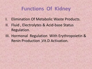
  • 1. Functions Of Kidney I. Elimination Of Metabolic Waste Products. II. Fluid , Electrolytes & Acid-base Status Regulation. III. Hormonal Regulation With Erythropoietin & Renin Production ,Vit.D Activation.
  • 3. I. Physical Examination 1) Total Volume Normal Volume:600-2000ml/Day a) Polyuria:urine Volume >2000ml/Day  Causes :-Osmotic Diuresis :-Polydipsia(diabetes Insipidus) :-Diuretics :-Chronic Renal Failure
  • 4. • Oliguria b) EXCRETION <400ML/DAY  CAUSES :-DEHYDRATION :-CHF :-SHOCK :-RENAL ARTERY EMBOLISM :-ACUTE GLOMERULONEPHRITIS :-ATN :-RENAL FAILURE :-GU TRACK OBSTRUCTION
  • 5. C) ANURIA :- EXCRETION <100ML/DAY Causes :-Bilateral Complete Urinary Tract Obstruction :-Acute Cortical Necrosis :-Necrotizing Glomerulonephritis
  • 6. 2) Specific Gravity: It Reflects The Degree Of Concentration Of Urine. Methods: 1)reagent Strips 2)refractometer 3)urinometer 4)falling Drop Method
  • 7. 1.Reagent Strips  Indirect Method  Reagent Area Contains 3 Main Ingredients:1)polyelectrolytes 2)indicator Substance 3)buffer  PRINCIPLE:IT IS BASED ON THE Pka CHANGE OF THE PRE-TREATED POLYELECTROLYTES IN RELATION TO THE IONIC CONC.Of Urine.When The Ionic Conc.IS HIGH ,Pka IS DECREASED AS IS THE Ph.The Indicator Substance Then Change Color Relative To The Ionic Conc & This Is Translated To The Specific Gravity Values.  Advantage:not Affected By High Amounts Of Glucose,protein Or Radiographic Contrast Media.
  • 8. 2)refractometer  Principle:refractive Index Of A Solution Is Related To The Content Of Dissolved Solids Present.  It Is Temperature Compensated Hand Model(60 To 100f)  Requires Only Few Drops Of Urine.
  • 9. 3)URINOMETER IT IS A HYDROMETER ADOPTED TO DIRECTLY MEASURE THE SPECIFIC GRAVITY OF URINE AT ROOM TEMP.
  • 10. 4)falling Drop Method  Direct Method  More Accurate Than Refractometer  More Precise Than Urinometer
  • 11. Interpretation: Normal-1.016 To 1.022/24 Hr Increased In:-proteinuria :-Diabetes Melitus :-Diuretics :-Antibiotics :-Acute Glomerulonephritis Decreased In:-renal Insufficiency
  • 12. Diurnal Variation:higher In The Morning &Lower In Afternoon Fixed Specific Gravity:sp.GRAVITY OF ANY SAMPLE OF URINE COLLECTED REMAINS FIXED E.G.1.010 Seen In Chronic Glomerulonephritis
  • 13. 3.Color COLOR CONDITIONS PALE YELLOW NORMAL URINE RED Hb ANILNE DYES PHENOPHTHELIN SMOKY RED OR BROWN BLOOD DRUGS LIKE LEVODOPA YELLOWISH ACRIFLAVIN YELLOWISH GREEN BILE PIGMENTS CAROTENE ACRIFLAVIN YELLOW BROWN BILIRUBIN BROWN BLACK HOMOGENTISIC ACID MELANIN METHEMOGLOBIN PORTWINE PHORPHYRINS
  • 14. Physical Examination 4) Appearnce • Normal:clear • Milky:lipiduria :Chyluria :PRESENCE OF Pmns • Cloudy:phosphates :Carbonates :Uric Acid :Bacteria :Sperm
  • 15. Physical Examination 5)odour • Normal:urinoid • Maple Syrup-maple Syrup Urine Disease • Mousy-phenylketonuria • Sweet-ketosis • Fishy-tyrosinemia • Sulfurous-cystinuria • Lack Of Odour- ARF(due To ATN)
  • 16. Chemical Examination 1)proteins Commonly Perfomed Methods Are: I) REAGENT STRIPS: MECHANISM:THIS METHOD TAKES ADVANTAGE OF THE PROTEIN ERROR OF Ph INDICATORS. PROTEINS CARRY A CHARGE AT PHYSIOLOGICAL Ph , THEIR PRESENCE WILL ELICIT A Ph CHANGE.
  • 17. Chemical Examination -In The Absence Of Protein The Strip Is Yellow. -30-60 Sec. Following Urine Application, Variable Shades Of Green Depending On The Type And Conc. Of Protein Present. -Results Read In A “Plus” System As: Negative Trace 1+ ( 30 Mg% ) 2+ ( 100 Mg%) 3+ (300mg%) 4+ (>2gm%)
  • 18. Chemical Examination Ii) HEAT & ACETIC ACID TEST • Principle: Proteins Are Precipitated When Boiled In Acid Solution. • Method: • -Fill A Test Tube With Urine Up To 2/3. • -Boil The Upper Portion By Holding The Tube Up At The Bottom
  • 19. Chemical Examination -Appearance Of Cloudiness Or Precipitates On Top;may Be Due To Proteins Or Phosphates. -Add 1 T0 3 Drops Of 10%acetic Acid & Boil The Top Portion Again. -Cloudiness Due To Phosphates Will Disappear But Not Due To Proteins.
  • 20. Chemical Examination Iii) SULPOSALICYLIC ACID TEST -Mix Equal Amounts Of Urine With 5% Salphosalicylic Acid In A Test Tube. -Grading Negative: No Turbidity(5mg/Dl Or Less) TRACE: PERCEPTIBLE Turbidity(20mg/Dl) 1+ :Distinct Turbidity,no Discrete Granulation(50mg/Dl) 2+ :Turbidity With Granulation,no Flocculation(200mg/Dl) 3+ :Turbidity With Granulation And Flocculation(500mg/Dl) 4+ :Clumps Of Precipitated Protein Or Solid Precipitate (1gm/Dl OR MORE)
  • 21.  Causes Of Proteinuria • It Is Divided In To: 1)pre-renal:-chf -Cerebral Injury -Severe Infections -Fever 2)renal:-glomerulonephritis -Nephrosclerosis -Diabetes Glomerulosclerosis -Nephrotic Syndrome -Pyelonephritis 3)post-renal:-inflammation Of The Pelvis Of Kidney, Ureter, Bladder
  • 22. Quantification Of Proteinuria • HEAVY Proteinuria(>4gm/Day) - Nephrotic Syndrome Associated With 1 Primary Renal Disease: Idiopathic ARPGN Ch. Gn 2 Systemic Disease With Renal Involvement Dm Sickle Cell Disease Sle 3 Others Severe CHF Renal Vein Thrombosis Malignant HT
  • 23. Quantification Of Proteinuria • MODERATE PROTEINURIA (1 To 4 Gm/Day) - Nephrosclerosis - Multiple Myeloma - Toxic Nephropathy - Presence Of Calculi • MINIMAL Proteinuria(<1gm/Day) - Chronic Pyelonephritis - Chronic Interstitial Nephritis - Congenital- Polycystic Kidney Disease - Medullary Cystic Disease
  • 24. Qualitative Proteinuria • It Requires Electrophoretic Separation Of Urine Proteins. 1. Glomerular Pattern: Less Selective Proteinuria Suggest Severe Glomerular Damage. 2. Tubular Pattern: Loss Of LMW Proteins E.G. B2microglobulin Light Chain Ig Tubular Proteinuria Seen In: Fanconi’s Syndrome Cystinosis Wilson’s Disease Pyelonephritis
  • 25. Qualitative Proteinuria 3. Overflow Proteinuria - Due To The Overflow Of Excess Levels Of A Protein In The Circulation. - Initially Not Associated With Glomerular Or Tubular Damage But Themselves Cause Renal Damage E.G. Myoglobin Causing ATN
  • 26. Qualitative Proteinuria 4.Bence- Jones Proteinuria - It Represents Either Kappa Or Lambda Ig Light Chain. • Causes -Multiple Myeloma -Monoclonal Gammopathy -Cryoglobulinemia -Primary Amyloidosis -Adult Fanconi’s Syndrome
  • 27. 1) Heat Method -To 10 Ml Of Fresh Clear Urine ,Add 2ml Of Saturated Sod. Chloride Soln To Prevent Precipitation Of Mucin. -Add Acetic Acid Soln Drop By Drop To Make Ph Slightly Acidic. -Heat The Urine In A Beaker Or Waterbath Controlling The Temp.With A Thermometer.
  • 28. -If B-J Proeins Are Present ,The Urine Becomes Cloudy Between 40 & 50C And A Flocculant Precipitate Appears To 50c. -On Raising The Temp.To 100c The Precipitate Dissolves Completely Or Partially & Reappears On Cooling.
  • 29. Qualitative Proteinuria 2) Electrophoresis -Presence Of B-J Globoulin Is Indicated By Single Sharp Peak In The Globulin Region On Electrophoresis. -More Specific Than Heat Method. -Positive Test For B-j Proteins By Heat Method Should Always Be Confirmed By Electrophoresis
  • 30. Qualitative Proteinuria 5. Micro Albuminuria - Presence Of Albumin In Urine Above The Normal Level But Below The Detectable Range Of Conventional Urine Dip Stick Method. - Measured By 1 Immunological Method 2 Nephelometric Method 3 Radioimmuno Assay *Causes: - Indicator Of Early & Possibly Reversible Glomerular Damage - Dm - Ht
  • 31. Chemical Examination  Othe Types Of Proteinuria 1)postural Proteinuria:first Morning Urine Before Arising Shows High Specific Gravity But No Protein.Protein Only Appears After Person Is Upright. -Usually <1.5gm/Day -Benign Condition -Slowly Disappears With Time -Occurs In 15% Of Apparently Healthy Persons -SEEN IN SOME Pts. With Resolving Acute Pyelonephritis Or Gn.
  • 32. Chemical Examination 2)transient Proteinuria:commonly Found In Routine Urinalysis Of Asymptomatic Healthy Children & Young Adults Initially Progressive Renal Disease Is Not Present. Usually < 2 Gm/Day Disappear With Recovery From Precipitating Cause. -Associated With High Fever -Chf -Hypertension -Stress -Exposure To Cold -Strenous Exercise -Seizures
  • 33. Chemical Examination 3)persistent Proteinuria A)glomerular : IDIOPATHIC o Membrenoproliferative Gn o Membrenous Glomerulopathy o Minimal Change Disease o Focal Segmental Glomerulosclerosis o Amyloidosis
  • 34. Chemical Examination  Secondary o Infections o Vascular o Drugs(nsaids,heroin,gold,penicillamine o Autoimmune o Neoplasia o Hereditary & Metabolic Ds. B)decreased Tubular Reabsorption  Acquired o Drugs o Heavy Metals o Sarcoidosis o Acute Tubular Necrosis
  • 35. Chemical Examination o Interstitial Nephritis o Acute & Chronic Pyelonephritis o Renal Graft Rejection  Congenital o Fanconi Syndrome o Oculo-cerebral-renal Syndrome  Hereditary o Wilson Ds. o Sickle Cell Ds. o Medullary Cystic Ds.
  • 36. Chemical Examination 2)sugar & Other Reducing Substances Methods I.Reagent Strips -Based On A Specific Glucose Oxidase & Peroxidase Method -Specific For Glucose -Doesn’t React With Other Reducing Substances • Interpretation -Negative -Trace ( 100mg%) -1+ (250 Mg%) -2+ (500mg%) -3+ (1 Gm% ) -4+ (> 2 Gm%)
  • 37.
  • 38.  False Positive Results -Exposed To Air -Peroxidase Contamination -Oxidising Agents  False Negative Results -ASCORBIC ACID >25mg% -Drugs(aspirin) -Specific Gravity >1.020 -HIGH Ph
  • 39. Chemical Examination Ii. Benedict’s Qualitative Test Principle:cupric Ions Are Reduced To Cuprous Form By Sugar & Other Reducing Sustances When Heated. Method : 1] To 5ml Of Qualitative Benedict’s Reagent,8 Drops Of Urine Are Added & Mixture Is Boiled For 2 Minutes 2] Examine After Cooling
  • 40. Chemical Examination  Interpretation :Test Is Positive If The Originally Blue Reagent Is Discoloured With The Formation Of Precipitate; Olive Green,yellowish,orange Or Brick Red(depending Upon The Amount Of The Reducing Substances Present)  Sugars Reducing The Benedict’s Reagent: -Glucose -Fructose -Galactose -Pentoses
  • 41. Chemical Examination Other Reducing Substances : -Homogentisic Acid -Gluconates -Creatinine -Uric Acid -Certain Drugs
  • 42. Chemical Examination  Causes 1)hereditary I. Galactosemia II. Galactokinase Deficiency III. Sever Liver Disease With Galactose Intolerance IV. Hereditary Fructose Intolerance V. Phenylketonuria 2) Due To Hyperglycemia I. Diabetes Melitus II. Cushing’s Syndrome III. Administration Of Hormones IV. Drugs(morphine,anesthetic Drugs)
  • 43. Chemical Examination 3)renal I. Tubular Origin(s.Glucose<180mg%,oral & INTRAVENOUS GLUCOSE TOLERANCE TESTS ARE NORMAL,KETOSIS IS ABSENT) II. Fanconi Syndrome III. Toxic Renal Tubular Acidosis IV. Inflammatory Renal Disease V. Glomerular Due To Increased Glomerular Filtration Rate Without Tubular Damage) 4)lactosuria -During Lactation & Late In Normal Pregnancy -Sepsis
  • 44. Chemical Examination 3)ketone Bodies Acetone,aceto-acetic Acid & 3-hydroxybutyrate Are 3 Ketonbodies Seen In Urine. Methods I.Reagent Strips -Based On A Nitroprusside Reaction For Ketones 1)chemistrip -Detects About 10mg /Dl Of Acetoacetic Acid & 70mg/Dl Of Acetone 2)multistix -Detects 5-10mg/Dl Of Aceto-acetic Acid -Doesn’t React With Acetone
  • 45. Chemical Examination Ii.Rothera’s Test For Acetone & Aceto-acetic Acid Method -Take 5 Ml Of Urine In A Test Tube -Add Approx.1gm OF AMMONIUM SULPHATE & 3 DROPS OF FRESHLY PREPARED SODIUM NITROPRUSSIDE SOLN. -Now Run Along The Side Of The Tube,liquor Ammonia So As To Form A Layer On The Top. -If Permangenate Colour Ring Appears At The Junction Of The Two Fluid,test Is Positive.
  • 46. Chemical Examination 3)gerhardt’s Test For Acetoacetic Acid -To 10 Ml Of Fresh Urine ,Add A Few Drops Of 10%ferric Chloride Drop By Drop Until A Precipitate Is Formed. -Filter The Precipitate & To Filtrate Add More Ferric Chloride Drop By Drop . -If Test Is Positive,a Violet Red Colour Develops.
  • 47. Causes Of Ketonuria -Metabolic Conditions E.G.Dm Renal Glycosuria -Dietary Conditions E.G.Starvation High Fat Diets -Increased Metabolic Requirements E.G.Hyperthyroidism Fever
  • 48. Chemical Examination 4)bile Salts & Bile Pigments Methods: A)gmelin’s Test For Bile Pigments -Take 3 Ml Of Conc. Nitric Acid In A Test Tube. -Place Equal Amount Of Urine On It. -Shake The Tube Gently From Side To Side. -Note The Color Change. -Interpretation: If Bile Pigments Are Present , There Is Play Of Colors: Yellow,red,violet,blue & Green.
  • 49. B) Harrison Fouchet’s Test For Bile Pigments - 10 Ml Urine + 5 Ml Bacl2 Soln. -It Gives Precipitates. -Filter It Through Whatman No.2 Filter Paper. -Gently Transfer Ppt. On Other Filter Paper. -Now Add 1 To 2 Drops Of Fouchet’s Reagent On It. * Interpretation : It Gives Green Or Blue Color If Bilirubin Is Present.
  • 50. c) Hay’s Sulphur Flower Test For Bile Salts -Take 5 Ml Of Urine In A Large Test Tube Or Beaker. -Sprinkle Sulphur Powder On The Surface. * Interpretation : If Bile Salts Are Present, Sulphur Particles Sink To Bottom Of The Tube. o Inreased In Jaundice.
  • 51. Microscopic Examination • 15ml Of Mixed Sample Of Urine Is Spun At 2000rpm For 10mins. • The Sediment Is Suspended In 1 To 2 Drops Of Urine & Coverslip Preparation Is Made For Microscopy. • It Is Performed To Detect Cells,casts & Crystals.
  • 52. Microscopic Examination 1}cells A)red Blood Cells -Pale Biconcave Discs. Normal: 0 To 2 Cells/Hpf Abnormal: >3 Cells/Hpf Causes: Trauma Gn Intestitial Nephritis Calculus Tumors Acute Infection Tb Atn
  • 53. • DYSMORPHIC Rbcs -Cells With Protusions Or Fragmentation -Their Presence Strongly Suggests Renal Glomerular Bleeding. B)leucocytes I)neutrophils:granular Sphere About 12um With Multilobulated Nuclei. Normal:<5 Leucocytes /Hpf Females May Show Higher Number
  • 54. • Pyuria-increased No.Of Leucocytes In Urine Causes:pyelonephritis Cystitis Prostatitis Urethritis Acute Urethral Syndrome Fever Physiological-following Exercise
  • 55. C)eosinophils -Not Normally Seen -Seen In -Tubuloiterstitial Ds Associated With Hypersensitivity To Drugse.G.Penicillin -Disorder Of GUT -Uti -Renal Transplant Rejection D)lymphocytes & Mononuclear Leucocytes -Small Lymphocytes Are Normally +Nt In Urine -Mononuclear Cells>30% Suggests Chronic Inflammation
  • 56. E)epithelial Cells I)squamous Epi.Cells -Seen In Normal Urine Ii)transitional Epi.Cells
  • 57. Iii)renal Tubular Epi.Cells -Small No.+Nt Normally -Increased In-atn -Drugs -Heavy Metals
  • 58. Iv)collecting Duct Cells -Increased In : -Renal Transplant Rejection -Atn -Ischemic Injuries -Malignant Nephrosclerosis -Acute GN With Tubular Damage -Drugs & Chemicals
  • 59. • 2}casts -These Are Cylindrical Structures With Parallel Edges, Basically These Are Precipitates Of Proteins Formed In DCT And Collecting Tubules. -Tamm-horsfall Protein,constituting About 1/3 Of Total Urinary Protein, Forms Matrix Of All Casts. -Classification Of Casts I.Matrix Ii.Cellular Iii.Inclusions Iv.Pigments
  • 60. I.Matrix Casts 1)hyaline Casts -0 To 2 Casts/Lpf Is Normal -Increased In-renal Ds. -Transiently Increased In-exercise -Heat Exposure -Dehydration -Fever -Chf -Diuretic Therapy
  • 61. 2)waxy Casts Seen In -Tubular Inflammation & Damage -Crf -Acute & Chronic Renal Allograft Rejection
  • 62. Ii.Cellular Casts 1)rbcs CASTS Seen In:agn Ig Nephropathy Lupus Nephritis Sabe,infarction
  • 63. 2)leucocyte Casts Seen In-tubulointerstitial Ds -Glomerular Ds. -Interstitial Nephritis. -Lupus Nephritis
  • 64. 3)renal Tubular Epithelial Cell Cast Seen In -ATN -Viral Ds E.G.Cmv -Exposure To Drugs -Heavy Metal Poisoning -Allograft Rejection 4)mixed Cells -Erythrocytes,neutrophils & Renal Tubular Cells
  • 65. Iii.Inclusions 1)granular Cast Seen In -Glomerular & Tubular Ds -Tubulointerstitial Ds. -Renal Allograft Rejection -Pyelonephritis -Viral Infections -Chronic Lead Poisoning
  • 66. 2)fatty Casts Seen In Nephrotic Syndrome 3)crystal Cast -Cast Containing Urate,ca Oxlate & Sulfonamides. 4)haemosiderin Granules 5)melanin Granules
  • 67. Iv.Pigmented Casts 1.Hb Cast Seen With -Erythrocyte Cast -Glomerular Ds. 2.Myoglobin Cast Seen In Myoglobinuria Arf 3.Bilirubin & Other Drug Casts Seen In Obstructive Jaundice. V.Broad Casts Seen In CRF Indicates Tubular Dilatation
  • 68. 3}crystals I.Crystals In Acidic Urine A) Ca.Oxlate Crystals -Oxaluria -Crohn’s Ds. -Oxalate Stones -Methoxyfurane Toxicity
  • 69. B) Amorphous Urate - Precipitate On Standing In Conc. Urine C) Crystalline Uric Acid Seen In Pt. On Chemotherapy Lesch-nyhan Syndrome Uric Acid Stones
  • 70. Ii.Crystals Found In Alkaline Urine A)triple Phosphate B)amorphous Phosphate C)ca Carbonate D)ammonium Biuret - All Are Seen In UTI
  • 71. Iii. Other Crystals In Urine A)cysteine Crystals -Cystinuria -Cystine Calculi
  • 72. B)tyrosine Crystals -Severe Liver Disease C)leucine Crystals -Severe Liver Disease D)sulfadiazine Crystals -Pt On Sulfonamide Therapy E)ampicillin (High Dose) F)radiographic Media -After I.V. Radiographic Studies
  • 73. 4) Abnormal Cells & Other Formed Elements I)tumor Cells -Exfoliated From Renal Pelvis,ureter,bladder Wall & Urethra Ii)viral Inclusion Cells Herpes –Synctial Giant Cells Containing Eosinophilic ,Intranuclear Inclusions CMV -Enlarged Cells With Basophilic Intranuclear Inclusions Polyoma –Dense,basophilic ,Homogenous Intanuclear Inclusions
  • 74. Iii) Platelets Up To 30,000/Ul Seen In HUS Iv)bacteria Significant When >100000 Organisms/Ml With Gram’s Stain V)fungi Candidia Yeast Vi) Parasites Commonly Found Parasites Are -Trichomonas Vaginalis -Schistosoma Haematobium -Entamoeba Histolytica
  • 75. Reference Values For The Urinary Sediment Considered As Abnormal • Erythrocytes>5 Cells At Hpf • Leukocytes>5 Cells At Hpf • Renal Tubular Cells>2 Cells At Hpf • Transitional Cells>5 Cells At Hpf • Squamous Cells Rarely Significant • Hyaline Casts>3 Cast At Lpf • Granular Casts>1 Cast At Lpf • Pathological Casts Any • Crystals Any
  • 76. Special Examination 1) Quantitative Sugar Estimation -By Quantitative Benedict’s Test -Having Prognostic Value In DM -But Now A Days Not Useful Because Of Availability Of Glycosylated Hb.
  • 77. 2) Quantitative Protein Estimation -For This 24 Hrs Urine Collection Sample Is Required A)esbach’s ALBUMINOMETER -Urine Is Filled Up To “U” Mark. -Esbach’s Reagent Is Filled Up To “R” Mark. -Keep It Overnight. -Next Day Precipitated Protein Form Whitish Clot Like Coagulant Will Be Read As Per Marking On Instrument. -Result Is Recorded In Gm/Lit/24 Hrs.
  • 78. • B) Biuret Method – Colorimetric Method – Less Time Consuming – More Specific – Routinely Used And More Reliable  PRINCIPLE :PEPTIDES OF PROTEIN REACT WITH ALKALINE COPPER TARTARATE SOLN.TO GIVE A VOILET COLOURED COMPLEX WHICH IS MEASURED COLORIMRTRICALLY AT 540nm WHICH IS DIRECTLY PROPORTIONAL TO THE CONC.Of Protein In Specimen. C)sulphosalicylic Acid /Tca Method – Turbidimetric & Semi-quantitative Methods
  • 79. 3)urobilinogen -Normally Present In Urine. -Unstable In Acid Urine -When Exposed To Light,reduced To Form A Pigment Urobilin,hence Urine Sample Is Collected In Amber Coloured Bottle. Method Principle:formation Of Red Coloured Azo Dye With Diazonium Compound -To 10ml Of Urine,add 1ml Of Ehrlich’s Reagent -If Intense Red Colour Develops,further Testing Is Done By Using Serial Dilution Of Urine . -Reading Is Recorded Up To The Test Tube Showing Faint Pink Colour.
  • 80. NORMAL: 1:10 To 1:30 Dilution Significance: *Hemolytic Jaundice-increased *Hepatocellular Jaundice-early Stage->increased Late Stage->decreased *Obstructive Jaundice-absent Or Decreased
  • 81. Biochemical Tests Perfomed In Blood  Blood Urea  Serum Creatinine
  • 82.  Blood Urea • End Product Of Proteins & Amino Acid Metabolism • Produced By Liver • Excreted Mainly By Kidneys • Measured By Following Methods
  • 83. I.Berthelot Method Principle:urea Is Converted To Ammonium By The Use Of Urease.Ammonium Ion Then Reacts With A Mixture Of Salicylate,sodium Nitroprusside & Hypochlorite To Yield A Blue – Green Chromophore.The Intensity Of The Color Formed Is Proportional To The Urea Concentration In The Sample. It Is Colorimetric End Point Test.
  • 84. Reagents Reagent I : Urea Enzyme Reagent Reagent II : Urea Color Developer Urea Standard : 50mg/Dl Sample Serum From Plain Bulb Plasma From Edta,citrate Bulb
  • 85.
  • 86. BLANK STANDARD SAMPLE REAGENT I 1 ml 1 ml 1 ml STANDARD - 10ul - SAMPLE - - 10ul MIX WELL & INCUBATE FOR 5 mins AT 37c OR 10 mins AT RT REAGENT II 1 ml 1 ml 1 ml MIX WELL & INCUBATE FOR 5 mins AT 37c OR 10 mins AT RT
  • 87. • Measure The Absorbance Of Standard & Test Against Reagent Blank At 578nm. • Calculation • Urea ( Mg/Dl)=(at-ab/AS-AB) X Conc.Of Std. • Normal Value: 15 – 40 Mg/Dl • Bun=urea X (M.W. Of Nitrogen /M.W.Of Urea) • Normal Value: 5 – 20 Mg/Dl
  • 88. Other Methods Ii.Nessler Method Principle : When Diluted Serum Is Incubated With Urease Powder ,It Produces Liquid Ammonia.After Depolarization With Zn(oh)2,color Is Produced By Ammonia With Nessler’s Reagent,which Is Read At Green Filter. Iii.Oxime Method Principle : On Heating Diacetyle Monoxime Give Hydroxylamine & Diacetyl Which React With Urea & Give Color Complex.
  • 89. • Limitation Of Urea Measurement 1)considerable Glomerular Damage Must Occur Before Urea Increases. 2)urea Depends On : i) Protein Intake In Diet ii) Catabolic State Of Protein iii) High Protein Load E.G.Upper G.I.Bleeding iv) Advanced Liver Disease v) State Of Hydration
  • 90. AZOTEMIA: Biochemical Abnormality Denoting Higher Level Of BUN In Blood. Uremia :Clinical Menifestation Comprised Of Increased Urea In Plasma,acidemia ,Electrolyte Imbalance Associated With Clinical Features Such As Vomiting,nausea,anaemia, Altered Mentation & Altered Hemostasis
  • 91.  Increased Level Of Urea Seen In : I)increased Tissue Protein Catabolism Seen In-fever -Diabetic Coma -Thyrotoxicosis -After A Major Operation Ii)excess Brekdown Of Blood Protein Seen In -Leukemia -Gastrointestinal Ds. Iii)diminished Excretion Of Urea -Commonest & Most Common Cause -Divided Into
  • 92. 1)pre-renal -Urine Is Hypertonic -Mild Proteinuria -Urine Sediment Contain Hyaline Or Granular Casts Due To Decreased Renal Blood Flow: -Shock -Chf -Dehydration -Haemorrhage -Renal Artery Stenosis
  • 93. 2)renal Due To Decreased Glomerular Filtration -Arf -Crf -Gn -Pyelonephritis -Tubular Necrosis -Interstitial Nephritis
  • 94. 3)post Renal Due To Obstruction In Urinary Tact -Ureteric Stone -Urethral Stone -Urethral Stricture -Bladder Stone -Prostatic Hypertrophy -Prostatic Tumor
  • 95.  Serum Creatinine • Creatinine Is Endogenous Substance Produced By The Muscle From Creatine & Creatine Phosphate By A Non-enzymatic Dehydration Process. • Most Widely Used As Marker Of Gfr Because I)endogenous Substance Of Fairly Constant Rate Of Production Ii)not Bound To Plasma Proteins ,Therefore Is Filtered Freely By The Glomerulus Iii)not Reabsorbed By The Renal Tubules & Only A Small Amount Is Secreted By The Tubules.
  • 96. I. Alkaline Picrate Method PRINCIPLE : In An Alkaline Medium ,Creatinine Reacts With Picrate To Produce An Orange Yellow Colour Which Is Proportionaal To The Creatinine Concentration In Sample. Reagents : 1.Picric Acid 2.Sodium Hydroxide 3.Creatinine Standard- 2mg/Dl Sample : Serum Or Plasma
  • 97.
  • 98.
  • 99.  Procedure -Reading Is Taken At 510nm TEST STANDARD BLANK PICRIC ACID 0.5% NaOH PICRIC ACID 0.5% NaOH DISTILLED WATER PICRIC ACID 0.5%NaOH DISTILLED WATER TEST STANDARD BLANK PICRIC ACID 500ul 500ul 500ul 0.5%NaOH 500ul 500ul 500ul DISTILLED WATER - - 50ul STANDARD - 50ul - SERUM 50ul - -
  • 100. Normal Values Male :0.6- 1.2 Mg% Female :0.5-1.0 Mg% Other Methods Ii.ENZYMATIC METHOD : Creatinine Is Hydrolysed By Creatinine Iminohydrolase To Ammonia & N- methylhydantoin.The Ammonia Then Combines With 2-oxoglutarate & NADH In The Presence Of Glutamate Dehydrogenase To Produce Glutamate & NAD+.The Consumption Of Nadh,measured As Decreased In Absorption At 340nm.
  • 101. Iii.High Perfomance Liquid Chromatography -Excellent Specificity -Requires Deproteinization Of Sample -Lengthy Procedure
  • 102. • Creatinine Vaues Increase In 1.Skeletal Muscle Causes -Trauma -Rapidly Progressive Muscular Dystrophy -Poliomyositis -Dermatomyositis -Myesthenia Gravis -Starvation -Sepsis -Major Surgery
  • 103. 2.Acute & Chronic Renal Diseases In Renal Ds.Creatinine Tends To Rise Slowly Than Urea. 3.Others -Diabetic Acidosis -Purperium
  • 104. Bun : Creatinine Ratio • Helps To Differentiate Pre-renal & Post-renal Azotemia From Renal Azotemia. • Normal Value : 12 To 16 1.Increased Ratio With Normal Creatinine A)pre Renal Azotemia(due To Decreased GFR) B)catabolic States With Increased Tissue Breakdown C)gi Hemorrhage
  • 105. D)high Protein Intake E)impaired Renal Function Plus- -Excess Protein Intake Or Tissue Breakdown -Urine Reabsorption (E.G.Ureterocolostomy) -Patients With Reduced Muscle Mass F)selective Increase In Plasma Urea During Use Of Loop Diuretics 2.Increased Ratio With Elevated Creatinine -Post Renal Azotemia E.G.Obstuctive Uropathy -Pre Renal Azotemia Superimposed On Renal Disease
  • 106. 3.Decreased Ratio With Decreased Bun -Atn -Low Protein Diet -Severe Liver Ds. -Repeated Dialysis -Inherited Deficiency Of Urea Cycle Enzymes -Pregnancy 4.Decreased Ratio With Increased Creatinine -Rhabdomyolysis -Phenacemide Therapy -Muscular Patients Developing Renal Failure
  • 107. Tests Measuring Gfr • GFR Is Measured By Clearance Tests. • Clearance Test Gives Relatively Accurate & Useful Measure Of The Gfr & Also The Excretory Capacity Of The Kidney. Substances Used For Measuring Gfr : I. Exogenous Substances I)inulin Ii)mannitol Iii)sodium Thiosulphate Iv)tc-dtpa( Diethylene Triamine Penta-acetic
  • 108. Ii.Endogenous Substances I) Creatinine Ii) Urea -It Requires 24 Hrs Urine Collection
  • 109. Equation For Clearance Is : Cx=uxv/Px Cx=clearance Of A Substance X Ux=conc.Of The Substance X In Urine Px=conc.Of Substance X In Plasma V=volume Of Urine Per Unit Time
  • 110. Interpretation o Good Estimate In Pt.With Reduced GFR. o Inaccurate If S.Creatinine Is Not Stable o Unreliable In Pts.With Extremes Of Age,body Size Or Composition o A Normal GFR In Association With Impaired Concentrating Ability May Be Found In Sickle Cell Anaemia,diabetes Insipidus,pyelonephritis. o Increased Gfr Is A Risk Factor For Progressive Nephropathy In Dm
  • 111. o If Urea Clearance Value Falls <20% - Severe Renal Failure <5% - Uremic Coma
  • 112. STAGE DESCRIPTION GFR (ml/min/1.73m2) STAGE 1 KIDNEY DAMAGE WITH NORMAL OR INCREASED GFR >90 STAGE 2 KIDNEY DAMAGE WITH MILD DECREASE IN GFR 60 - 89 STAGE 3 MODERATE DECREASE IN GFR 30 - 59 STAGE 4 SEVER DECREASE IN GFR 15 - 29 STAGE 5 RENAL FAILURE < 15 OR DIALYSIS
  • 113. Determination Of Acute Renal Failure • To Differntiate Pre-renal Azotemia & Acute Tubular Necrosis. • Fractional Excretion : Is The Quantity Of A Substance Excreted In Urine Expressed As A Fraction Of The Filtered Load Of The Same Substance. Fe(sod.) =( Usod./Psod.) X (Pcreat/Ucreat) • If It Is < 0.01 – Pre Renal Azotemia • If It Is >0.01 - Atn
  • 114. Urine & Serum Diagnostic Indices PRE RENAL RENAL POST RENAL URINE SODIUM <20 mEq/ L >20 mEq/ L >20 mEq /L URINE CHLORIDE <20 mEq/L >20 mEq /L -------- FENa < 1 % > 2% >2% FEUrea < 35% ------ ------ URINE OSMOLARITY >500 mg% < 350 mg% < 350 mg% URINE/SERUM CREATININE > 40 < 20 < 20 URINE/SERUM UREA > 8 < 3 < 3 RENAL FAILURE INDEX < 1% > 1% >1% BUN : CREATININE > 20 ~ 10 ~10
  • 115. Renal Biopsy • Should Be Preceded By I)confirmation That Two Kidneys Are Present Ii)no Renal Infection Is Present Iii)there Is No Bleeding Disorder • Examination Should Include I)histology –Stained By H &E,trichome,pas,silver Ii)immunofluorescence-with Antisera Specific For Igg,iga,igm,c1q,c3,c4,fibrinogen,albumin,kappa & Lambda Light Chains Iii)electronmicroscopy –Alport Syndrome ,Thin Basement Membrane Nephropathy
  • 116. • Indications For Biopsy I)acute Renal Allograft Dysfunction Ii)persistent Or Reccurent Haematuria With Proteinuria Iii)nephrotic Syndrome To Distinguish Etiology Iv)proteinuria > 1gm/Day Or With Abnormal Urine Sediment V)nephrotic Syndrome- If Unresponsive To Therapy Or Before Therapy Vi)non-nephrotic Proteinuria With Progressive Disease Vii)evaluation Of Collagen Ds.(Sle)
  • 117. • Contraindications For Biopsy I)haemorrhagic Ds Ii)solitary Kidney Iii)active Kidney Infection Iv)renal Artery Vasculitis With Aneurysm V)hydronephrosis Vi)uncontrolled Severe HT Vii)uncooperative Pt.
  • 118. References Henry’s Clinical Diagnosis & Management By Laboratory Methods Interpretation Of Diagnostic Tests,jacques Wallach. Textbook Of Medical Laboratory Technology,godkar Www.Google.Com