Liver cirrhosis
•Descargar como PPTX, PDF•
343 recomendaciones•134,850 vistas

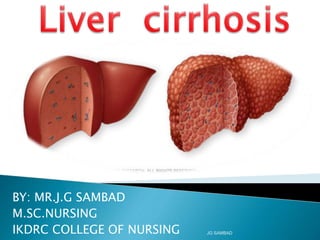
1. Cirrhosis is a consequence of chronic liver disease characterized by replacement of liver tissue with scar tissue. It impairs liver function and can result from various causes like chronic alcoholism or hepatitis. 2. Symptoms may include jaundice, ascites, bruising easily, and confusion. Complications arise as the disease progresses and can affect other organs. 3. Diagnosis involves blood tests, imaging, and biopsy showing nodular regeneration of liver tissue and fibrosis. Treatment focuses on managing complications and their underlying causes.

Recomendados
Más contenido relacionado
La actualidad más candente
La actualidad más candente (20)
Similar a Liver cirrhosis
Similar a Liver cirrhosis (20)
Más de MR. JAGDISH SAMBAD
Más de MR. JAGDISH SAMBAD (20)
Último
Último (20)
Liver cirrhosis
- 1. BY: MR.J.G SAMBAD M.SC.NURSING IKDRC COLLEGE OF NURSING JG SAMBAD
- 2. "With ordinary talent and extraordinary perseverance, all things are attainable." - Thomas E. Buxton "Achievement is connected with action, not in genes..…!” - Conrad Hilton JG SAMBAD
- 3. 1.5 kg, wedge shape 4 lobes, Right, left, Caudate, Quadrate. Double blood supply Hepatic arteries Portal – Venous blood Acini / Portal triad. Lobules – central. V JG SAMBAD
- 4. JG SAMBAD
- 5. JG SAMBAD
- 6. Metabolism – Carbohydrate, Fat & Protein Secretory – bile, Bile acids, salts & pigments Excretory – Bilirubin, drugs, toxins Synthesis – Albumin, coagulation factors Storage – Vitamins, carbohydrates etc. Detoxification – toxins, ammonia, etc. JG SAMBAD
- 7. •Blood •Conjugated & Conjugated •Urine – Urobilinogen •Stool – Stercobilin JG SAMBAD
- 8. JG SAMBAD
- 9. The word "cirrhosis" is a neologism that derives from Greek kirrhos, meaning "tawny" (the orange-yellow colour of the diseased liver). While the clinical entity was known before, it was René Laennec who gave it the name "cirrhosis" in his 1819 work in which he also describes the stethoscope. Cirrhosis is a consequence of chronic liver disease characterized by replacement of liver tissue by fibrous scar tissue as well as regenerative nodules (lumps that occur as a result of a process in which damaged tissue is regenerated, leading to progressive loss of liver function. JG SAMBAD
- 10. Cirrhosis of liver is a chronic, progressive disease characterized by widespread fibrosis (scaring) & nodule formation. Cirrhosis occurs when the normal flow of blood, bile, & hepatic metabolites is altered by fibrosis. JG SAMBAD
- 11. JG SAMBAD
- 12. 1. Alcoholic cirrhosis- Most common, due to chronic alcoholism. Scar tissue characteristically surrounds the portal area. 2. Postnecrotic cirrhosis- There are broad bands of scar tissue due to late results of acute viral hepatitis, postintoxication with industrial chemicals. 3. Biliary cirrhosis- Scaring occurs around bile duct in liver, Results from chronic biliary obstruction & infection. 4. Cardiac cirrhosis- Associated with severe right sided long term heart failure, fairly rare. JG SAMBAD
- 13. Primary event is injury to hepatocellular elements Initiates inflammatory response with cytokine release->toxic substances Destruction of hepatocytes, bile duct cells, vascular endothelial cells Repair through cellular proliferation and regeneration Formation of fibrous scar JG SAMBAD
- 14. JG SAMBAD
- 15. JG SAMBAD
- 16. Cirrhosis has many possible causes; sometimes more than one cause is present in the same patient. In the Western World, chronic alcoholism and hepatitis C are the most common causes. 1. Alcoholic liver disease (ALD). Alcoholic cirrhosis develops in 15% of individuals who drink heavily for more than a decade. There is great variability in the amount of alcohol needed to cause cirrhosis (as little as 3-4 drinks a day in some men and 2-3 in some women). Alcohol seems to injure the liver by blocking the normal metabolism of protein, fats, and carbohydrates. 2 . Chronic hepatitis C. Infection with this virus causes inflammation of and low grade damage to the liver that over several decades can lead to cirrhosis. JG SAMBAD
- 17. 3. Chronic hepatitis B. The hepatitis B virus is probably the most common cause of cirrhosis worldwide, especially South-East Asia, but it is less common in the United States and the Western world. Hepatitis B causes liver inflammation and injury that over several decades can lead to cirrhosis. Hepatitis D is dependent on the presence of hepatitis B, but accelerates cirrhosis in co-infection. JG SAMBAD
- 18. 4.Non-alcoholic steatohepatitis (NASH) In NASH, fat builds up in the liver and eventually causes scar tissue. This type of hepatitis appears to be associated with diabetes, protein malnutrition, obesity, coronary artery disease, and treatment with corticosteroid medications. This disorder is similar to that of alcoholic liver disease but patient does not have an alcohol history 5.Primary biliary cirrhosis May be asymptomatic or complain of fatigue, pruritus, and non-jaundice skin hyperpigmentation with hepatomegaly. There is prominent alkaline phosphatase elevation as well as elevations in cholesterol and bilirubin. JG SAMBAD
- 19. 6. Primary sclerosing cholangitis PSC is a progressive cholestatic disorder presenting with pruritus, steatorrhea, fat soluble vitamin deficiencies, and metabolic bone disease. There is a strong association with inflammatory bowel disease (IBD). 7. Autoimmune hepatitis This disease is caused by the immunologic damage to the liver causing inflammation and eventually scarring and cirrhosis 8. Hereditary hemochromatosis Usually presents with family history of cirrhosis, skin hyperpigmentation, diabetes mellitus, pseudogout, and/or cardiomyopathy, all due to signs of iron overload JG SAMBAD
- 20. 9. Wilson's disease Autosomal recessive disorder characterized by low serum ceruloplasmin and increased hepatic copper content on liver biopsy 10. Alpha 1-antitrypsin deficiency (AAT) Autosomal recessive disorder. Patients may also have COPD, especially if they have a history of tobacco smoking. Serum AAT levels are low. 11. Cardiac cirrhosis Due to chronic right sided heart failure which leads to liver congestion. JG SAMBAD
- 21. Galactosemia Cystic fibrosis Drugs or toxins Certain parasitic infections (such as schistosomiasis) JG SAMBAD
- 22. Some of the following signs and symptoms may occur in the presence of cirrhosis or as a result of the complications of cirrhosis. Many are nonspecific and may occur in other diseases and do not necessarily point to cirrhosis. Likewise, the absence of any does not rule out the possibility of cirrhosis. Spider angiomata or spider nevi. Vascular lesions consisting of a central arteriole surrounded by many smaller vessels due to an increase in estradiol. These occur in about 1/3 of cases. Palmar erythema Exaggerations of normal speckled mottling of the palm, due to altered sex hormone metabolism. JG SAMBAD
- 23. Gynecomastia Benign proliferation of glandular tissue of male breasts presenting with a rubbery or firm mass extending concentrically from the nipples. This is due to increased estradiol and can occur in up to 66% of patients. Hypogonadism Manifested as impotence, infertility, loss of sexual drive, and testicular atrophy due to primary gonadal injury or suppression of hypothalamic or pituitary function. Liver size. Can be enlarged, normal, or shrunken. Splenomegaly (increase in size of the spleen). Due to congestion of the red pulp as a result of portal hypertension. JG SAMBAD
- 24. Ascites Accumulation of fluid in the peritoneal cavity giving rise to flank dullness (needs about 1500 mL to detect flank dullness). It may be associated with hydrocele and penile flomation (swelling of the penile shaft) in men. Caput medusa In portal hypertension, the umbilical vein may open. Blood from the portal venous system may be shunted through the periumbilical veins into the umbilical vein and ultimately to the abdominal wall veins, manifesting as caput medusa. JG SAMBAD
- 25. Cruveilhier-Baumgarten murmur Venous hum heard in epigastric region (on examination by stethoscope) due to collateral connections between portal system and the remnant of the umbilical vein in portal hypertension. Fetor hepaticus Musty odor in breath due to increased dimethyl sulfide. Jaundice Yellow discoloring of the skin, eye, and mucus membranes due to increased bilirubin (at least 2-3 mg/dL or 30 mmol/L). Urine may also appear dark. Asterixis Bilateral asynchronous flapping of outstretched, dorsiflexed hands seen in patients with hepatic encephalopathy. Other Weakness, fatigue, anorexia, weight loss. JG SAMBAD
- 26. Nail changes. ◦ Muehrcke's nails - paired horizontal bands separated by normal color due to hypoalbuminemia (low production of albumin). ◦ Terry's nails - proximal two thirds of the nail plate appears white with distal one-third red, also due to hypoalbuminemia ◦ Clubbing - angle between the nail plate and proximal nail fold > 180 degrees JG SAMBAD
- 27. Hypertrophic osteoarthropathy Chronic proliferative periostitis of the long bones that can cause considerable pain. Dupuytren's contracture Thickening and shortening of palmar fascia that leads to flexion deformities of the fingers. Thought to be due to fibroblastic proliferation and disorderly collagen deposition. It is relatively common (33% of patients). JG SAMBAD
- 28. JG SAMBAD
- 29. JG SAMBAD
- 30. JG SAMBAD
- 31. JG SAMBAD
- 32. JG SAMBAD
- 33. JG SAMBAD
- 34. JG SAMBAD
- 35. The gold standard for diagnosis of cirrhosis is a liver biopsy Histologically cirrhosis can be classified as micronodular, macronodular, or mixed, but this classification has been abandoned since it is nonspecific to the etiology JG SAMBAD
- 36. Lab findings The following findings are typical in cirrhosis: Aminotransferases - AST and ALT are moderately elevated, with AST > ALT. However, normal aminotransferases do not preclude cirrhosis. Alkaline phosphatase - usually slightly elevated. GGT -- correlates with AP levels. Typically much higher in chronic liver disease from alcohol. Bilirubin - may elevate as cirrhosis progresses. Albumin - levels fall as the synthetic function of the liver declines with worsening cirrhosis since albumin is exclusively synthesized in the liver JG SAMBAD
- 37. Prothrombin time- increases since the liver synthesizes clotting factors. Globulins - increased due to shunting of bacterial antigens away from the liver to lymphoid tissue. Serum sodium - hyponatremia due to inability to excrete free water resulting from high levels of ADH and aldosterone. Thrombocytopenia - due to both congestive splenomegaly as well as decreased thrombopoietin from the liver. However this rarely results in platelet count < 50,000/mL. Leukopenia and neutropenia- due to splenomegaly with splenic margination. Coagulation defects - the liver produces most of the coagulation factors and thus coagulopathy correlates with worsening liver disease. JG SAMBAD
- 38. Other laboratory studies performed in newly diagnosed cirrhosis may include: Serology for hepatitis viruses, autoantibodies (ANA, anti-smooth muscle, anti-mitochondria, anti-LKM) Ferritin and transferrin saturation (markers of iron overload), copper and ceruloplasmin (markers of copper overload) Immunoglobulin levels (IgG, IgM, IgA) - these are non-specific but may assist in distinguishing various causes Cholesterol and glucose Alpha 1-antitrypsin JG SAMBAD
- 39. Imaging Ultrasound is routinely used in the evaluation of cirrhosis, where it may show a small and nodular liver in advanced cirrhosis along with increased echogenicity with irregular appearing areas. Ultrasound may also screen for hepatocellular carcinoma, portal hypertension and Budd-Chiari syndrome (by assessing flow in the hepatic vein). Other tests performed in particular circumstances include abdominal CT and liver/bile duct MRI (MRCP). Endoscopy Gastroscopy (endoscopic examination of the esophagus, stomach and duodenum is performed in patients with established cirrhosis to exclude the possibility of esophageal varices. If these are found, prophylactic local therapy may be applied (sclerotherapy or banding) and beta blocker treatment may be commenced. Rarely diseases of the bile ducts, such as as primary sclerosing, can be causes of cirrhosis. Imaging of the bile ducts, such as ERCP or MRCP (MRI of biliary tract and pancreas) can show abnormalities in these patients, and may aid in the diagnosis. JG SAMBAD
- 40. JG SAMBAD
- 41. As the disease progresses, complications may develop. In some people, these may be the first signs of the disease. Bruising and bleeding due to decreased production of coagulation factors. Jaundice due to decreased processing of bilirubin. Itching (pruritus) due to bile products deposited in the skin. Hepatic encephalopathy - the liver does not clear ammonia and related nitrogenous substances from the blood, which are carried to the brain, affecting cerebral functioning: neglect of personal appearance, unresponsiveness, forgetfulness, trouble concentrating, or changes in sleep habits. Sensitivity to medication due to decreased metabolism of the active compounds. Hepatocellular carcinoma is primary liver cancer, a frequent complication of cirrhosis. It has a high mortality rate. JG SAMBAD
- 42. Portal hypertension - blood normally carried from the intestines and spleen through the hepatic portal vein flows more slowly and the pressure increases; this leads to the following complications: ◦ Ascites - fluid leaks through the vasculature into the abdominal cavity. ◦ Esophageal varices - collateral portal blood flow through vessels in the stomach and esophagus. These blood vessels may become enlarged and are more likely to burst. JG SAMBAD
- 43. Problems in other organs. ◦ Cirrhosis can cause immune system dysfunction, leading to infection. Signs and symptoms of infection may be aspecific are more difficult to recognize (e.g. worsening encephalopathy but no fever). ◦ Fluid in the abdomen (ascites) may become infected with bacteria normally present in the intestines (spontaneous bacterial peritonitis). ◦ Hepatorenal syndrome - insufficient blood supply to the kidneys, causing acute renal failure. This complication has a very high mortality (over 50%). ◦ Hepatopulmonary syndrome - blood bypassing the normal lung circulation (shunting), leading to cyanosis and dyspnea (shortness of breath), characteristically worse on sitting up. ◦ Portopulmonary hypertension - increased blood pressure over the lungs as a consequence of portal hypertension. JG SAMBAD
- 44. As cirrhosis of the liver becomes severe, signals are sent to the kidneys to retain salt and water in the body. The excess salt and water first accumulates in the tissue beneath the skin of the ankles and legs because of the effect of gravity when standing or sitting. This accumulation of fluid is called edema or pitting edema. (Pitting edema refers to the fact that pressing a fingertip firmly against an ankle or leg with edema causes an indentation in the skin that persists for some time after release of the pressure. Actually, any type of pressure, such as from the elastic band of a sock, may be enough to cause pitting.) The swelling often is worse at the end of a day after standing or sitting and may lessen overnight as a result of the loss of the effects of gravity when lying down. As cirrhosis worsens and more salt and water are retained, fluid also may accumulate in the abdominal cavity between the abdominal wall and the abdominal organs. This accumulation of fluid (called ascites) causes swelling of the abdomen, abdominal discomfort, and increased weight. JG SAMBAD
- 45. JG SAMBAD
- 46. Fluid in the abdominal cavity (ascites) is the perfect place for bacteria to grow. Normally, the abdominal cavity contains a very small amount of fluid that is able to resist infection well, and bacteria that enter the abdomen (usually from the intestine) are killed or find their way into the portal vein and to the liver where they are killed. In cirrhosis, the fluid that collects in the abdomen is unable to resist infection normally. In addition, more bacteria find their way from the intestine into the ascites. Therefore, infection within the abdomen and the ascites, referred to as spontaneous bacterial peritonitis or SBP, is likely to occur. SBP is a life- threatening complication. Some patients with SBP have no symptoms, while others have fever, chills, abdominal pain and tenderness, diarrhea, and worsening ascites. JG SAMBAD
- 47. In the cirrhotic liver, the scar tissue blocks the flow of blood returning to the heart from the intestines and raises the pressure in the portal vein (portal hypertension). When pressure in the portal vein becomes high enough, it causes blood to flow around the liver through veins with lower pressure to reach the heart. The most common veins through which blood bypasses the liver are the veins lining the lower part of the esophagus and the upper part of the stomach. As a result of the increased flow of blood and the resulting increase in pressure, the veins in the lower esophagus and upper stomach expand and then are referred to as esophageal and gastric varices; the higher the portal pressure, the larger the varices and the more likely a patient is to bleed from the varices into the esophagus or stomach. Bleeding from varices usually is severe and, without immediate treatment, can be fatal. Symptoms of bleeding from varices include vomiting blood (the vomitus can be red blood mixed with clots or "coffee grounds" in appearance, the latter due to the effect of acid on the blood), passing stool that is black and tarry due to changes in the blood as it passes through the intestine (melena), and orthostatic dizziness or fainting (caused by a drop in blood pressure especially when standing up from a lying position). Bleeding also may occur from varices that form elsewhere in the intestines, for example, the colon, but this is rare. For reasons yet unknown, patients hospitalized because of actively bleeding esophageal varices have a high risk of developing spontaneous bacterial peritonitis. JG SAMBAD
- 48. JG SAMBAD
- 49. Some of the protein in food that escapes digestion and absorption is used by bacteria that are normally present in the intestine. While using the protein for their own purposes, the bacteria make substances that they release into the intestine. These substances then can be absorbed into the body. Some of these substances, for example, ammonia, can have toxic effects on the brain. Ordinarily, these toxic substances are carried from the intestine in the portal vein to the liver where they are removed from the blood and detoxified. As previously discussed, when cirrhosis is present, liver cells cannot function normally either because they are damaged or because they have lost their normal relationship with the blood. In addition, some of the blood in the portal vein bypasses the liver through other veins. The result of these abnormalities is that toxic substances cannot be removed by the liver cells, and, instead, the toxic substances accumulate in the blood. JG SAMBAD
- 50. When the toxic substances accumulate sufficiently in the blood, the function of the brain is impaired, a condition called hepatic encephalopathy. Sleeping during the day rather than at night (reversal of the normal sleep pattern) is among the earliest symptoms of hepatic encephalopathy. Other symptoms include irritability, inability to concentrate or perform calculations, loss of memory, confusion, or depressed levels of consciousness. Ultimately, severe hepatic encephalopathy causes coma and death. The toxic substances also make the brains of patients with cirrhosis very sensitive to drugs that are normally filtered and detoxified by the liver. Doses of many drugs that normally are detoxified by the liver have to be reduced to avoid a toxic buildup in cirrhosis, particularly sedatives and drugs that are used to promote sleep. Alternatively, drugs may be used that do not need to be detoxified or eliminated from the body by the liver, for example, drugs that are eliminated by the kidneys. JG SAMBAD
- 51. BRAIN LIVER Toxic N2 metabolites From Intestines Porta systemic shunts JG SAMBAD
- 52. Patients with worsening cirrhosis can develop the hepatorenal syndrome. This syndrome is a serious complication in which the function of the kidneys is reduced. It is a functional problem in the kidneys, that is, there is no physical damage to the kidneys. Instead, the reduced function is due to changes in the way the blood flows through the kidneys themselves. The hepatorenal syndrome is defined as progressive failure of the kidneys to clear substances from the blood and produce adequate amounts of urine even though some other important functions of the kidney, such as retention of salt, are maintained. If liver function improves or a healthy liver is transplanted into a patient with hepatorenal syndrome, the kidneys usually begin to work normally. This suggests that the reduced function of the kidneys is the result of the accumulation of toxic substances in the blood when the liver fails. There are two types of hepatorenal syndrome. One type occurs gradually over months. The other occurs rapidly over a week or two. JG SAMBAD
- 53. Traditionally, liver damage from cirrhosis cannot be reversed, but treatment could stop or delay further progression and reduce complications. A healthy diet is encouraged, as cirrhosis may be an energy-consuming process. Close follow-up is often necessary. Antibiotics will be prescribed for infections, and various medications can help with itching. Laxatives, such as lactulose, decrease risk of constipation; their role in preventing encephalopathy is limited. JG SAMBAD
- 54. Alcoholic cirrhosis caused by alcohol abuse is treated by abstaining from alcohol. Treatment for hepatitis-related cirrhosis involves medications used to treat the different types of hepatitis, such as interferon for viral hepatitis and corticosteroids for autoimmune hepatitis. Cirrhosis caused by Wilson's disease, in which copper builds up in organs, is treated with chelation therapy (e.g. penicillamine) to remove the copper JG SAMBAD
- 55. Treatment of cirrhosis includes 1) preventing further damage to the liver, 2) treating the complications of cirrhosis, 3) preventing liver cancer or detecting it early, and 4) liver transplantation. JG SAMBAD
- 56. Consume a balanced diet and one multivitamin daily. Patients with PBC with impaired absorption of fat soluble vitamins may need additional vitamins D and K. Avoid drugs (including alcohol) that cause liver damage. All patients with cirrhosis should avoid alcohol. Most patients with alcohol induced cirrhosis experience an improvement in liver function with abstinence from alcohol. Even patients with chronic hepatitis B and C can substantially reduce liver damage and slow the progression towards cirrhosis with abstinence from alcohol. Avoid nonsteroidal antiinflammatory drugs (NSAIDs, e.g., ibuprofen). Patients with cirrhosis can experience worsening of liver and kidney function with NSAIDs. Eradicate hepatitis B and hepatitis C virus by using anti-viral medications. Not all patients with cirrhosis due to chronic viral hepatitis are candidates for drug treatment. Some patients may experience serious deterioration in liver function and/or intolerable side effects during treatment. Thus, decisions to treat viral hepatitis have to be individualized, after consulting with doctors experienced in treating liver diseases (hepatologists). JG SAMBAD
- 57. Remove blood from patients with hemochromatosis to reduce the levels of iron and prevent further damage to the liver. In Wilson's disease, medications can be used to increase the excretion of copper in the urine to reduce the levels of copper in the body and prevent further damage to the liver. Suppress the immune system with drugs such as prednisone and azathioprine (Imuran) to decrease inflammation of the liver in autoimmune hepatitis. Treat patients with PBC with a bile acid preparation, ursodeoxycholic acid (UDCA), also called ursodiol (Actigall). Results of an analysis that combined the results from several clinical trials showed that UDCA increased survival among PBC patients during 4 years of therapy. The development of portal hypertension also was reduced by the UDCA. It is important to note that despite producing clear benefits, UDCA treatment primarily retards progression and does not cure PBC. Other medications such as colchicine and methotrexate also may have benefit in subsets of patients with PBC. Immunize patients with cirrhosis against infection with hepatitis A and B to prevent a serious deterioration in liver function. There are currently no vaccines available for immunizing against hepatitis C. JG SAMBAD
- 58. Edema and ascites. Retention of salt and water can lead to swelling of the ankles and legs (edema) or abdomen (ascites) in patients with cirrhosis. Doctors often advise patients with cirrhosis to restrict dietary salt (sodium) and fluid to decrease edema and ascites. The amount of salt in the diet usually is restricted to 2 grams per day and fluid to 1.2 liters per day. In most patients with cirrhosis, however, salt and fluid restriction is not enough, and diuretics have to be added. Diuretics are medications that work in the kidneys to promote the elimination of salt and water into the urine. A combination of the diuretics spironolactone (Aldactone) and furosemide can reduce or eliminate the edema and ascites in most patients. During treatment with diuretics, it is important to monitor the function of the kidneys by measuring blood levels of blood urea nitrogen (BUN) and creatinine to determine if too much diuretic is being used. Too much diuretic can lead to kidney dysfunction that is reflected in elevations of the BUN and creatinine levels in the blood. Sometimes, when the diuretics do not work (in which case the ascites is said to be refractory), a long needle or catheter is used to draw out the ascitic fluid directly from the abdomen, a procedure called abdominal paracentesis. It is common to withdraw large amounts (liters) of fluid from the abdomen when the ascites is causing painful abdominal distension and/or difficulty breathing because it limits the movements of the diaphragms. Another treatment for refractory ascites is a procedure called transjugular intravenous portosystemic shunting (TIPS, see below). JG SAMBAD
- 59. If large varices develop in the esophagus or upper stomach, patients with cirrhosis are at risk for serious bleeding due to rupture of these varices. Once varices have bled, they tend to rebleed and the probability that a patient will die from each bleeding episode is high (30%-35%). Therefore, treatment is necessary to prevent the first (initial) bleeding episode as well as rebleeding. Treatments include medications and procedures to decrease the pressure in the portal vein and procedures to destroy the varices. Propranolol (Inderal), a beta blocker, is effective in lowering pressure in the portal vein and is used to prevent initial bleeding and rebleeding from varices in patients with cirrhosis. Another class of oral medications that lowers portal pressure is the nitrates, for example, isosorbide dinitrate ( Isordil). Nitrates often are added to propranolol if propranolol alone does not adequately lower portal pressure or prevent bleeding. Octreotide (Sandostatin) also decreases portal vein pressure and has been used to treat variceal bleeding. JG SAMBAD
- 60. During upper endoscopy (EGD), either sclerotherapy or band ligation can be performed to obliterate varices and stop active bleeding and prevent rebleeding. Sclerotherapy involves infusing small doses of sclerosing solutions into the varices. The sclerosing solutions cause inflammation and then scarring of the varices, obliterating them in the process. Band ligation involves applying rubber bands around the varices to obliterate them. (Band ligation of the varices is analogous to rubber banding of hemorrhoids.) Complications of sclerotherapy include esophageal ulcers, bleeding from the esophageal ulcers, esophageal perforation, esophageal stricture (narrowing due to scarring that can cause dysphagia), mediastinitis (inflammation in the chest that can cause chest pain), pericarditis (inflammation around the heart that can cause chest pain), and peritonitis (infection in the abdominal cavity). Studies have shown that band ligation may be slightly more effective with fewer complications than sclerotherapy. JG SAMBAD
- 61. Transjugular intrahepatic portosystemic shunt (TIPS) is a non- surgical procedure to decrease the pressure in the portal vein. TIPS is performed by a radiologist who inserts a stent (tube) through a neck vein, down the inferior vena cava and into the hepatic vein within the liver. The stent then is placed so that one end is in the high pressure portal vein and the other end is in the low pressure hepatic vein. This tube shunts blood around the liver and by so doing lowers the pressure in the portal vein and varices and prevents bleeding from the varices. TIPS is particularly useful in patients who fail to respond to beta blockers, variceal sclerotherapy, or banding. (TIPS also is useful in treating patients with ascites that do not respond to salt and fluid restriction and diuretics.) TIPS can be used in patients with cirrhosis to prevent variceal bleeding while the patients are waiting for liver transplantation. The most common side effect of TIPS is hepatic encephalopathy. Another major problem with TIPS is the development of narrowing and occlusion of the stent, causing recurrence of portal hypertension and variceal bleeding and ascites. The estimated frequency of stent occlusion ranges from 30%-50% in 12 months. Fortunately, there are methods to open occluded stents. Other complications of TIPS include bleeding due to inadvertent puncture of the liver capsule or a bile duct, infection, heart failure, and liver failure JG SAMBAD
- 62. A surgical operation to create a shunt (passage) from the high-pressure portal vein to veins with lower pressure can lower blood flow and pressure in the portal vein and prevent varices from bleeding. One such surgical procedure is called distal splenorenal shunt (DSRS). It is appropriate to consider such a surgical shunt for patients with portal hypertension who have early cirrhosis. (The risks of major shunt surgery in these patients is less than in patients with advanced cirrhosis.) During DSRS, the surgeon detaches the splenic vein from the portal vein, and attaches it to the renal vein. Blood then is shunted from the spleen around the liver, lowering the pressure in the portal vein and varices and preventing bleeding from the varices. JG SAMBAD
- 63. . Patients with an abnormal sleep cycle, impaired thinking, odd behavior, or other signs of hepatic encephalopathy usually should be treated with a low protein diet and oral lactulose. Dietary protein is restricted because it is a source of the toxic compounds that cause hepatic encephalopathy. Lactulose, which is a liquid, traps the toxic compounds in the colon. Consequently, they cannot be absorbed into the blood stream and cause encephalopathy. To be sure that adequate lactulose is present in the colon at all times, the patient should adjust the dose to produce 2-3 semiformed bowel movements a day. (Lactulose is a laxative, and the adequacy of treatment can be judged by loosening or increasing frequency of stools.) If symptoms of encephalopathy persist, oral antibiotics such as neomycin or metronidazole (Flagyl), can be added to the treatment regimen. Antibiotics work by blocking the production of the toxic compounds by the bacteria in the colon. JG SAMBAD
- 64. Several types of liver disease that cause cirrhosis are associated with a particularly high incidence of liver cancer, for example, hepatitis B and C, and it would be useful to screen for liver cancer since early surgical treatment or transplantation of the liver can cure the patient of cancer. The difficulty is that the methods available for screening are only partially effective, identifying at best only 50% of patients at a curable stage of their cancer. Despite the partial effectiveness of screening, most patients with cirrhosis, particularly hepatitis B and C, are screened yearly or every six months with ultrasound examination of the liver and measurements of cancer- produced proteins in the blood, e.g. alpha fetoprotein. JG SAMBAD
- 65. Cirrhosis is irreversible. Many patients' liver function will gradually worsen despite treatment and complications of cirrhosis will increase and become difficult to treat. Therefore, when cirrhosis is far advanced, liver transplantation often is the only option for treatment. Recent advances in surgical transplantation and medications to prevent infection and rejection of the transplanted liver have greatly improved survival after transplantation. On average, more than 80% of patients who receive transplants are alive after five years. Not everyone with cirrhosis is a candidate for transplantation. Furthermore, there is a shortage of livers to transplant, and there usually is a long (months to years) wait before a liver for transplanting becomes available. Therefore, measures to retard the progression of liver disease and treat and prevent complications of cirrhosis are vitally important. If complications cannot be controlled or when the liver ceases functioning, liver transplantation is necessary. Survival from liver transplantation has been improving over the 1990s, and the five-year survival rate is now around 80%, depending largely on the severity of disease and other medical problems in the recipient. Transplantation necessitates the use of immune suppressants (ciclosporin or tacrolimus). JG SAMBAD
- 66. Assessment- History Physical Examination Mental Status Nutritional status JG SAMBAD
- 67. Activity intolerance R/T fatigue Altered nutrition Impaired skin integrity Risk for injury JG SAMBAD
- 68. Promoting rest Improving nutritional status Providing skin care Reducing risk of injury Monitoring & managing Potential complications Bleeding & hemorrhage Hepatic encephalopathy Fluid Volume excess Promoting home & self care Client teaching. JG SAMBAD
- 69. Learn from the mistakes of others. You can't live long enough to make them all yourself…! JG SAMBAD
- 70. JG SAMBAD