
Recomendados
Más contenido relacionado
La actualidad más candente
La actualidad más candente (14)
Similar a 03 biochemistry
Similar a 03 biochemistry (20)
Más de Hazel Joy Chong
Más de Hazel Joy Chong (20)
Último
Último (20)
03 biochemistry
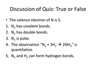
- 1. Discussion of Quiz: True or False • The valence electron of N is 5. 1. N2 has covalent bonds. 2. N2 has double bonds. 3. N2 is polar. 4. The observation “N2 + 3H2 2NH3” is quantitative. 5. N2 and H2 can form hydrogen bonds.
- 2. Quiz 1. Draw all 9 isomers of C7H16. 2. Identify the molecule as cis-trans and/or E-Z. 3. Identify the functional group(s) in the molecule. CH3 CH3 H F O O OHOH SH
- 3. Tips for Today’s Experiment • Unless specified, the maximum number of drops before recording “no change” is 10 drops. • The recording of data is similar to the previous experiment. • Some test tubes will become hot due to certain reactions. Be careful in handling those. • If the experiment says “note any change”, most likely (but not always) there is a change. • Make sure that all your group mates have seen the changes before proceeding to the next step. • The concentrations of the chemicals are much higher than the previous experiment. Please take extra care in handling these chemicals.
- 4. Tips for Today’s Experiment • As much as possible, do the experiments with salicylic acid first, as the reagent quickly crystallizes back out of the solvent. • Because the litmus paper may interfere with the reactions, a stirring rod and watch glass will be provided. Do not dip the litmus paper into the reactions. • The colors of the original solutions must be recorded to see if any change has really occurred.
- 5. Biochemistry
- 6. Proteins • Amino acids are the monomers and contain an amino group, a carboxyl group, and a side chain. • The amino group of one amino acid reacts with the carboxyl group of another amino acid by a dehydration reaction, forming a peptide bond. • Polypeptides are polymers of any combination of the 20 amino acids. • Proteins are three-dimensional biologically functional molecules composed of one or more polypeptides.
- 7. Functions of Proteins • Enzymes speed up chemical reactions. (Example: digestive enzymes) • Defense proteins protect against disease. (Example: antibodies) • Storage proteins store amino acids. (Example: casein in milk, ovalbumin in egg white) • Transport proteins carry substances. (Example: hemoglobin in blood)
- 8. Functions of Proteins • Hormones coordinate activities. (Example: insulin) • Contractile and motor proteins help move the body. (Example: actin and myosin in muscle) • Receptors help cells respond to stimuli. (Example: neurotransmitters in nerve cells) • Structural proteins provide support. (Example: keratin in hair, collagen in connective tissues, silk in spider webs and cocoons)
- 9. Types of Amino Acids Based on Side Chain • Hydrophobic nonpolar: glycine, alanine, valine, leucine, isoleucine, methionine, phenylalanine, tryptophan, proline • Hydrophilic polar: serine, threonine, cysteine, tyrosine, asparagine, glutamine • Acidic: aspartic acid, glutamic acid • Basic: lysine, arginine, histidine • Cysteine has sulfhydryl groups.
- 10. Levels of Structure in Proteins • Primary structure is the linear sequence of amino acids. • Secondary structure is the combination of α- helices, β-pleated sheets, triple helix, and/or β- turns formed by hydrogen bonding in the polypeptide backbone. • Tertiary structure is the three-dimensional shape formed by the combination of hydrophobic interactions, disulfide bridges, and electrostatic attractions in the side chains. • Quaternary structure is the association of multiple tertiary structures, forming a functional protein.
- 11. Tertiary Structure • Hydrophobic interactions are due to van der Waals interactions of hydrophobic amino acids. • Disulfide bridges are covalent bonds formed between sulfhydryl groups of amino acids. • Electrostatic attractions are due to the ionic charges of acidic and basic amino acids.
- 12. Question • Determine the type of tertiary structure that will dominate in the following polypeptides. 1. A polypeptide with many valine and leucine amino acids 2. A polypeptide with many glutamic acid and lysine amino acids 3. A polypeptide with many cysteine amino acids
- 13. Protein Structure • Denaturation is a permanent change in the shape of the protein due to changes in the environment (pH, salt concentration, temperature). • Chaperonins or chaperone proteins are molecules that assist in the proper folding of proteins. • X-ray crystallography is the technique to determine the three-dimensional structure of a protein. • NMR (nuclear magnetic resonance) spectroscopy may also help in the determination of structure. • Bioinformatics may be used to predict structure.
- 14. Technique: X-Ray Crystallography • The technique is only possible for proteins that can be crystallized. • The technique cannot show dynamic motion of a protein inside the cell. 1. A crystallized protein is placed in front of an X-ray beam. 2. The protein will scatter the X-rays into an orderly pattern, a phenomenon known as diffraction.
- 15. Technique: X-Ray Crystallography 3. The diffraction pattern is recorded as spots by a digital detector. 4. Based on the pattern and the primary structure of the protein, a three-dimensional model may be constructed by computer software. 5. The computer software makes an electron- density map of the spots using a mathematical technique known as Fourier transform.
- 16. Technique: How to Grow Crystals A. Microbatch Method • The mother liquor contains all components needed for crystal formation, including the buffer and the precipitating agent. • The precipitating agent is the material that will cause the protein to precipitate and may be a salt, polymer, organic solvent, or combinations of them. • The mother liquor and proteins are mixed at high concentrations to cause precipitation of the protein as a crystal. • The method is done in oil to prevent evaporation.
- 17. Technique: How to Grow Crystals B. Vapor-Diffusion Method 1. The undersaturated protein solution is mixed with a drop of the mother liquor. 2. The solution is equilibrated (put into chemical equilibrium) with solution containing only mother liquor. The difference in concentration causes water to evaporate into the mother liquor, leaving the drop more concentrated. 3. The supersaturation of the protein and the higher concentration of the precipitating agent causes the protein to precipitate as a crystal.
- 18. Technique: NMR Spectroscopy • The technique relies on nuclear spin angular momentum of nuclei, which is only possible for certain elements like 1H, 13C, 15N, 19F, and 31P. • 1H is typically used because of its high sensitivity and natural abundance, but proteins contain a lot of 1H molecules, requiring two-dimensional NMR techniques. 1. A strong, static magnetic field is applied to the protein. 2. Nuclear spin generates a magnetic dipole that can be either parallel (low energy) or antiparallel (high energy).
- 19. Technique: NMR Spectroscopy 3. A short pulse of electromagnetic energy is applied at right angles to the nuclei in the magnetic field, causing some low-energy nuclei to become high-energy nuclei. 4. The change is recorded as an absorption spectrum, and data from several repeated experiments are averaged to minimize errors. 5. Nuclear spins are then measured by atoms through space (nuclear Overhauser effect spectroscopy or NOESY) and by covalent bonds (total correlation spectroscopy or TOCSY). 6. A computer software combines these data to form a three-dimensional model.
- 20. Nucleic Acids • Nucleotides are the monomers and are composed of a nitrogen-containing base, a pentose sugar, and one or more phosphate groups. • Nucleosides occur when the phosphate groups are removed. • Nucleic acids or polynucleotides are the polymers formed by phosphodiester linkages, which are composed of a phosphate group joining the sugars of two nucleotides. • DNA (deoxyribonucleic acid) stores the genetic information of an organism that is passed from generation to generation. • RNA (ribonucleic acid) copies the DNA and is converted into protein.
- 21. Main Features of DNA and RNA • The structure of DNA is a double helix, two strands that spiral around an imaginary axis. • The two strands are antiparallel, one is 5’3’, the other is 3’5’. • In DNA, A pairs with T, and C pairs with G always. • When making a copy of RNA, the same pairing is followed, except U is used instead of T.
- 22. Question • Suppose there is a strand 5’-ATCGGCTA-3’. 1. What is the sequence of the antiparallel strand in DNA? 2. If this were copied in RNA, what will be the sequence?
- 23. Question • Suppose that a particular protein is bound to many fatty acids. This particular protein probably contains A. Leucine B. Tyrosine C. Aspartic acid D. Histidine
- 24. Technique: Spectrophotometry • This technique may be used to quantify the amount of biomolecule present in a sample. 1. Use one of the colorful tests that is currently being used to identify biomolecules. 2. Perform the colorful test in a cuvette with a positive sample. 3. Using a machine known as spectrophotometer, determine the wavelength at which maximum absorption occurs.
- 25. Technique: Spectrophotometry 4. Perform the test using samples of known and differing concentrations at the determined wavelength. 5. Plot the results into a straight line, which is known as the calibration curve. 6. Determine the concentration of the unknown sample by measuring at the determined wavelength and comparing with the calibration curve.
- 26. The Microscope
- 27. Light Microscopy • The microscope is an instrument that magnifies objects that are too small to be seen by the eye. • Microscopy is the science of studying objects using a microscope. • Light microscopy uses transmitted light to view objects. 1. A simple light microscope such as magnifying lens uses a single lens. 2. A compound light microscope uses a set of lenses or lens systems to magnify objects and different colorful stains to provide image contrast.
- 28. Mechanical Parts of a Compound Microscope • The base is the bottom-most part that supports the entire microscope. • The pillar is the part above the base that supports the entire microscope. • The inclination joint allows for tilting of the microscope for the convenience of the user. • The arm is the curved or slanted part which is held while carrying the microscope. • The stage is the platform where object to be examined is placed and secured by stage clips and may be moveable.
- 29. Mechanical Parts of a Compound Microscope • The eye is the circular opening through the central part of the stage and is used to allow light to pass up through the stage. • The mechanical stage control knobs are two knobs that are usually located below the stage. One knob controls forward/reverse movement, and the other controls right/left movement. • The body tube is attached to the arm and bears the lenses. • The draw tube is the cylindrical structure on top of the body tube that holds the ocular. • The nosepiece is a rotating disc where the objectives are attached. When moving the nosepiece, the fingers should be placed on the disc and not the objectives.
- 30. Mechanical Parts of a Compound Microscope • The dust shield lies atop the nosepiece and keeps dust particles from settling on the objectives. • The coarse adjustment knob is geared to the body tube and elevates or lowers it when rotated, bringing the object into approximate focus. • The fine adjustment knob is a smaller knob for delicate focusing bringing the object into perfect focus. • The condenser adjustment knob elevates or lowers the condenser to regulate the intensity of light. • The iris diaphragm lever is a lever in front of the condenser that may be moved horizontally to open and close the diaphragm.
- 31. Optical Parts of a Compound Microscope • The mirror is located beneath the stage and has concave and plane surfaces to gather and direct light to illuminate the object. Some microscope models have no mirror and have built-in lamp instead, mounted below the sub-stage for illumination. • The sub-stage consists of two components. 1. The iris diaphragm regulates the amount of light necessary to obtain a clearer view of the object. 2. The condenser is a set of lenses between the mirror and the stage that concentrates light rays on the specimen. • The eyepiece or ocular is a set of lens found on top of the body tube which functions to further magnify the image produced by the objective lenses.
- 32. Optical Parts of a Compound Microscope • The objectives are metal cylinders attached below the nosepiece which contain specially ground and polished lenses. There are usually three objectives in use. 1. The low power or 16mm objective (LPO) gives the lowest magnification (10x) and is used to locate the specimen as it possesses a relatively large field of vision. 2. The high power or 4mm objective (HPO) gives higher magnification, usually 40x or 43x, and is used to focus on the finer details of the specimen. 3. The oil immersion objective (OIO) gives the highest magnification, usually 97x or 100x, and is used wet either with cedar wood oil or synthetic immersion oil.
- 33. Proper Handling of a Compound Microscope • A microscope may be seriously damaged if dropped or bumped against a hard object. • The microscope should always be carried with both hands in a straight upright position, one under the base and the other on the arm. The eyepieces are not attached and will fall out if the microscope is carried at an angle or upside down. • Place the microscope at least 5cm from the edge of the table to prevent it from falling. • The clips must be tightly clipped on the stage, and the mirror must be in vertical position. • Do not touch the mirror with your fingers. Handle it by the frame to tip it at any desired angle.
- 34. Proper Handling of a Compound Microscope • Always keep the stage dry and clean. • Never allow liquids, particularly acids and alcohol, to come in contact with any part of the microscope. Do not incline the microscope when examining fresh mounts. • After using the microscope, the LPO should be in position, and the coarse adjustment knob should be moved down until it stops. • The student should always report immediately to the instructor any defects (missing or damaged part) of the microscope.
- 35. Some Terminologies • The magnifying power of an objective is shown by a figure engraved on the sleeve. • The numerical aperture (NA) is also engraved on the sleeve, next to the magnification (0.30 on the LPO, 0.65 on the HPO, and 1.30 on the OIO). • The resolution limit or resolving power is related to the NA and is the ability to reveal closely adjacent details as separate and distinct. It is a function of the wavelength of light and the design of the condenser. • The shortest wavelengths of visible light provide the maximum resolution. The maximum resolving power of a good medical laboratory microscope is 0.25μm (the resolving power of the human eye is 0.25mm).
- 36. Some Terminologies • The field of view is the entire area that can be seen through the microscope. • The larger the magnifying power of an objective lens, the smaller the field of view is. • The working distance of a particular objective is the distance between the front lens of an objective and the object on the slide when it is in focus. • In most microscopes, when a new objective is brought into use, the specimen should be in focus though enlarged and blurred (parfocal) and in the same position in relation to the field of view as in the previous objective (parcentral).
- 37. Some Terminologies • An optical section consists of the field that is in sharp focus at any plane below the surface of a more or less transparent specimen when seen through the microscope. • Depth of field is the thickness of the object that is in sharp focus. • The magnification of an object is the degree to which an object is enlarged or reduced using a system of lenses. • The linear magnification is the product of the magnifying powers of the objective and the ocular.
- 38. How to View Specimens Under a Compound Light Microscope • Clean the mirrors and lenses with lens paper or lint free cloth. Avoid using tissue paper as it may scratch the delicate lenses. • When properly used, the microscope should cause no eye strain. • Try to keep both eyes open when working the monocular microscope (microscope with only one eyepiece), and use the dominant eye to look through the ocular. • If you wear glasses, it will not be necessary to use them with the microscope, since the microscope automatically corrects for this.
- 39. How to View Specimens Under a Compound Light Microscope • Before placing any prepared slide on the stage, make it a habit to examine the specimen with the eye. • Look at the label which describes the material mounted on the center of the slide. • If the slide is dirty, clean it by rubbing lightly with a soft cloth or paper towel. • Bring the objective as far away from the stage as possible, and place a prepared slide on the stage, centering the objective on the focus over the hole. • Secure the slide with stage clips. Take care that the clip is never on the cover slip; otherwise, the specimen will be squashed.
- 40. How to View Specimens Under a Compound Light Microscope • Adjust the mirror so that the entire field is evenly illuminated. • The diaphragm should be opened as wide as possible in both cases, and the condenser should be as near the stage as it can move. • Try readjusting the mirror and the diaphragm to get the most effective illumination. • When using higher magnifications, more light is needed than when using lower magnifications. • For most microscopic work, it is best to keep the condenser at its highest level. Only rarely is it desirable to lower it slightly. When the condenser is used at a lowered position, the resolving power is greatly reduced.
- 41. How to View Specimens Under a Compound Light Microscope • The amount of light may be decreased by lowering the condenser or closing the diaphragm. • Start by bringing the LPO as close as possible to the slide, looking from the side to make sure that the objective does not touch the slide. Make sure the side with the cover slip is up. • Look into the eyepiece, and slowly move the object away from the slide until the image comes into view. Focus until the image becomes distinct. • Use the fine adjustment knob to make smaller adjustments and sharpen the focus. • The image, as seen under the microscope, is inverted so that the right side of the object is seen at the left, and the top is seen at the bottom and vice-versa for both circumstances.
- 42. How to View Specimens Under a Compound Light Microscope • If it is necessary to move the image in one direction to center it, practice moving the specimen in the opposite direction. • Once the specimen is in focus under LPO, and the portion to be examined further is at the center of the field of vision, rotate the nosepiece so that the HPO is in position. • Again, watch from the side taking care that the objective does not run into the slide. • Focus, but this time, use only the fine adjustment knob.
- 43. How to View Specimens Under a Compound Light Microscope • When you have finished viewing a slide and wish to view another, use the following procedure: 1. Be sure the LPO is in position. Loosen stage clips. 2. Remove the slide by slipping it horizontally forward (never up, as it might touch the lens in the objective). 3. Return the slide to the instructor, and take another slide.