SlideShare una empresa de Scribd logo
1 de 104
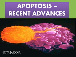
APOPTOSIS –
RECENT ADVANCES
EKTA JAJODIA
•German scientist Carl Vogt -
Principle of apoptosis (1842)
•Walther Flemming – Process of
programmed cell death (1845)
John Foxton Ross Kerr –
Distinguished apoptosis from
traumatic cell death (1962)
INTRODUCTION
Apoptosis is an energy dependent programmed cell
death for removal of unwanted cells
DEFINITION
Cell death mechanisms
Death by suicide Death by injury
sclero dinesh
APOPTOSIS NECROSIS
NATURAL YES NO
EFFECTS BENEFICIAL DETRIMENTAL
Physiological or
pathological
Always pathological
Single cells Sheets of cells
Energy dependent Energy independent
Cell shrinkage Cell swelling
Membrane integrity
maintained
Membrane integrity lost
APOPTOSIS NECROSIS
Role for mitochondria and
cytochrome C
No role for mitochondria
No leak of lysosomal enzymes Leak of lysosomal enzymes
Characteristic nuclear
changes
Nuclei lost
Apoptotic bodies form Do not form
DNA cleavage No DNA cleavage
Activation of specific
proteases
No activation
Regulatable process Not regulated
Evolutionarily conserved Not conserved
Dead cells ingested by
neighboring cells
Dead cells ingested by
neutrophils and macrophages
Apoptosis in physiologic situations
Apoptosis in pathologic situations
APOPTOSIS
APOPTOSIS IN
PHYSIOLOGICAL
CONDITIONS
Formation of
free and
independent
digits
Development
of the brain
Development
of
reproductive
organs
APOPTOSIS
DURING
EMBRYOGENESIS
Apoptosis in bud
formation during
which many
interdigital cells
die. They are
stained black by a
TUNEL method
Incomplete
differentiation in two
toes due to lack of
apoptosis
Involution of
hormone-dependent
tissues upon
hormone withdrawal
•Endometrial cell breakdown during menstrual cycle
•Ovarian follicular atresia in menopause
•Regression of the lactating breast after weaning
•Prostatic atrophy after castration
Cell loss in
proliferating cell
populations
•Immature lymphocytes in bone marrow and thymus
that fail to express useful antigen receptors
•B lymphocytes in germinal centers
•Epithelial cells in intestinal crypts
•So as to maintain a constant number
Elimination of
potentially harmful
self-reactive
lymphocytes
Death of host cells
that have served
their useful purpose
•Neutrophils in an a/c inflammatory response
•Lymphocytes at the end of an immune response
APOPTOSIS IN
PATHOLOGICAL
CONDITIONS
DNA
damage
Accumulation
of mis-folded
proteins
Cell injury in
certain
infections
Pathological
atrophy in
parenchymal
organs after
duct obstruction
• Defective apoptosis
and increased cell
survival
TOO
LITTLE
• Increased apoptosis
and excess cell
death
TOO
MUCH
Unfolded protein
response(UPR) and
Endoplasmic
reticulum(ER) stress
PLAYERS
1) Sensors – BAD , BIM , BID,
noxa, puma
2) Proapoptotic- BAX , BAK
3) Cytochrome C
4) Apaf-1 [ apoptosis activating
factor – 1 ]
5) Caspases
DEFENDERS
1) Antiapoptotic -
BCL 2,
BCL XL,
MCL-1
2) IAPS: [ inhibitors of
apoptosis
The fundamental events in apoptosis is the activation of
enzymes called CASPASES
• Cysteine proteases
• Cysteine-dependent
ASPartate-specific
proteASES
Caspases
CASPASES
• There are 14 different caspase enzymes
Active cysteine residue in the
catalytic site
Synthesized as inactive
zymogens (PROCASPASES)
• Inflammatory Caspases: 1, 4, 5 and 13
• Initiator Caspases: 2, 8, 9, and 10
– Long N-terminal domain
– Interact with effector caspases
• Effector Caspases: 3, 6, 7 and 14
– Little to no N-terminal domain
– Initiate cell death
3 TYPES OF
CASPASES
Procasp 2 & 9
have CARD
domain
Procasp 8 &
10 have DED
domain
In intrinsic
pathway
Procasp 9
Procasp 9
activated by
apoptosome
in extrinsic
pathway
Procasp 8 &
10
Procasp 8 &
10 activated
by death
receptors
2
8
9
10
CARD – caspase recruitment domain
DED – death effector domain
3
6
7
procaspase 3 and 7
activated by granzyme B
14
BCL-2
PROTEINS
Proapoptotic
proteins
• BH123 protein
• BAX
• BAK
Antiapoptotic
proteins
Bcl2
BclXL
Mcl-1
Sensors/proapop
totic proteins
• Bad
• Bid
• Bim
• Puma
• Noxa
DAMAGE Physiological death signals
DEATH SIGNAL
PROAPOPTOTIC
PROTEINS
ANTIAPOPTOTIC
PROTEINS
INHIBITORS OF
APOPTOTIC PROTEINS
(IAPs)
•1st identified in baculovirus
•8 types are described
•IAPs are characterised by BIR domains (baculoviral IAP
repeats)
•These BIR domains bind to caspases and inhibit them
•IAPs are regulated by IAP binding proteins – Smac/DIABLO
(second mitochondrial activator of caspases)
NAIP – neuronal apoptosis inhibitor protein
Cellular IAP1
Cellular IAP2
XIAP – X-linked inhibitor of apoptosis
Survivin
Ts-IAP – testis specific IAP
BRUCE – BIR containing ubiquitin conjugating enzyme
Livin
Normally Smac/DIABLO resides in mitochondria(intermitochondrial
space)
When apoptotic stimulus triggers
Smac/DIABLO is proteolytically cleaved and released into cytosol
Bind to IAPs and inhibit them
Causes apoptosis
XIAP has been best described
Inhibits apoptosis in 2 ways
Direct inhibition of
apoptosis by binding
of BIR domains to
caspases
Proteosome dependent
degradation of caspases
• Mitochondrial protein present at inter mitochondrial
space
• Can activate caspase cascade
• Binds to APAF-1 and form apoptosome
CYTOCHROME C
[ apoptosis activating factor – 1 ]
• Present in cytosol
• Forms apoptosome after combining with
cytochrome c
APAF-1
Initiation
• Intrinsic pathway
• Extrinsic Pathway
Execution
• Cleavage of DNA
Phagocyt--
osis
• Engulfment of cell
PATHWAYS OF
APOPTOSIS
INTRINSIC PATHWAY
(MITOCHONDRIAL)
INTRINSIC PATHWAY [MITOCHONDRIAL PATHWAY]
Radiation , Toxins, Free radicals
misfolded proteins/ DNA damage
ER STRESS Sensing of double strand DNA break
Double strand DNA break is sensed by 2
proteins
ATM (Ataxia telangiectasia
mutated)
ATR (ataxia telangiectasia and
RAD3 related protein
Phosphorylates and activates CHK1 and CHK2
(checkpoint kinase 1 and 2)
Phosphorylates and activates P53
P53 form tetramers with other p53 molecules and
cause increase expression of the following genes
Genes involved in DNA repair
P21 genes
Proapoptotic proteins/sensors
(puma/noxa)
Activation of proapoptotic proteins (BAX
and BAK)
Oligomerisation and increase in outer
mitochondrial membrane permeability
Leakage of cytochrome c
into cytosol
Release of Smac/DIABLO into
cytosol which binds to IAPs and
inhibits them
Cytochrome c in cytosol + APAF 1
Form Hexamer ( Apoptosome )
Binds to procaspase- 9
( critical initiator caspase)
Enzyme cleaves the adjacent caspase 9 molecules
i.e . Autoamplification process
Execution phase
EXTRINSIC
PATHWAY
Death Receptors
Members of TNF receptor family which contain a
cytoplasmic domain involved in protein-protein
interactions .i.e called Death Domain .
2 types of death receptors are there :
• TNFr (tumour necrosis factor receptor )
• FasR (fatty acid synthetase receptor )
The ligand for Fas is called FasL
The ligand for TNFr is TNFα
Adaptor Proteins
also contain death domain
a. FADD [ Fas associated death domain ]
b. TRADD [ TNF receptor associated death domain]
The death
receptor
pathway
• Extrinsic
pathway
Inhibited by FLIP- binds to
procaspase8 but cannot cleave
and activate it
EXECUTION PHASE
• Final phase of apoptosis
• Mediated by proteolytic cascade
• After initiator caspases ( 2, 8, 9 and 10) are cleaved to
generate its active form, the enzymatic death program
is set in motion by rapid sequential activation of the
executioner caspases – 3,6
• Caspase -3 activates DNase which causes degradation
of chromosomal DNA within the nuclei and causes
chromatin condensation.
• Caspase -3 induces cytoskeletal reorganisation and
disintegration of cell into apoptotic bodies.
REMOVAL OF DEAD CELLS
Factors by which apoptotic cells attracts
phagocytes towards them :
• Apoptotic bodies- “bite size”-edible for
phagocytes
• Phosphatidyl serine “flips” out from inner to
outer layer –recognized by macrophages
receptors
• Some apoptotic bodies express thrombospondin
recognized by phagocytes
APOPTOSIS
ASSAYS- detection
and methods
Cell Viability and Death
• Functional assay
• DNA labeling assay
• Morphological assay
• Reproductive assay
• Membrane integrity assay
• Exclusion dyes
• Flourescent dyes
• annexinV
• LDH assay
MEMBRANE
INTEGRITY ASSAY
• MTT, XTT assay
• Crystal violet/ Acid
• phosphatase(AP) assay
• Alamar Blue oxidation-Reduction
assay
• Neutral red assay
FUNCTIONAL
ASSAY
• Fluorescent conjugates
DNA LABELLING
ASSAY
• Microscopic observation
• -Caspase 3 detection
• -PARP cleavage assay
MORPHOLOGICAL
and MECHANISM
BASED ASSAY
• Colony formation assay
REPRODUCTIVE
ASSAY
• annexinV
• Caspase3
• p53
IMMUNOHISTOCHEMIS
TRY
MEMBRANE
INTEGRITY ASSAY
•Exclusion dyes
•Flourescent dyes
•AnnexinV
•LDH assay
EXCLUSION
DYES
• Viable cells : small, round and refractile
• Non-viable cells : swollen, larger, dark blue
• Tryphan blue dye exclusion methods
Principle
Trypan Blue dye Exclusion Methods
• Ethidium bromide (EtBr) and propidium iodide (PI)
• PI binds to nucleic acids upon membrane damage
• PI is impermeable to intact plasma membrane.
• Intercalates with DNA or RNA  red
FLUORESCENT
DYES
Fluorescein diacetate (FDA) is a nonpolar ester which
passes through plasma membranes and is hydrolyzed by
intracellular esterases to produce free fluorescein
 the polar fluorescein is confined within cells which have
an intact plasma membrane and can be observed under
appropriate excitation conditions
Undamaged cell : highly fluorescent fluorescein dye
Damaged cell : fluoresce only weakly
 greenish-yellow at 450-480 nm
Intact cell –
PI and FDA is added
Fluorescein in
intact cells
Schematic illustration of the principle of
PI/FDA cell viability assay
● FDA (Fluorescein diacetate)
● PI (Propidium iodide)
Plasma membrane is damaged
; fluorescein leaks out
PI enters and stains
nucleic acids
ANNEXIN V
ASSAY
• Annexin V: An Early Marker of Apoptosis
• One of the earliest indications of apoptosis is the
translocation of the membrane phospholipid
phosphatidylserine (PS) from the inner to the outer
leaflet of the plasma membrane.
• Once exposed to the extracellular environment, binding
sites on PS become available for Annexin V, a 35-36 kDa,
Ca 2+-dependent, phospholipid binding protein with a
high affinity for PS.
• The translocation of PS precedes other apoptotic
processes such as loss of plasma membrane integrity,
DNA fragmentation, and chromatin condensation.
• As such, Annexin V can be conjugated to biotin or to a
fluorochrome such as FITC, PE, APC, Cy5, or Cy5.5, and
used for the easy, flow cytometric identification of cells
in the early stages of apoptosis
• Because PS translocation also occurs during necrosis,
Annexin V is not an absolute marker of apoptosis.
• Therefore, it is often used in conjunction with vital dyes
such as 7-amino-actinomysin (7-AAD) or propidium
iodide (PI), which bind to nucleic acids, but can only
penetrate the plasma membrane when membrane
integrity is breached, as occurs in the later stages of
apoptosis or in necrosis
• Result
• annexin-/PI-, annexin +/PI-, annexin+/PI+
No Apoptosis = Cell Viability
Cells that are negative for both Annexin V and the vital
dye have no indications of apoptosis: PS translocation has
not occurred and the plasma membrane is still intact.
Early Apoptosis
Cells that are Annexin V-positive and vital dye-negative,
however, are in early apoptosis as PS translocation has
occurred, yet the plasma membrane is still intact.
Late Apoptosis or Cell Death
Cells that are positive for both Annexin V and the vital dye
are either in the late stages of apoptosis or are already
dead, as PS translocation has occurred and the loss of
plasma membrane integrity is observed.
When measured over time, Annexin V and a vital dye can
be used to monitor the progression of apoptosis: from cell
viability, to early-stage apoptosis, and finally to late-stage
apoptosis and cell death.
LDH LEAKAGE
ASSAY
Test principle
The assay is based on consideration that tumor cells
possess high concentration of intracellular LDH and the
cleavage of a tetrazolium salt when LDH is present in the
culture supernatant.
• LDH catalyzes the reduction of NAD+ to NADH and H+ by
oxidation of lactate to pyruvate. In the second step of
the reaction, diaphorase uses the newly-formed NADH
and H+ to catalyze the reduction of a tetrazolium salt
(INT) to highly-colored formazan which absorbs strongly
at 490-520 nm.
FUNCTIONAL
ASSAY
•MTT, XTT assay
•Crystal violet/ Acid
•phosphatase(AP) assay
•Alamar Blue oxidation- Reduction assay
• Neutral red assay
MTT ASSAY
Introduction
 This assay is a sensitive, quantitative and reliable
colorimetric assay that measures viability, proliferation
and activation of cells.
 The assay is based on the capacity of mitochondrial
dehydrogenase enzymes in living cells to convert the
yellow water-soluble substrate dimethylthiazol
tetrazolium bromide (MTT) into a dark blue formazan
product which is insoluble in water
 The amount of formazan produced is directly proportional
to number of viable cells present in the sample.
metabolically active Cell
MTT
Formazan
Insoluble
Compare with MTT assay and XTT assay
Culture cells in a MTP
for a certain period of time (37℃)
MTT assay XTT assay
Prepare labeling mixture
Incubate cells (0.5-4 h, 37℃)
Add solubilizing solution
(Isopropanol) and incubate
Measure absorbance using an ELISA reader
Add XTT labeling mixtureAdd MTT labeling reagent
Insoluble formazan Soluble formazan
DNA LABELLING ASSAY
•Fluorescent conjugates
TUNEL ASSAY
•The single cell gel electrophoresis (SCGE), the Comet
Assay, is a fairly simple procedure by using a micro gel
and electrophoresis to detect DNA damage
•The damage in DNA looks like a comet, so that’s why it is
known as Comet Assay
• This technique is further developed and introduced the
use of high alkaline conditions. This step increased the
ability of the assay to detect not only the double strand
breaks but also the single strand breaks.
COMET ASSAY
MORPHOLOGICAL
MECHANISM BASED
ASSAY
•Microscopic observation
•-Caspase 3 detection
•-PARP cleavage assay
•Endonuclease assay
Principle
During apoptosis the endonuclease enzymes are activated
to cleave the genomic DNA in to many smaller fragments
ENDONUCLEASE
ASSAY
Steps
1. Preparation of agarose gel containing
Ethidium bromide and genomic DNA
2. Making well
3. Preparation of cell lysate
4. Incubation in humidified atmosphere
after loading cell lysate
5. Observe the DNA degradation
MORPHOLOGY OF CELLS IN APOPTOSIS
• Cell Shrinkage and dense cytoplasm
• Chromatin condensation to periphery and later
fragmentation.
• Plasma membrane is intact
• Formation of cytoplasmic blebs and apoptotic
bodies.
• Structurally altered so that the apoptotic cell
becomes “tasty” for phagocytosis
• Phagocytosis of apoptotic bodies by macrophages
• Dead cell is rapidly cleared before contents are
leaked out
• No inflammatory reaction
MORPHOLOGY OF APOPTOSIS
Progressive cell shrinkage
Chromatin condensation
Plasma membrane blebbing
Apoptotic bodies
Phagocytosis - no inflammation
MORPHOLOGY OF CELLS IN APOPTOSIS
Various methods available for
detection of apoptotic cells
Light microscopy
• In H&E the apoptotic cell appears as round or
oval mass of intensely eosinophilic cytoplasm
with fragments of dense nuclear chromatin
MICROSCOPY
Apoptosis in neutrophils
Councilman bodies- liver tissue
Electron microscopy
Defines subcellular changes
like
• Chromatin condensation
• Plasma membrane
blebbing
Immunohistochemistry
Immunohistochemical
detection of apoptotic cells
using antibodies against a
wide range of substrates
most importantly :
• Caspase 3
• P53
• Annexin V
NECROPTOSIS
•Mechanistically resembles apoptosis
•Morphologically resembles necrosis
•Also known as programmed necrosis
•CASPASES ARE NOT ACTIVATED
•RIp1 and RIP3 complex formed
•Inflammation present
PYROPTOSIS
Thank u….

Más contenido relacionado

La actualidad más candente

Apoptosis (Intrinsic And Extrinsic Pathway with assays)
Apoptosis (Intrinsic And Extrinsic Pathway with assays)Apoptosis (Intrinsic And Extrinsic Pathway with assays)
Apoptosis (Intrinsic And Extrinsic Pathway with assays)227777222an
 
Apoptosis (presentation)
Apoptosis (presentation)Apoptosis (presentation)
Apoptosis (presentation)Leo Vang
 
Apoptosis (Programm of cell death)
Apoptosis (Programm of cell death)Apoptosis (Programm of cell death)
Apoptosis (Programm of cell death)Shiv Patel
 
Apoptosis basics + microphotographs
Apoptosis basics + microphotographsApoptosis basics + microphotographs
Apoptosis basics + microphotographsashish223
 
PROGRAMMED CELL DEATH (APOPTOSIS )
PROGRAMMED CELL DEATH (APOPTOSIS ) PROGRAMMED CELL DEATH (APOPTOSIS )
PROGRAMMED CELL DEATH (APOPTOSIS ) Amany Elsayed
 
Apoptosis,biological changes,pathways extrinsic, intrinsic, carcinogenecity,n...
Apoptosis,biological changes,pathways extrinsic, intrinsic, carcinogenecity,n...Apoptosis,biological changes,pathways extrinsic, intrinsic, carcinogenecity,n...
Apoptosis,biological changes,pathways extrinsic, intrinsic, carcinogenecity,n...Bahaudin zakriya university multan pakistan
 
Intrinsic and Extrinsic Pathway of Apoptosis
Intrinsic and Extrinsic  Pathway of ApoptosisIntrinsic and Extrinsic  Pathway of Apoptosis
Intrinsic and Extrinsic Pathway of ApoptosisAnantha Kumar
 
Apoptosis(program cell death)
Apoptosis(program cell death)Apoptosis(program cell death)
Apoptosis(program cell death)PrashantSharma807
 
Apoptosis in health and diseases
Apoptosis in health and diseasesApoptosis in health and diseases
Apoptosis in health and diseasesmeenuev
 
Apoptosis
ApoptosisApoptosis
Apoptosisjinx11
 
Apoptosis, cell death, and proliferation
Apoptosis, cell death, and proliferationApoptosis, cell death, and proliferation
Apoptosis, cell death, and proliferationsilene25
 

La actualidad más candente (20)

Apoptosis.ppt
Apoptosis.pptApoptosis.ppt
Apoptosis.ppt
 
Apoptosis
ApoptosisApoptosis
Apoptosis
 
Apoptosis (Intrinsic And Extrinsic Pathway with assays)
Apoptosis (Intrinsic And Extrinsic Pathway with assays)Apoptosis (Intrinsic And Extrinsic Pathway with assays)
Apoptosis (Intrinsic And Extrinsic Pathway with assays)
 
Apoptosis
ApoptosisApoptosis
Apoptosis
 
Apoptosis (presentation)
Apoptosis (presentation)Apoptosis (presentation)
Apoptosis (presentation)
 
Apoptosis
ApoptosisApoptosis
Apoptosis
 
Apoptosis (Programm of cell death)
Apoptosis (Programm of cell death)Apoptosis (Programm of cell death)
Apoptosis (Programm of cell death)
 
Apoptosis basics + microphotographs
Apoptosis basics + microphotographsApoptosis basics + microphotographs
Apoptosis basics + microphotographs
 
Apoptosis
ApoptosisApoptosis
Apoptosis
 
Apoptosis
ApoptosisApoptosis
Apoptosis
 
Apoptosis
ApoptosisApoptosis
Apoptosis
 
PROGRAMMED CELL DEATH (APOPTOSIS )
PROGRAMMED CELL DEATH (APOPTOSIS ) PROGRAMMED CELL DEATH (APOPTOSIS )
PROGRAMMED CELL DEATH (APOPTOSIS )
 
Apoptosis,biological changes,pathways extrinsic, intrinsic, carcinogenecity,n...
Apoptosis,biological changes,pathways extrinsic, intrinsic, carcinogenecity,n...Apoptosis,biological changes,pathways extrinsic, intrinsic, carcinogenecity,n...
Apoptosis,biological changes,pathways extrinsic, intrinsic, carcinogenecity,n...
 
Apoptosis seminar
Apoptosis seminarApoptosis seminar
Apoptosis seminar
 
Intrinsic and Extrinsic Pathway of Apoptosis
Intrinsic and Extrinsic  Pathway of ApoptosisIntrinsic and Extrinsic  Pathway of Apoptosis
Intrinsic and Extrinsic Pathway of Apoptosis
 
Apoptosis(program cell death)
Apoptosis(program cell death)Apoptosis(program cell death)
Apoptosis(program cell death)
 
Apoptosis in health and diseases
Apoptosis in health and diseasesApoptosis in health and diseases
Apoptosis in health and diseases
 
Apoptosis
ApoptosisApoptosis
Apoptosis
 
Apoptosis, cell death, and proliferation
Apoptosis, cell death, and proliferationApoptosis, cell death, and proliferation
Apoptosis, cell death, and proliferation
 
Apoptosis
ApoptosisApoptosis
Apoptosis
 

Similar a Seminar- recent advances in apoptosis

Apoptosis definition, mechanism , Apoptosis vs necrosis, Assays of apoptosis,...
Apoptosis definition, mechanism , Apoptosis vs necrosis, Assays of apoptosis,...Apoptosis definition, mechanism , Apoptosis vs necrosis, Assays of apoptosis,...
Apoptosis definition, mechanism , Apoptosis vs necrosis, Assays of apoptosis,...MuhammadKhizar39
 
apopwqgdcbhuredgbcfyhhvhcctosis (1).pptx
apopwqgdcbhuredgbcfyhhvhcctosis (1).pptxapopwqgdcbhuredgbcfyhhvhcctosis (1).pptx
apopwqgdcbhuredgbcfyhhvhcctosis (1).pptxSarithaRani4
 
Apoptosis - programmed cell death that occurs in multicellular organisms
Apoptosis - programmed cell death that occurs in multicellular organismsApoptosis - programmed cell death that occurs in multicellular organisms
Apoptosis - programmed cell death that occurs in multicellular organismsMeethuRappai1
 
Apoptosis
ApoptosisApoptosis
ApoptosisTKANHAR
 
Apoptosis roohit
Apoptosis roohitApoptosis roohit
Apoptosis roohitroohith
 
physiology of apoptosis first year mbbs medical.pdf
physiology of apoptosis first year mbbs medical.pdfphysiology of apoptosis first year mbbs medical.pdf
physiology of apoptosis first year mbbs medical.pdfDratoshKatiyar
 

Similar a Seminar- recent advances in apoptosis (20)

Apoptosis
ApoptosisApoptosis
Apoptosis
 
APOPTOSIS
APOPTOSISAPOPTOSIS
APOPTOSIS
 
Apoptosis
ApoptosisApoptosis
Apoptosis
 
Apaptosis
Apaptosis Apaptosis
Apaptosis
 
Apoptosis definition, mechanism , Apoptosis vs necrosis, Assays of apoptosis,...
Apoptosis definition, mechanism , Apoptosis vs necrosis, Assays of apoptosis,...Apoptosis definition, mechanism , Apoptosis vs necrosis, Assays of apoptosis,...
Apoptosis definition, mechanism , Apoptosis vs necrosis, Assays of apoptosis,...
 
APOPTOSIS MBBS.pdf
APOPTOSIS MBBS.pdfAPOPTOSIS MBBS.pdf
APOPTOSIS MBBS.pdf
 
apopwqgdcbhuredgbcfyhhvhcctosis (1).pptx
apopwqgdcbhuredgbcfyhhvhcctosis (1).pptxapopwqgdcbhuredgbcfyhhvhcctosis (1).pptx
apopwqgdcbhuredgbcfyhhvhcctosis (1).pptx
 
Appoptosis -pro.pptx
Appoptosis -pro.pptxAppoptosis -pro.pptx
Appoptosis -pro.pptx
 
Apoptosis - programmed cell death that occurs in multicellular organisms
Apoptosis - programmed cell death that occurs in multicellular organismsApoptosis - programmed cell death that occurs in multicellular organisms
Apoptosis - programmed cell death that occurs in multicellular organisms
 
Apoptosis
ApoptosisApoptosis
Apoptosis
 
Apoptosis.pptx
Apoptosis.pptxApoptosis.pptx
Apoptosis.pptx
 
Apoptosis
ApoptosisApoptosis
Apoptosis
 
Apoptosis
ApoptosisApoptosis
Apoptosis
 
Apoptosis
ApoptosisApoptosis
Apoptosis
 
Apoptosis
ApoptosisApoptosis
Apoptosis
 
Apoptosis roohit
Apoptosis roohitApoptosis roohit
Apoptosis roohit
 
APoptosis
APoptosisAPoptosis
APoptosis
 
Apoptosis
ApoptosisApoptosis
Apoptosis
 
physiology of apoptosis first year mbbs medical.pdf
physiology of apoptosis first year mbbs medical.pdfphysiology of apoptosis first year mbbs medical.pdf
physiology of apoptosis first year mbbs medical.pdf
 
Slide share
Slide shareSlide share
Slide share
 

Más de Ekta Jajodia

Pitfalls in minor salivary gland neoplasms
Pitfalls in minor salivary gland neoplasmsPitfalls in minor salivary gland neoplasms
Pitfalls in minor salivary gland neoplasmsEkta Jajodia
 
Data set for cutaneous melanoma reporting
Data set for cutaneous melanoma reportingData set for cutaneous melanoma reporting
Data set for cutaneous melanoma reportingEkta Jajodia
 
Seminar primary immunodeficiency syndrome
Seminar primary immunodeficiency syndromeSeminar primary immunodeficiency syndrome
Seminar primary immunodeficiency syndromeEkta Jajodia
 
The human eosinophils
The human eosinophilsThe human eosinophils
The human eosinophilsEkta Jajodia
 
principle of Immunohistochemistry and its use in diagnostics
principle of Immunohistochemistry and its use in diagnosticsprinciple of Immunohistochemistry and its use in diagnostics
principle of Immunohistochemistry and its use in diagnosticsEkta Jajodia
 
Liver function tests
Liver function testsLiver function tests
Liver function testsEkta Jajodia
 
lymphoepithelial lesion
 lymphoepithelial lesion lymphoepithelial lesion
lymphoepithelial lesionEkta Jajodia
 
IMWG updates on plasma cell dyscrasias
IMWG updates on plasma cell dyscrasias IMWG updates on plasma cell dyscrasias
IMWG updates on plasma cell dyscrasias Ekta Jajodia
 
Autoimmune bullous lesions of skin
Autoimmune bullous lesions of skinAutoimmune bullous lesions of skin
Autoimmune bullous lesions of skinEkta Jajodia
 
Special stains in histopathology
Special stains in histopathologySpecial stains in histopathology
Special stains in histopathologyEkta Jajodia
 
Recent advances in lung tumors and tumor like lesions
Recent advances in lung tumors and tumor like lesionsRecent advances in lung tumors and tumor like lesions
Recent advances in lung tumors and tumor like lesionsEkta Jajodia
 

Más de Ekta Jajodia (13)

Pitfalls in minor salivary gland neoplasms
Pitfalls in minor salivary gland neoplasmsPitfalls in minor salivary gland neoplasms
Pitfalls in minor salivary gland neoplasms
 
Data set for cutaneous melanoma reporting
Data set for cutaneous melanoma reportingData set for cutaneous melanoma reporting
Data set for cutaneous melanoma reporting
 
Seminar primary immunodeficiency syndrome
Seminar primary immunodeficiency syndromeSeminar primary immunodeficiency syndrome
Seminar primary immunodeficiency syndrome
 
The human eosinophils
The human eosinophilsThe human eosinophils
The human eosinophils
 
principle of Immunohistochemistry and its use in diagnostics
principle of Immunohistochemistry and its use in diagnosticsprinciple of Immunohistochemistry and its use in diagnostics
principle of Immunohistochemistry and its use in diagnostics
 
Liver function tests
Liver function testsLiver function tests
Liver function tests
 
lymphoepithelial lesion
 lymphoepithelial lesion lymphoepithelial lesion
lymphoepithelial lesion
 
IMWG updates on plasma cell dyscrasias
IMWG updates on plasma cell dyscrasias IMWG updates on plasma cell dyscrasias
IMWG updates on plasma cell dyscrasias
 
Microtomy
MicrotomyMicrotomy
Microtomy
 
Autoimmune bullous lesions of skin
Autoimmune bullous lesions of skinAutoimmune bullous lesions of skin
Autoimmune bullous lesions of skin
 
Apheresis
ApheresisApheresis
Apheresis
 
Special stains in histopathology
Special stains in histopathologySpecial stains in histopathology
Special stains in histopathology
 
Recent advances in lung tumors and tumor like lesions
Recent advances in lung tumors and tumor like lesionsRecent advances in lung tumors and tumor like lesions
Recent advances in lung tumors and tumor like lesions
 

Último

Book Call Girls in Kasavanahalli - 7001305949 with real photos and phone numbers
Book Call Girls in Kasavanahalli - 7001305949 with real photos and phone numbersBook Call Girls in Kasavanahalli - 7001305949 with real photos and phone numbers
Book Call Girls in Kasavanahalli - 7001305949 with real photos and phone numbersnarwatsonia7
 
Call Girl Bangalore Nandini 7001305949 Independent Escort Service Bangalore
Call Girl Bangalore Nandini 7001305949 Independent Escort Service BangaloreCall Girl Bangalore Nandini 7001305949 Independent Escort Service Bangalore
Call Girl Bangalore Nandini 7001305949 Independent Escort Service Bangalorenarwatsonia7
 
VIP Call Girls Lucknow Nandini 7001305949 Independent Escort Service Lucknow
VIP Call Girls Lucknow Nandini 7001305949 Independent Escort Service LucknowVIP Call Girls Lucknow Nandini 7001305949 Independent Escort Service Lucknow
VIP Call Girls Lucknow Nandini 7001305949 Independent Escort Service Lucknownarwatsonia7
 
Call Girl Lucknow Mallika 7001305949 Independent Escort Service Lucknow
Call Girl Lucknow Mallika 7001305949 Independent Escort Service LucknowCall Girl Lucknow Mallika 7001305949 Independent Escort Service Lucknow
Call Girl Lucknow Mallika 7001305949 Independent Escort Service Lucknownarwatsonia7
 
Call Girls Jp Nagar Just Call 7001305949 Top Class Call Girl Service Available
Call Girls Jp Nagar Just Call 7001305949 Top Class Call Girl Service AvailableCall Girls Jp Nagar Just Call 7001305949 Top Class Call Girl Service Available
Call Girls Jp Nagar Just Call 7001305949 Top Class Call Girl Service Availablenarwatsonia7
 
call girls in green park DELHI 🔝 >༒9540349809 🔝 genuine Escort Service 🔝✔️✔️
call girls in green park  DELHI 🔝 >༒9540349809 🔝 genuine Escort Service 🔝✔️✔️call girls in green park  DELHI 🔝 >༒9540349809 🔝 genuine Escort Service 🔝✔️✔️
call girls in green park DELHI 🔝 >༒9540349809 🔝 genuine Escort Service 🔝✔️✔️saminamagar
 
College Call Girls Vyasarpadi Whatsapp 7001305949 Independent Escort Service
College Call Girls Vyasarpadi Whatsapp 7001305949 Independent Escort ServiceCollege Call Girls Vyasarpadi Whatsapp 7001305949 Independent Escort Service
College Call Girls Vyasarpadi Whatsapp 7001305949 Independent Escort ServiceNehru place Escorts
 
Call Girls Service Nandiambakkam | 7001305949 At Low Cost Cash Payment Booking
Call Girls Service Nandiambakkam | 7001305949 At Low Cost Cash Payment BookingCall Girls Service Nandiambakkam | 7001305949 At Low Cost Cash Payment Booking
Call Girls Service Nandiambakkam | 7001305949 At Low Cost Cash Payment BookingNehru place Escorts
 
Call Girl Service Bidadi - For 7001305949 Cheap & Best with original Photos
Call Girl Service Bidadi - For 7001305949 Cheap & Best with original PhotosCall Girl Service Bidadi - For 7001305949 Cheap & Best with original Photos
Call Girl Service Bidadi - For 7001305949 Cheap & Best with original Photosnarwatsonia7
 
See the 2,456 pharmacies on the National E-Pharmacy Platform
See the 2,456 pharmacies on the National E-Pharmacy PlatformSee the 2,456 pharmacies on the National E-Pharmacy Platform
See the 2,456 pharmacies on the National E-Pharmacy PlatformKweku Zurek
 
Russian Call Girls in Pune Riya 9907093804 Short 1500 Night 6000 Best call gi...
Russian Call Girls in Pune Riya 9907093804 Short 1500 Night 6000 Best call gi...Russian Call Girls in Pune Riya 9907093804 Short 1500 Night 6000 Best call gi...
Russian Call Girls in Pune Riya 9907093804 Short 1500 Night 6000 Best call gi...Miss joya
 
College Call Girls Pune Mira 9907093804 Short 1500 Night 6000 Best call girls...
College Call Girls Pune Mira 9907093804 Short 1500 Night 6000 Best call girls...College Call Girls Pune Mira 9907093804 Short 1500 Night 6000 Best call girls...
College Call Girls Pune Mira 9907093804 Short 1500 Night 6000 Best call girls...Miss joya
 
Call Girls ITPL Just Call 7001305949 Top Class Call Girl Service Available
Call Girls ITPL Just Call 7001305949 Top Class Call Girl Service AvailableCall Girls ITPL Just Call 7001305949 Top Class Call Girl Service Available
Call Girls ITPL Just Call 7001305949 Top Class Call Girl Service Availablenarwatsonia7
 
Housewife Call Girls Bangalore - Call 7001305949 Rs-3500 with A/C Room Cash o...
Housewife Call Girls Bangalore - Call 7001305949 Rs-3500 with A/C Room Cash o...Housewife Call Girls Bangalore - Call 7001305949 Rs-3500 with A/C Room Cash o...
Housewife Call Girls Bangalore - Call 7001305949 Rs-3500 with A/C Room Cash o...narwatsonia7
 
Asthma Review - GINA guidelines summary 2024
Asthma Review - GINA guidelines summary 2024Asthma Review - GINA guidelines summary 2024
Asthma Review - GINA guidelines summary 2024Gabriel Guevara MD
 
Call Girls Kanakapura Road Just Call 7001305949 Top Class Call Girl Service A...
Call Girls Kanakapura Road Just Call 7001305949 Top Class Call Girl Service A...Call Girls Kanakapura Road Just Call 7001305949 Top Class Call Girl Service A...
Call Girls Kanakapura Road Just Call 7001305949 Top Class Call Girl Service A...narwatsonia7
 
Call Girls Hebbal Just Call 7001305949 Top Class Call Girl Service Available
Call Girls Hebbal Just Call 7001305949 Top Class Call Girl Service AvailableCall Girls Hebbal Just Call 7001305949 Top Class Call Girl Service Available
Call Girls Hebbal Just Call 7001305949 Top Class Call Girl Service Availablenarwatsonia7
 
Call Girls Service In Shyam Nagar Whatsapp 8445551418 Independent Escort Service
Call Girls Service In Shyam Nagar Whatsapp 8445551418 Independent Escort ServiceCall Girls Service In Shyam Nagar Whatsapp 8445551418 Independent Escort Service
Call Girls Service In Shyam Nagar Whatsapp 8445551418 Independent Escort Serviceparulsinha
 

Último (20)

Book Call Girls in Kasavanahalli - 7001305949 with real photos and phone numbers
Book Call Girls in Kasavanahalli - 7001305949 with real photos and phone numbersBook Call Girls in Kasavanahalli - 7001305949 with real photos and phone numbers
Book Call Girls in Kasavanahalli - 7001305949 with real photos and phone numbers
 
Call Girl Bangalore Nandini 7001305949 Independent Escort Service Bangalore
Call Girl Bangalore Nandini 7001305949 Independent Escort Service BangaloreCall Girl Bangalore Nandini 7001305949 Independent Escort Service Bangalore
Call Girl Bangalore Nandini 7001305949 Independent Escort Service Bangalore
 
VIP Call Girls Lucknow Nandini 7001305949 Independent Escort Service Lucknow
VIP Call Girls Lucknow Nandini 7001305949 Independent Escort Service LucknowVIP Call Girls Lucknow Nandini 7001305949 Independent Escort Service Lucknow
VIP Call Girls Lucknow Nandini 7001305949 Independent Escort Service Lucknow
 
Escort Service Call Girls In Sarita Vihar,, 99530°56974 Delhi NCR
Escort Service Call Girls In Sarita Vihar,, 99530°56974 Delhi NCREscort Service Call Girls In Sarita Vihar,, 99530°56974 Delhi NCR
Escort Service Call Girls In Sarita Vihar,, 99530°56974 Delhi NCR
 
Call Girl Lucknow Mallika 7001305949 Independent Escort Service Lucknow
Call Girl Lucknow Mallika 7001305949 Independent Escort Service LucknowCall Girl Lucknow Mallika 7001305949 Independent Escort Service Lucknow
Call Girl Lucknow Mallika 7001305949 Independent Escort Service Lucknow
 
Call Girls Jp Nagar Just Call 7001305949 Top Class Call Girl Service Available
Call Girls Jp Nagar Just Call 7001305949 Top Class Call Girl Service AvailableCall Girls Jp Nagar Just Call 7001305949 Top Class Call Girl Service Available
Call Girls Jp Nagar Just Call 7001305949 Top Class Call Girl Service Available
 
call girls in green park DELHI 🔝 >༒9540349809 🔝 genuine Escort Service 🔝✔️✔️
call girls in green park  DELHI 🔝 >༒9540349809 🔝 genuine Escort Service 🔝✔️✔️call girls in green park  DELHI 🔝 >༒9540349809 🔝 genuine Escort Service 🔝✔️✔️
call girls in green park DELHI 🔝 >༒9540349809 🔝 genuine Escort Service 🔝✔️✔️
 
College Call Girls Vyasarpadi Whatsapp 7001305949 Independent Escort Service
College Call Girls Vyasarpadi Whatsapp 7001305949 Independent Escort ServiceCollege Call Girls Vyasarpadi Whatsapp 7001305949 Independent Escort Service
College Call Girls Vyasarpadi Whatsapp 7001305949 Independent Escort Service
 
sauth delhi call girls in Bhajanpura 🔝 9953056974 🔝 escort Service
sauth delhi call girls in Bhajanpura 🔝 9953056974 🔝 escort Servicesauth delhi call girls in Bhajanpura 🔝 9953056974 🔝 escort Service
sauth delhi call girls in Bhajanpura 🔝 9953056974 🔝 escort Service
 
Call Girls Service Nandiambakkam | 7001305949 At Low Cost Cash Payment Booking
Call Girls Service Nandiambakkam | 7001305949 At Low Cost Cash Payment BookingCall Girls Service Nandiambakkam | 7001305949 At Low Cost Cash Payment Booking
Call Girls Service Nandiambakkam | 7001305949 At Low Cost Cash Payment Booking
 
Call Girl Service Bidadi - For 7001305949 Cheap & Best with original Photos
Call Girl Service Bidadi - For 7001305949 Cheap & Best with original PhotosCall Girl Service Bidadi - For 7001305949 Cheap & Best with original Photos
Call Girl Service Bidadi - For 7001305949 Cheap & Best with original Photos
 
See the 2,456 pharmacies on the National E-Pharmacy Platform
See the 2,456 pharmacies on the National E-Pharmacy PlatformSee the 2,456 pharmacies on the National E-Pharmacy Platform
See the 2,456 pharmacies on the National E-Pharmacy Platform
 
Russian Call Girls in Pune Riya 9907093804 Short 1500 Night 6000 Best call gi...
Russian Call Girls in Pune Riya 9907093804 Short 1500 Night 6000 Best call gi...Russian Call Girls in Pune Riya 9907093804 Short 1500 Night 6000 Best call gi...
Russian Call Girls in Pune Riya 9907093804 Short 1500 Night 6000 Best call gi...
 
College Call Girls Pune Mira 9907093804 Short 1500 Night 6000 Best call girls...
College Call Girls Pune Mira 9907093804 Short 1500 Night 6000 Best call girls...College Call Girls Pune Mira 9907093804 Short 1500 Night 6000 Best call girls...
College Call Girls Pune Mira 9907093804 Short 1500 Night 6000 Best call girls...
 
Call Girls ITPL Just Call 7001305949 Top Class Call Girl Service Available
Call Girls ITPL Just Call 7001305949 Top Class Call Girl Service AvailableCall Girls ITPL Just Call 7001305949 Top Class Call Girl Service Available
Call Girls ITPL Just Call 7001305949 Top Class Call Girl Service Available
 
Housewife Call Girls Bangalore - Call 7001305949 Rs-3500 with A/C Room Cash o...
Housewife Call Girls Bangalore - Call 7001305949 Rs-3500 with A/C Room Cash o...Housewife Call Girls Bangalore - Call 7001305949 Rs-3500 with A/C Room Cash o...
Housewife Call Girls Bangalore - Call 7001305949 Rs-3500 with A/C Room Cash o...
 
Asthma Review - GINA guidelines summary 2024
Asthma Review - GINA guidelines summary 2024Asthma Review - GINA guidelines summary 2024
Asthma Review - GINA guidelines summary 2024
 
Call Girls Kanakapura Road Just Call 7001305949 Top Class Call Girl Service A...
Call Girls Kanakapura Road Just Call 7001305949 Top Class Call Girl Service A...Call Girls Kanakapura Road Just Call 7001305949 Top Class Call Girl Service A...
Call Girls Kanakapura Road Just Call 7001305949 Top Class Call Girl Service A...
 
Call Girls Hebbal Just Call 7001305949 Top Class Call Girl Service Available
Call Girls Hebbal Just Call 7001305949 Top Class Call Girl Service AvailableCall Girls Hebbal Just Call 7001305949 Top Class Call Girl Service Available
Call Girls Hebbal Just Call 7001305949 Top Class Call Girl Service Available
 
Call Girls Service In Shyam Nagar Whatsapp 8445551418 Independent Escort Service
Call Girls Service In Shyam Nagar Whatsapp 8445551418 Independent Escort ServiceCall Girls Service In Shyam Nagar Whatsapp 8445551418 Independent Escort Service
Call Girls Service In Shyam Nagar Whatsapp 8445551418 Independent Escort Service
 

Seminar- recent advances in apoptosis

  • 2. •German scientist Carl Vogt - Principle of apoptosis (1842) •Walther Flemming – Process of programmed cell death (1845)
  • 3. John Foxton Ross Kerr – Distinguished apoptosis from traumatic cell death (1962)
  • 5. Apoptosis is an energy dependent programmed cell death for removal of unwanted cells DEFINITION
  • 6. Cell death mechanisms Death by suicide Death by injury sclero dinesh
  • 7. APOPTOSIS NECROSIS NATURAL YES NO EFFECTS BENEFICIAL DETRIMENTAL Physiological or pathological Always pathological Single cells Sheets of cells Energy dependent Energy independent Cell shrinkage Cell swelling Membrane integrity maintained Membrane integrity lost
  • 8. APOPTOSIS NECROSIS Role for mitochondria and cytochrome C No role for mitochondria No leak of lysosomal enzymes Leak of lysosomal enzymes Characteristic nuclear changes Nuclei lost Apoptotic bodies form Do not form DNA cleavage No DNA cleavage Activation of specific proteases No activation Regulatable process Not regulated Evolutionarily conserved Not conserved Dead cells ingested by neighboring cells Dead cells ingested by neutrophils and macrophages
  • 9. Apoptosis in physiologic situations Apoptosis in pathologic situations APOPTOSIS
  • 11. Formation of free and independent digits Development of the brain Development of reproductive organs APOPTOSIS DURING EMBRYOGENESIS
  • 12. Apoptosis in bud formation during which many interdigital cells die. They are stained black by a TUNEL method Incomplete differentiation in two toes due to lack of apoptosis
  • 13. Involution of hormone-dependent tissues upon hormone withdrawal •Endometrial cell breakdown during menstrual cycle •Ovarian follicular atresia in menopause •Regression of the lactating breast after weaning •Prostatic atrophy after castration
  • 14. Cell loss in proliferating cell populations •Immature lymphocytes in bone marrow and thymus that fail to express useful antigen receptors •B lymphocytes in germinal centers •Epithelial cells in intestinal crypts •So as to maintain a constant number
  • 16. Death of host cells that have served their useful purpose •Neutrophils in an a/c inflammatory response •Lymphocytes at the end of an immune response
  • 18. DNA damage Accumulation of mis-folded proteins Cell injury in certain infections Pathological atrophy in parenchymal organs after duct obstruction
  • 19. • Defective apoptosis and increased cell survival TOO LITTLE • Increased apoptosis and excess cell death TOO MUCH
  • 21.
  • 22.
  • 23. PLAYERS 1) Sensors – BAD , BIM , BID, noxa, puma 2) Proapoptotic- BAX , BAK 3) Cytochrome C 4) Apaf-1 [ apoptosis activating factor – 1 ] 5) Caspases DEFENDERS 1) Antiapoptotic - BCL 2, BCL XL, MCL-1 2) IAPS: [ inhibitors of apoptosis
  • 24. The fundamental events in apoptosis is the activation of enzymes called CASPASES • Cysteine proteases • Cysteine-dependent ASPartate-specific proteASES Caspases CASPASES
  • 25. • There are 14 different caspase enzymes Active cysteine residue in the catalytic site Synthesized as inactive zymogens (PROCASPASES)
  • 26.
  • 27. • Inflammatory Caspases: 1, 4, 5 and 13 • Initiator Caspases: 2, 8, 9, and 10 – Long N-terminal domain – Interact with effector caspases • Effector Caspases: 3, 6, 7 and 14 – Little to no N-terminal domain – Initiate cell death 3 TYPES OF CASPASES
  • 28. Procasp 2 & 9 have CARD domain Procasp 8 & 10 have DED domain In intrinsic pathway Procasp 9 Procasp 9 activated by apoptosome in extrinsic pathway Procasp 8 & 10 Procasp 8 & 10 activated by death receptors 2 8 9 10 CARD – caspase recruitment domain DED – death effector domain
  • 29. 3 6 7 procaspase 3 and 7 activated by granzyme B 14
  • 30. BCL-2 PROTEINS Proapoptotic proteins • BH123 protein • BAX • BAK Antiapoptotic proteins Bcl2 BclXL Mcl-1 Sensors/proapop totic proteins • Bad • Bid • Bim • Puma • Noxa
  • 31.
  • 32.
  • 33.
  • 34.
  • 35. DAMAGE Physiological death signals DEATH SIGNAL PROAPOPTOTIC PROTEINS ANTIAPOPTOTIC PROTEINS
  • 36. INHIBITORS OF APOPTOTIC PROTEINS (IAPs) •1st identified in baculovirus •8 types are described •IAPs are characterised by BIR domains (baculoviral IAP repeats) •These BIR domains bind to caspases and inhibit them •IAPs are regulated by IAP binding proteins – Smac/DIABLO (second mitochondrial activator of caspases)
  • 37. NAIP – neuronal apoptosis inhibitor protein Cellular IAP1 Cellular IAP2 XIAP – X-linked inhibitor of apoptosis Survivin Ts-IAP – testis specific IAP BRUCE – BIR containing ubiquitin conjugating enzyme Livin
  • 38. Normally Smac/DIABLO resides in mitochondria(intermitochondrial space) When apoptotic stimulus triggers Smac/DIABLO is proteolytically cleaved and released into cytosol Bind to IAPs and inhibit them Causes apoptosis
  • 39. XIAP has been best described Inhibits apoptosis in 2 ways Direct inhibition of apoptosis by binding of BIR domains to caspases Proteosome dependent degradation of caspases
  • 40. • Mitochondrial protein present at inter mitochondrial space • Can activate caspase cascade • Binds to APAF-1 and form apoptosome CYTOCHROME C
  • 41. [ apoptosis activating factor – 1 ] • Present in cytosol • Forms apoptosome after combining with cytochrome c APAF-1
  • 42. Initiation • Intrinsic pathway • Extrinsic Pathway Execution • Cleavage of DNA Phagocyt-- osis • Engulfment of cell PATHWAYS OF APOPTOSIS
  • 44. INTRINSIC PATHWAY [MITOCHONDRIAL PATHWAY] Radiation , Toxins, Free radicals misfolded proteins/ DNA damage ER STRESS Sensing of double strand DNA break
  • 45. Double strand DNA break is sensed by 2 proteins ATM (Ataxia telangiectasia mutated) ATR (ataxia telangiectasia and RAD3 related protein Phosphorylates and activates CHK1 and CHK2 (checkpoint kinase 1 and 2) Phosphorylates and activates P53
  • 46. P53 form tetramers with other p53 molecules and cause increase expression of the following genes Genes involved in DNA repair P21 genes Proapoptotic proteins/sensors (puma/noxa)
  • 47.
  • 48.
  • 49. Activation of proapoptotic proteins (BAX and BAK) Oligomerisation and increase in outer mitochondrial membrane permeability Leakage of cytochrome c into cytosol Release of Smac/DIABLO into cytosol which binds to IAPs and inhibits them
  • 50. Cytochrome c in cytosol + APAF 1 Form Hexamer ( Apoptosome ) Binds to procaspase- 9 ( critical initiator caspase) Enzyme cleaves the adjacent caspase 9 molecules i.e . Autoamplification process Execution phase
  • 52.
  • 53. Death Receptors Members of TNF receptor family which contain a cytoplasmic domain involved in protein-protein interactions .i.e called Death Domain . 2 types of death receptors are there : • TNFr (tumour necrosis factor receptor ) • FasR (fatty acid synthetase receptor ) The ligand for Fas is called FasL The ligand for TNFr is TNFα Adaptor Proteins also contain death domain a. FADD [ Fas associated death domain ] b. TRADD [ TNF receptor associated death domain]
  • 54. The death receptor pathway • Extrinsic pathway Inhibited by FLIP- binds to procaspase8 but cannot cleave and activate it
  • 55.
  • 56. EXECUTION PHASE • Final phase of apoptosis • Mediated by proteolytic cascade • After initiator caspases ( 2, 8, 9 and 10) are cleaved to generate its active form, the enzymatic death program is set in motion by rapid sequential activation of the executioner caspases – 3,6 • Caspase -3 activates DNase which causes degradation of chromosomal DNA within the nuclei and causes chromatin condensation. • Caspase -3 induces cytoskeletal reorganisation and disintegration of cell into apoptotic bodies.
  • 57.
  • 58. REMOVAL OF DEAD CELLS Factors by which apoptotic cells attracts phagocytes towards them : • Apoptotic bodies- “bite size”-edible for phagocytes • Phosphatidyl serine “flips” out from inner to outer layer –recognized by macrophages receptors • Some apoptotic bodies express thrombospondin recognized by phagocytes
  • 60. Cell Viability and Death • Functional assay • DNA labeling assay • Morphological assay • Reproductive assay • Membrane integrity assay
  • 61. • Exclusion dyes • Flourescent dyes • annexinV • LDH assay MEMBRANE INTEGRITY ASSAY • MTT, XTT assay • Crystal violet/ Acid • phosphatase(AP) assay • Alamar Blue oxidation-Reduction assay • Neutral red assay FUNCTIONAL ASSAY • Fluorescent conjugates DNA LABELLING ASSAY
  • 62. • Microscopic observation • -Caspase 3 detection • -PARP cleavage assay MORPHOLOGICAL and MECHANISM BASED ASSAY • Colony formation assay REPRODUCTIVE ASSAY • annexinV • Caspase3 • p53 IMMUNOHISTOCHEMIS TRY
  • 64. EXCLUSION DYES • Viable cells : small, round and refractile • Non-viable cells : swollen, larger, dark blue • Tryphan blue dye exclusion methods
  • 65. Principle Trypan Blue dye Exclusion Methods
  • 66. • Ethidium bromide (EtBr) and propidium iodide (PI) • PI binds to nucleic acids upon membrane damage • PI is impermeable to intact plasma membrane. • Intercalates with DNA or RNA  red FLUORESCENT DYES
  • 67. Fluorescein diacetate (FDA) is a nonpolar ester which passes through plasma membranes and is hydrolyzed by intracellular esterases to produce free fluorescein  the polar fluorescein is confined within cells which have an intact plasma membrane and can be observed under appropriate excitation conditions Undamaged cell : highly fluorescent fluorescein dye Damaged cell : fluoresce only weakly  greenish-yellow at 450-480 nm
  • 68. Intact cell – PI and FDA is added Fluorescein in intact cells Schematic illustration of the principle of PI/FDA cell viability assay ● FDA (Fluorescein diacetate) ● PI (Propidium iodide) Plasma membrane is damaged ; fluorescein leaks out PI enters and stains nucleic acids
  • 70. • Annexin V: An Early Marker of Apoptosis • One of the earliest indications of apoptosis is the translocation of the membrane phospholipid phosphatidylserine (PS) from the inner to the outer leaflet of the plasma membrane. • Once exposed to the extracellular environment, binding sites on PS become available for Annexin V, a 35-36 kDa, Ca 2+-dependent, phospholipid binding protein with a high affinity for PS. • The translocation of PS precedes other apoptotic processes such as loss of plasma membrane integrity, DNA fragmentation, and chromatin condensation.
  • 71. • As such, Annexin V can be conjugated to biotin or to a fluorochrome such as FITC, PE, APC, Cy5, or Cy5.5, and used for the easy, flow cytometric identification of cells in the early stages of apoptosis • Because PS translocation also occurs during necrosis, Annexin V is not an absolute marker of apoptosis.
  • 72. • Therefore, it is often used in conjunction with vital dyes such as 7-amino-actinomysin (7-AAD) or propidium iodide (PI), which bind to nucleic acids, but can only penetrate the plasma membrane when membrane integrity is breached, as occurs in the later stages of apoptosis or in necrosis • Result • annexin-/PI-, annexin +/PI-, annexin+/PI+
  • 73. No Apoptosis = Cell Viability Cells that are negative for both Annexin V and the vital dye have no indications of apoptosis: PS translocation has not occurred and the plasma membrane is still intact. Early Apoptosis Cells that are Annexin V-positive and vital dye-negative, however, are in early apoptosis as PS translocation has occurred, yet the plasma membrane is still intact.
  • 74. Late Apoptosis or Cell Death Cells that are positive for both Annexin V and the vital dye are either in the late stages of apoptosis or are already dead, as PS translocation has occurred and the loss of plasma membrane integrity is observed. When measured over time, Annexin V and a vital dye can be used to monitor the progression of apoptosis: from cell viability, to early-stage apoptosis, and finally to late-stage apoptosis and cell death.
  • 75.
  • 76.
  • 77. LDH LEAKAGE ASSAY Test principle The assay is based on consideration that tumor cells possess high concentration of intracellular LDH and the cleavage of a tetrazolium salt when LDH is present in the culture supernatant.
  • 78. • LDH catalyzes the reduction of NAD+ to NADH and H+ by oxidation of lactate to pyruvate. In the second step of the reaction, diaphorase uses the newly-formed NADH and H+ to catalyze the reduction of a tetrazolium salt (INT) to highly-colored formazan which absorbs strongly at 490-520 nm.
  • 79.
  • 80. FUNCTIONAL ASSAY •MTT, XTT assay •Crystal violet/ Acid •phosphatase(AP) assay •Alamar Blue oxidation- Reduction assay • Neutral red assay
  • 81. MTT ASSAY Introduction  This assay is a sensitive, quantitative and reliable colorimetric assay that measures viability, proliferation and activation of cells.  The assay is based on the capacity of mitochondrial dehydrogenase enzymes in living cells to convert the yellow water-soluble substrate dimethylthiazol tetrazolium bromide (MTT) into a dark blue formazan product which is insoluble in water
  • 82.  The amount of formazan produced is directly proportional to number of viable cells present in the sample. metabolically active Cell MTT Formazan Insoluble
  • 83. Compare with MTT assay and XTT assay Culture cells in a MTP for a certain period of time (37℃) MTT assay XTT assay Prepare labeling mixture Incubate cells (0.5-4 h, 37℃) Add solubilizing solution (Isopropanol) and incubate Measure absorbance using an ELISA reader Add XTT labeling mixtureAdd MTT labeling reagent Insoluble formazan Soluble formazan
  • 86.
  • 87. •The single cell gel electrophoresis (SCGE), the Comet Assay, is a fairly simple procedure by using a micro gel and electrophoresis to detect DNA damage •The damage in DNA looks like a comet, so that’s why it is known as Comet Assay • This technique is further developed and introduced the use of high alkaline conditions. This step increased the ability of the assay to detect not only the double strand breaks but also the single strand breaks. COMET ASSAY
  • 88.
  • 89. MORPHOLOGICAL MECHANISM BASED ASSAY •Microscopic observation •-Caspase 3 detection •-PARP cleavage assay •Endonuclease assay
  • 90. Principle During apoptosis the endonuclease enzymes are activated to cleave the genomic DNA in to many smaller fragments ENDONUCLEASE ASSAY
  • 91. Steps 1. Preparation of agarose gel containing Ethidium bromide and genomic DNA 2. Making well 3. Preparation of cell lysate 4. Incubation in humidified atmosphere after loading cell lysate 5. Observe the DNA degradation
  • 92. MORPHOLOGY OF CELLS IN APOPTOSIS • Cell Shrinkage and dense cytoplasm • Chromatin condensation to periphery and later fragmentation. • Plasma membrane is intact • Formation of cytoplasmic blebs and apoptotic bodies. • Structurally altered so that the apoptotic cell becomes “tasty” for phagocytosis • Phagocytosis of apoptotic bodies by macrophages • Dead cell is rapidly cleared before contents are leaked out • No inflammatory reaction
  • 93. MORPHOLOGY OF APOPTOSIS Progressive cell shrinkage Chromatin condensation Plasma membrane blebbing Apoptotic bodies Phagocytosis - no inflammation
  • 94. MORPHOLOGY OF CELLS IN APOPTOSIS
  • 95. Various methods available for detection of apoptotic cells Light microscopy • In H&E the apoptotic cell appears as round or oval mass of intensely eosinophilic cytoplasm with fragments of dense nuclear chromatin
  • 96.
  • 99. Electron microscopy Defines subcellular changes like • Chromatin condensation • Plasma membrane blebbing
  • 100. Immunohistochemistry Immunohistochemical detection of apoptotic cells using antibodies against a wide range of substrates most importantly : • Caspase 3 • P53 • Annexin V
  • 101. NECROPTOSIS •Mechanistically resembles apoptosis •Morphologically resembles necrosis •Also known as programmed necrosis •CASPASES ARE NOT ACTIVATED •RIp1 and RIP3 complex formed •Inflammation present
  • 102.

Notas del editor

  1. Role of mitochondria in apoptosis Mitochondria play an important role in the regulation of cell death. They contain many pro-apoptotic proteins such as Apoptosis Inducing Factor (AIF), Smac/DIABLO and cytochrome C. These factors are released from the mitochondria following the formation of a pore in the mitochondrial membrane called the Permeability Transition pore, or PT pore. These pores are thought to form through the action of the pro-apoptotic members of the bcl-2 family of proteins, which in turn are activated by apoptotic signals such as cell stress, free radical damage or growth factor deprivation. Mitochondria also play an important role in amplifying the apoptotic signalling from the death receptors, with receptor recruited caspase 8 activating the pro-apoptotic bcl-2 protein, Bid. Role of Bcl-2 proteins The bcl-2 proteins are a family of proteins involved in the response to apoptosis. Some of these proteins (such as bcl-2 and bcl-XL) are anti-apoptotic, while others (such as Bad, Bax or Bid) are pro-apoptotic. The sensitivity of cells to apoptotic stimuli can depend on the balance of pro- and anti-apoptotic bcl-2 proteins. When there is an excess of pro-apoptotic proteins the cells are more sensitive to apoptosis, when there is an excess of anti-apoptotic proteins the cells will tend to be more resistant. An excess of pro-apoptotic bcl-2 proteins at the surface of the mitochondria is thought to be important in the formation of the PT pore. An animation illustrating the general principles is shown below. The pro-apoptotic bcl-2 proteins are often found in the cytosol where they act as sensors of cellular damage or stress. Following cellular stress they relocate to the surface of the mitochondria where the anti-apoptotic proteins are located. This interaction between pro- and anti-apoptotic proteins disrupts the normal function of the anti-apoptotic bcl-2 proteins and can lead to the formation of pores in the mitochondria and the release of cytochrome C and other pro-apoptotic molecules from the intermembrane space. This in turn leads to the formation of the apoptosome and the activation of the caspase cascade. The release of cytochrome C from the mitochondria is a particularly important event in the induction of apoptosis. Once cytochrome C has been released into the cytosol it is able to interact with a protein called Apaf-1. This leads to the recruitment of pro-caspase 9 into a multi-protein complex with cytochrome C and Apaf-1 called the apoptosome. Formation of the apoptosome leads to activation of caspase 9 and the induction of apoptosis. The role of mitochondria in the induction of apoptosis is summarised in the figure below.