Pleural empyema dr.tinku joseph
•Download as PPT, PDF•
173 likes•26,572 views

PowerPoint presentation on Pleural Empyema by Dr.Tinku Joseph

Recommended
More Related Content
What's hot
What's hot (20)
Viewers also liked
Similar to Pleural empyema dr.tinku joseph
Similar to Pleural empyema dr.tinku joseph (20)
More from Dr.Tinku Joseph
More from Dr.Tinku Joseph (20)
Recently uploaded
Recently uploaded (20)
Pleural empyema dr.tinku joseph
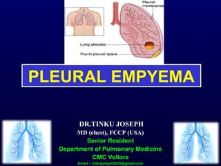
- 1. PLEURAL EMPYEMAPLEURAL EMPYEMA DR.TINKU JOSEPH MD (chest), FCCP (USA) Senior Resident Department of Pulmonary Medicine CMC Vellore Email -: tinkujoseph2010@gmail.com
- 3. Pleural Empyema / Pyothorax / Purulent Pleuritis / Empyema Thoracis Accumulation of Pus in the Pleural cavity.
- 4. Etiology (Introduction of infection) EMPYEMA NON TRAUMATIC TRAUMATIC THORACIC SEPSIS EXTRATHORACIC SEPSIS IATROGENIC NON- IATROGENIC SUBPHRENIC ABSCESS, HEPATIC ABSCESS MEDIASTINITIS PULMONARY DISEASE OSTEOMYELI TIS LUNG RESECTION, OESOPHAGEAL TEARS, PARACETESIS THORACIS, LIVER BIOPSY STABBINGS,G UNSHOT WOUNDS,ETC PNEUMONIA, TB, BRONCHIECTASIS ,LUNG ABCESS STERNUM, VERTEBRAE, RIBS
- 5. Bacteriological data. • Streptococcus pneumoniae: 15-20% –Increased resistance • Staphylococcus:15-30% • Streptococcus spp • Gram Negative: 20-50% –Klebsiella, Enterobacter, Pseudomonas, Hemophilus, E.Coli • Anaerobes: –Fusobacterium, Bacteroides fragilis
- 6. Influence of predisposing factors In adults – empyema arises as a complication of CAP,often pneumococcal. Frank aspiration – anaerobes. Aerobic gram negative bacilli infection likely to affect pleura – from below diaphragm or as a result of oesophageal instrumentation. Mycobacteria and fungi more common in immunocompromised.
- 7. Uncommon microbial causes Tuberculous Fungal – Aspergillous,Cryptococcus,Blastomyces, Histoplasmosis. Actinomyces – aerobic gram negative filamentous bacteria. Clostridia – anaerobic organism. Hydatid disease – Echinococcus. Lung fluke – Paragonimus westermani. Protozoa – Trichomonas,Entamoeba histolytica.
- 8. Pathology-Stages Acute (exudative) stage: Approximately in 7 days. Infected by pathogenic organism, pleural membranes becomes edematous and produce exudation of proteinaceous fluid that starts to fill the pleural cavity. Pleura fills with thin serous fluid that shows relatively low white cell count. Visceral pleura and underlying lung remains mobile.
- 9. Stages cont, Transitional (Fibrinopurulent) stage: From day 7 to 21 day. If infection proceeds unchecked by antimicrobials. Thick, Opaque fluid with positive culture (pus) and Deposition of thin fibrin layer over the pleura.(mainly parietal pleura). Empyema fluid now becomes more thicker and turbid. Higher white cell count. Lung movements in this later stages become increasingly restricted. Progressive loculation and formation of pouches in the pleura.
- 10. Stages cont, Stage of vascularization: Fibrinous layers starts to organize as collagen. Becomes vascularized by ingrowth of capillaries.
- 11. Stages cont, Organizing (chronic) Stage: after 21 days. Usually 4-6 weeks. Empyema cavity becomes surrounded by a cortex. Contains frank pus. Inner layers shows inflammatory cells. Outer layers gets fibrous – exerts restrictive effect. Compressing the underlying lung (trapped lung effect). Draws the ribs together producing chest deformity. Later on gets calcified – fibrothorax.
- 12. Symptoms & signs Depends on nature of infecting organism competence of patients immune system. Ranges from complete absence of symptoms to a severe illness with all usual manifestations of systemic toxicity. Fever Cough & Expectoration. Pleuretic chest pain. Dyspnoea Easy fatiguability. Loss of weight. Night sweating.
- 13. • Finger clubbing. • Signs of pleural effusion. • Empyema necessitants.
- 14. Complications Rupture into the lung; BronchoPleural fistula. Spread to the subcutaneous tissue; Empyema necessitans. Septicaemia & septic shock.
- 15. Diagnosis LRTI – possibility of complicating empyema. History and physical findings may be suggestive. CXR,USG,CT. Thoracentesis- PH < 7.4 Glucose <40 mg/dl LDH> 1000 iu/dl Protein > 2.5 gm/dl Sp.gravity >1.018 Other findings (non specific):neutrophil leucocytosis and hypoalbuminaemia.
- 16. Chest x ray In early stages same as uncomplicated pleural effusion. As time passes, fibrosis develops around empyema cavity. Fluid contained in one location. Homogenous shadow extending upwards.
- 19. • Lateral cxr – opacity convex anteriorly. • Tapering at its upper and lower ends • Extending into the thorax. • D – shaped shadow.
- 20. Other findings on cxr Air fluid level – pneumothorax,BPF,Iatrogenic, gas forming organisms such as clostridia. Underlying pulmonary shadowing -: delayed resolution of pneumonia,lung abcess,tumours of lung,mediastinum,pleura. Hydatid cyst,partial lung collapse. USG – pockets of fluid. CT thorax – similarly helpful,inflamatory lymphadenopathy
- 22. Post pneumonectomy empyema nessitants.
- 24. Goals of the treatment Treat the infection. Drain the purulent effusion adequately and completely. Re-expand the lung to fill the pleural space. Eliminate complications and avoid chronicity.
- 25. Antimicrobial Therapy Choice of antibiotic – microbiological C/S testing. Anaerobes- may be treated with Benzylpenicillin. If resistant – add metronidazole. Better response – Clindamycin + Penicillin ( active against Bacteroids fragilis and other penicillin- resistant anaerobes.(Arch Intern med 1995,150:1700). Amoxyclav.(Beta lactam inhibitors). Ampicillin+Sulbactam. Piperacillin,Ticarcillin.(combined with tazobactum). Imipenam,meropenam(anaerobes as well as Gram +/-)
- 26. Cefoxitin – most potent cephalosporin. Chloramphenicol- very effective,but not routinely given due to fear of bone marrow suppression. Macrolides,Quinolones not so effective. Pneumococcus Responds to high dose benzylpenicillin initially,continuing with oral phenoxy methyl penicillin(penicillin V) or amoxycillin. Alternatives for penicillin allergic individuals- Cefradin or Clarithromycin.
- 27. Staphylococcus aureus Dicloxacillin,oxacillin for parenteral use. First generation cephalosporins – cefradine. MRSA- vancomycin,Linezolid. Gram negative aerobes Serious aerobic infections may be treated with the combination of a third generation cephalosporin – Ceftazidime and an amynoglycoside such as gentamycin. Mixed infection,including anaerobes – piperacillin.
- 28. Adults with empyema who are admitted from the community, and in whom infecting organism have not yet been identified may be treated initially with a combination that includes co-amoxyclav,metronidazole and flucloxacillin. This regimen is modified in the light of cultures and the patients clinical response. Duration of therapy is likely to be several weeks. It can be continued for at least 3 weeks after all drainage has ceased.
- 29. BTS guidelines for the management of empyema Origin of infection Intravenous antibiotic treatment Oral antibiotic treatment Community acquired culture negative pleural infection Cefuroxime 1.5 g tds iv + metronidazole 400 mg tds orally or 500 mg tds iv Amoxycillin 1 g tds + clavulanic acid 125 mg tds Benzyl penicillin 1.2 g qds iv + ciprofloxacin 400 mg bd iv Amoxycillin 1 g tds + metronidazole 400 mg tds Meropenem 1 g tds iv + metronidazole 400 mg tds orally or 500 mg tds iv Clindamycin 300 mg qds Hospital acquired culture negative pleural infection Piperacillin + tazobactam 4.5 g qds iv Not applicable Ceftazidime 2 g tds iv, Meropenem 1 g tds iv ± metronidazole 400 mg tds orally or 500 mg tds iv
- 30. Tuberculous Empyema Rare entity. Purulent fluid loaded with tuberculous organisms. Usually develops in fibrous scar tissue resulting from pleurisy, artificial pneumothorax or thoracoplasty. Underlying pleura is heavily calcified. Sub acute or chronic illness Fatigue, low grade fever and weight loss. Radigraphically – obvious pleural effusion, pleural thickening. CT scan – thick calcified pleural rind and rib thickening surrounded by loculated pleural fluid.
- 31. Tuberculous Empyema Diagnosis – thoracentesis, AFB smear and culture. Treatment – intensive chemotherapy coupled with serial thoracentesis can be curative at times. Multiple drug regimen at their maximal tolerated dosages. Strong tendency to develop resistant organisms. ATT frequently do not reach there normal levels in the pleural space owing to the thick, fibrous and often calcified pleura. VATS/Decortication.
- 32. Aspergillus – amphotericin or itraconazole. Cryptococcosis- in immunocompromised patient is treated with amphotericin and flucytosine Blstomycosis,Coccidiomycosis – amphotericin/Itraconazole. Actinimycosis – high doze Benzyl penicillin followed by prolonged therapy with oral amoxycillin. Nocardia – co-trimoxazole,sulfadiazine or imipenam. Clostridial empyemas- high dose benzyl penicillin/ metronidazole or imipenam. Salmonella – ciprofloxacin or ceftazidime.
- 33. Hydatid infection – surgical excision with albendazole cover. Paragonimus westermani – praziquantel. Trichomonas – metronidazole. Empyema complicating amoebiasis – metronidazole and diloxanide furoate.
- 34. Primary treatment options • Antibiotics alone; • Recurrent thoracocentesis • Insertion of chest drain alone or in combination with fibrinolytics • VATS. • Open decortication
- 35. Thoracocenthesis Big caliber needle. Repeated aspiration is carried out. Use of Abrams punch biopsy needle is useful initially. Wide callibre allow easy aspiration and also permits pleural biopsy. Mostly diagnosis technique Therapeutically used if the liquid remains fluid Helps in pleural lavage also.
- 36. Chest Tube Closed tube thoracostomy. As soon as the fluid is thick. Localization loculated: Chest imaging using ultrasonography and/or computed tomography Size: 20 - 28 F Passed under USG guidance,helps in breaking fibrinous septa and pus rapidly gets removed Bedside
- 37. Pleural Lavage Isotonic saline +/- Noxyflex (noxytioline) Modalités 3 way stopcock Directly through the CT: 250 to 500 ml Cautiously if suspicion of broncho-pleural fistula Timing: Immediately after CT placement+++ Once a day until the liquid is clear
- 38. Fibrinolytics Intrapleural Streptokinase; • Indications Acute or fibrino purulent stage Presence of loculations. Incomplete drainage after tube insertion • Contraindications: Chronic stage Post-operative empyema Empyema with BPF.
- 39. Fibrinolytics Was reported in 1949. Then was abandoned due to allergic reactions,but taken up again due to availability of purer forms of streptokinase,urokinase. (Davies RJO,Trail ZC Thorax 1997; 52:416.) Urokinase: 100 000 or 300 000 IU . Streptokinase: 250000 IU . 250.000 IU in 10-20 ml isotonic saline. Don’t evacuate before 24 to 48 hours. Constantly associated with fever (38-39°C). Then evacuate.
- 40. Local antibiotics Intrapleural instillation of antibiotics, especially metronidazole,Colimycin. Still debated. Do not replace systemic treatment.
- 41. Video-assisted thoracic surgery VATS. If closed drainage does not result in prompt re-expansion of the lung and especially if loculi have been identified by USG. Decision to intervene early is made. Debridement and drainage. Breakage of loculi,evacuating pus,debris and freeing lung. Helps in re expansion of lung.
- 42. Compare Chest Tube + Streptokinase (n=9) vs VATS (n=11) Wait et al, Chest 1997
- 43. Bronchoscopy Recommended following the successful conclusion of closed drainage. In order to exclude any endobronchial causes of obstruction, such as tumour or foreign body.
- 44. Open drainage If empyema persists both clinically and radiologically. In whom closed drainage has proved unsuccessful. If VATS unavailable, unsuccessful or considered inappropriate. Rib Resection Drainage. Eloesser Flap .
- 45. Rib Resection Drainage Performed under LA/GA when the pus is thick and loculated. Resection of the lowest rib above and below which pus can be aspirated. Open all the intact cyst that leads to conversion of empyema with free pus. Then place wide bore tube surrounded by gauze in the unsutured wound. Daily dressing. Gradual obliteration of empyema space. Antibiotics should continue for 6 weeks.
- 47. An earlier form of treatment involved surgical removal of most of the ribs on the infected side of the thorax, causing a permanent collapse of the lung and obliteration of the infected pleural space. This would leave the patient with a large portion of the upper chest removed, giving the impression that the shoulder had been detached from the body. Rarely performed today, the surgery was common during WORLD WAR 1
- 48. Eloesser Flap Drainage When drainage is protracted. Patient remains too ill. Unsuitable for thoracotomy. Then a more permanent fenestration or open window thoracostomy may be performed.
- 49. Once the ribs have been resected, the skin overlying the thoracostomy is marsupialized to the parietal pleura to permit packing and open pleural drainage. Pleurocutaneous fistula. Stoma may be closed if the underlying lung re-expands or may be left permanently open with daily dressing changes.
- 51. Open chest drainage (Eloesser flap).a) Photograph shows a right Eloesser flap 8 months .b) after creation that was closed with a muscle flap
- 52. Decortication Elective surgical procedure. Unsuitable for patients who are ill and toxic. Fibrous wall of the empyema cavity,reffered to as cortex is exposed at thoracotomy is stripped off and adjacent visceral and parietal pleura may be left intact. Indications Closed drainage/thoracoscopic methods have been unsuccessful. Patients who has entered a chronic phase in which underlying lung does not expand because of failure of cortex to become reabsorbed.
- 53. There is no optimal time for decortication. Some surgeons arguing for early intervention and others opting for a conservative approach. Early surgical intervention in pleural empyema.thorac cardiovascular surg 1985.
- 54. Decortication
- 56. Thoracoplasty Rarely used now a days. To obliterate an empyema space following pneumonectomy. Manage a persistent empyema cavity in patients where extensive ipsilateral parenchymal disease would prevent re expansion after decortication.
- 57. Physiotherapy Key to a correct evolution After CT removal Often and for a long time….. Decrease surgery Decrease long term pain and functional limitations.
- 58. Indications Thoracocentesis Clear liquid Not clear or purulent effusion pH>7.20 pH<7.20 No intervention Not loculated Loculated Drainage Pleural lavage Fibrinolytics 24-48h Drainage Fibrinolytics Pleural lavage VATS Drainage Pleural lavage Failure VATS Surgery Failure Surgery Reccurent thoracocentesis Hamm et al, ERJ 1997