SlideShare una empresa de Scribd logo
1 de 68
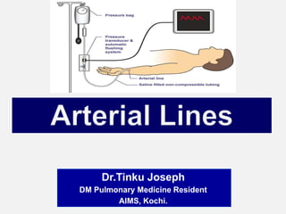
Dr.Tinku Joseph
DM Pulmonary Medicine Resident
AIMS, Kochi.
 Indications
 Preparation & Equipments
 Positioning
 Insertion
 Complications
 Mechanism of Action
 Troubleshooting
 An arterial line is an
invasive method to
measure BP.
1.Continuous, beat-to-beat blood pressure
measurement.
- Hemodynamically unstable pts /ICU pts requiring inotropic
support
- Patients undergoing major surgery
2.Frequent arterial blood gas analysis
-pts with respiratory failure on ventilator
-severe acid/base disturbance.
3. Facilitation of reliable titration
of vasoactive medications.
4. Unable to obtain non invasive
BP.
1) Known deficiencies in collateral
circulation: Raynauds phenomenon,
Thromboangitis obliterans, Brachial
artery insufficiency.
2) Infection of the site.
3) Trauma to the proposed site.
4) Excessive anticoagulation.
Advantages of IBP measurement
 Continuous blood pressure recording
 Accurate blood pressure recording even
when patients are profoundly hypotensive vs
NIBP which is difficult or inaccurate
 Real time Visual Display
Disadvantages of IBP measurement
 Potential complications
 Skilled technique reqd
 Expensive
 The radial artery has low complication
rates compared with other sites.
 It is a superficial artery which aids
insertion, and also makes it compressible
for haemostasis
 The ulnar, brachial, axillary, dorsalis
pedis, posterial tibial, femoral arteries are
alternatives.
The idea here is to figure out if the
ulnar artery will supply the hand with
enough blood, if the radial artery is
blocked with an a-line.
 Allen’s test is recommended
before the insertion of a radial
arterial line.
 This is used to determine
collateral circulation between the
ulnar and radial arteries to the
hand
 If ulnar perfusion is poor and a
cannula occludes the radial
artery, blood flow to the hand
may be reduced.
 The test is performed by asking
the patient to clench their hand.
The ulnar and radial arteries are
occluded with digital pressure.
 The hand is unclenched and
pressure over the ulnar artery is
released. If there is good
collateral perfusion, the palm
should flush in less than 6
seconds.
Arterial cannula
 Made from polytetrafluoroethylene (‘Teflon’) to minimize
the risk of clot formation
 20G (pink) cannula - adult patients
22G (blue)- paediatrics
24G (yellow) - neonates and small babies
 Larger gauge cannulae increase the risk of thrombosis,
smaller cannulae cause damping of the signal.
 The cannula is connected to an arterial giving set.
 ARTERIAL SET
- Specialized plastic tubing, short and stiff to reduce resonance,
connected to a 500 ml bag of saline.
 SALINE BAG
-500 ml 0.9% saline pressurized to 300 mmHg using a
pressure bag, i.e. a pressure higher than arterial systolic
pressure to prevent backflow from the cannula into the giving
set.
-The arterial set and pressurized saline bag with 2500units
Heparin incorporate a continuous slow flushing system of 3–4
ml per hour to keep the line free from clots.
-The arterial set and arterial line should be free from air
bubbles.
- The line is attached to a transducer.
 DO NOT ALLOW THE SALINE BAG TO EMPTY
–To maintain patency of arterial cannula.
–To prevent air embolism
–To maintain accuracy of blood pressure reading
–To maintain accuracy of fluid balance chart
–To prevent backflow of blood
 TRANSDUCER, AMPLIFIER AND ELECTRICAL
RECORDING EQUIPMENT.
-The transducer is zeroed and placed level with the heart.
 Tape and/or steri-strips
 An arm board or towel roll
 Opsite or Tegaderm cover dressing
 Local anesthetic (1% or 2% lidocaine ,lidocaine
cream)
 Suture material for femoral arterial line placement
(2.0 silk)
 Scissors
 Monitor cable for transducing arterial waveform.
 Benzoin solution
1. Ensure that all pre-procedure steps are taken
2. Pressure tubing with transducer is connected to
bedside monitor.
3. Perform the Allen’s test to ensure adequate collateral
blood flow.
4. Wash hands and wear gloves
5. For the radial artery, the arm is restrained, palm up,
with an arm board to hold the wrist dorsiflexed
For the radial artery, the most
common insertion site, the arm is
restrained, palm up, with an
armboard to hold the wrist
dorsiflexed
6. Apply anesthetic agent (local lidocaine 1-2% or
lidocaine cream).
7. Locate pulsating artery via palpation.
8. Cleanse area selected for arterial line
placement.
9. Prepare patient for puncture.
10. Stabilize artery by pulling skin taut.
11. Puncture skin at 45-60 degree angle for radial
artery; 90 degrees for femoral artery.
12. Advance catheter when flash of blood is observed in
catheter.
13. Connect to pressure I.V. tubing and check for arterial
waveform on bedside monitor.
14. Cleanse area of any blood and allow site to dry.
15. Apply Benzoin to cleansed area and allow to dry.
16. Secure arterial line with tape and cover with a
Tegaderm dressing.
17. Secure I.V. tubing to prevent it from being caught and
pulling on arterial catheter. If a femoral arterial line is
placed, it should be secured with a suture.
18. Properly dispose of the I.V. sharps and other used
materials.
 1. Direct cannulation
 2. Transfixation
 3. Guidewire (Seldinger)
technique
 Haemorrhage may occur if there are leaks in
the system. Connections must be tightly
secured.
 Emboli. Air or thrombo emboli may occur.
Care should be taken to aspirate air bubbles
 Accidental drug injection may cause severe,
irreversible damage to the hand.
-No drugs should be injected via an
arterial line
- The line should be labelled (in red) to reduce
the likelihood of this occurring
 Arterial vasospasm
 Partial occlusion due to large cannula width,
multiple attempts at insertion and long duration
of use
 Permanent total occlusion
 Sepsis or bacteraemia secondary to infected
radial arterial lines is very rare (0.13%);
-local infection is more common.
-if the area looks inflamed the line site should be
changed.
 Concentration of a drug
into the tissues served by
the cannulated artery can
result in cell death
 Skin necrosis, severe
gangrene, limb ischemia,
amputation & permanent
disabilities
Mechanism of
action
 A transducer is a device
that reads the fluctuations
in pressure – it doesn’t
matter if it’s arterial, or
central venous, or PA
 The column of saline in the
arterial set transmits the
pressure changes to the
diaphragm in the transducer
 The transducer reads the changing pressure, and
changes it into an electrical signal that goes up and
down as the pressure does which is displayed as an
arterial waveform.
 The transducer connects to the bedside monitor with
a cable, and the wave shows up on the screen, going
from left to right.
 The transducer has to sit in a “transducer holder” – this
is the white plastic plate that screws onto the rolling pole
that holds the whole setup.
 The transducer has to be leveled correctly-to make sure
that it’s at the fourth intercostal space, at the mid-
axillary line (Phlebostatic axis)
 Make sure there’s no air in the line before you
hook it up to the patient – use the flusher to
clear bubbles out of the tubing.
 Zero the line to atmospheric pressure properly
 Choose a screen scale that lets you see the
waveform clearly.
 To ensure accuracy of readings
 Flush the device & turn it off to patient but open to
atmosphere
 This exerts pressure on transducer
 This pressure is called zero
 Zero once per shift or if values are questionable
 Ensure flush bag is pumped up
 Once inserted, an
arterial waveform trace
should be displayed at
all times
 This confirms that the
invasive arterial BP
monitoring is set up
correctly, and
minimizes problems.
 The highest point - systolic
pressure,
-the lowest is the diastolic.
Everybody see the little
notch on the diastolic down
slope? – there’s one in each
beat.
 A little after the beginning
of diastole – the start of the
downward wave – the aortic
valve flips closed,
generating a little back-
pressure bump: called the
“dicrotic notch”..
 Now we know how the arterial pressure monitoring
system works, we need to be able to decide
whether or not the trace (and BP in
numerical format) is accurate.
 Failure to notice this may lead to unnecessary, or
missed treatments for our patients.
 There are 2 main abnormal tracing problems that
can occur once the monitor gain is set correctly.
 Dampened trace
 Dampening occurs due to:
• air bubbles
• overly compliant, distensible tubing
• catheter kinks
• clots
• injection ports
• low flush bag pressure or no fluid in the flush bag
• Improper scaling
• Severe hypotension if everything else is ruled out
 This type of trace Under estimate SBP, over estimate
DBP
 Resonant trace
 Resonance occurs due to:
• long tubing
• overly stiff, non-compliant tubing
• increased vascular resistance
• reverberations in tubing causing harmonics that distort
the trace (i.e. high systolic and low diastolic)
• not-fully opened stopcock valve
 This type of trace:
 Over estimate SBP, under estimate DBP
 Arterial lines measure systolic BP approximately 5
mmHg higher and the diastolic BP
approximately 8 mmHg lower compared to non-
invasive BP (NIBP) measurement.
 “It takes a year just to learn which way to turn the
stopcocks!”
This is really true: some stopcocks point to where
they’re open, and some point to where they’re closed
– it just takes some time to learn which is which.
 The trick is remembering which way to turn the
stopcock, and avoiding a mess.
 Don’t forget to clear the stopcock, recap, and then
flush the line.
 Keep things nice and sterile.
 This probably means that the artery being monitored
gone into spasm.
 You need to think about things that might make this
happen:
-Is the patient very cold?
-Are his extremities poorly perfused?
-Is he on a “shipload” of pressors, making his
arterial bed tighten up –
- Is he “dry” as well?
 Sometimes arteries become unhappy with catheters in
them, and you just have to convince the team that the
patient needs a new one placed in another site.
The first thing to think about is:
1.Is the arterial catheter still in place? Yes? Try drawing
with a 3cc syringe from the stopcock – if it draws normally,
then you’ve got a hardware problem
2.Cables become loose?
3.Did the screen scale get accidentally set to, say, 40,
instead of 150 or 200mm of pressure?- you’ll only see a flat
line.
4.Is it a transducer setup Failure – try a new setup.
 If the line doesn’t draw –
-Is there a clot in the hub?
-Try taking the site dressing down – is the catheter kinked
going into the patient?
- Sometimes art-lines just fail – the artery spasms and
won’t open up – time for a new site.
 The routine now is 96 hours – make sure that you
label the line setup when you hang it. Obviously,
change the line setup if it is contaminated in any
way.
 Usually this will be pretty obvious:
the pulse will diminish, or go away altogether. The
hand may look dusky, or be cold, or lose some
sensation – remember to assess for coloring,
sensation, motion, and capillary refill.
 If you think that the a-line is threatening the
patient’s hand, let the team know right away, and be
ready to set up for another insertion somewhere else if
the line is still necessary.
 Compress the site with a sterile 4x4 for at least 5
minutes, or longer if the patient is anticoagulated.
 Assess the perfusion of the hand.
 Make sure you put the patient on the non-
invasive cuff at meaningful intervals while you
talk to the team about replacing the line
 This is usually pretty obvious – the patient is
hemodynamically stable, needs only one or two
blood draws in a day, no more need for ABGs
 Disconnect the cable from the monitor which will
automatically turn off the alarms.
 Take out the sutures in the usual way with a fresh sterile
kit.
 Have a gauzepiece ready, pull the catheter, and manually
compress the site for at least 3 to 5 minutes.
 Make sure the patient’s hand is still perfused.
 Check for hematoma or bleeding, put a compression
dressing on the site (not too tight!), which you can then
take off after about an hour.
 Recheck the site hourly for a few hours afterwards – a
hematoma could still form, and since there isn’t a whole lot
of room in a wrist, you’d definitely want to know!
Arterial lines by Dr.Tinku Joseph

Más contenido relacionado

La actualidad más candente

Central venous catheterization
Central venous catheterizationCentral venous catheterization
Central venous catheterizationMominul Haider
 
Pulmonary artery pressure monitoring
Pulmonary artery pressure monitoringPulmonary artery pressure monitoring
Pulmonary artery pressure monitoringPrincy Francis M
 
Arterial line analysis
Arterial line analysisArterial line analysis
Arterial line analysissamirelansary
 
33)Esophageal Tracheal Combitube
33)Esophageal Tracheal Combitube33)Esophageal Tracheal Combitube
33)Esophageal Tracheal Combitubephant0m0o0o
 
Endotracheal intubation
Endotracheal intubationEndotracheal intubation
Endotracheal intubationAgrawal N.K
 
Invasive blood pressure_monitoring
Invasive blood pressure_monitoringInvasive blood pressure_monitoring
Invasive blood pressure_monitoringUbaidur Rahaman
 
Hemodynamic monitoring ppt
Hemodynamic monitoring pptHemodynamic monitoring ppt
Hemodynamic monitoring pptUma Binoy
 
Pulmonary artery catheter
Pulmonary artery catheterPulmonary artery catheter
Pulmonary artery catheterrajkumarsrihari
 
Supraglottic airway device
Supraglottic airway deviceSupraglottic airway device
Supraglottic airway deviceDebojyoti Dutta
 
Neurological monitoring(1)
Neurological monitoring(1)Neurological monitoring(1)
Neurological monitoring(1)Manu Jacob
 
Mechanical ventilator for nurses 08.02.19
Mechanical ventilator for nurses 08.02.19Mechanical ventilator for nurses 08.02.19
Mechanical ventilator for nurses 08.02.19Johny Wilbert
 
Endotracheal tubes
Endotracheal tubesEndotracheal tubes
Endotracheal tubesPratik Kumar
 
Capnography
CapnographyCapnography
Capnographylarryide
 

La actualidad más candente (20)

Central venous catheterization
Central venous catheterizationCentral venous catheterization
Central venous catheterization
 
Pulmonary artery pressure monitoring
Pulmonary artery pressure monitoringPulmonary artery pressure monitoring
Pulmonary artery pressure monitoring
 
Arterial line analysis
Arterial line analysisArterial line analysis
Arterial line analysis
 
33)Esophageal Tracheal Combitube
33)Esophageal Tracheal Combitube33)Esophageal Tracheal Combitube
33)Esophageal Tracheal Combitube
 
Central Venous Access
Central Venous AccessCentral Venous Access
Central Venous Access
 
Hemodynamic monitoring
Hemodynamic monitoringHemodynamic monitoring
Hemodynamic monitoring
 
Endotracheal intubation
Endotracheal intubationEndotracheal intubation
Endotracheal intubation
 
Invasive blood pressure_monitoring
Invasive blood pressure_monitoringInvasive blood pressure_monitoring
Invasive blood pressure_monitoring
 
Central line
Central line Central line
Central line
 
Cardioversion
Cardioversion Cardioversion
Cardioversion
 
Hemodynamic monitoring ppt
Hemodynamic monitoring pptHemodynamic monitoring ppt
Hemodynamic monitoring ppt
 
Oxygen delivery systems
Oxygen delivery systemsOxygen delivery systems
Oxygen delivery systems
 
Pulmonary artery catheter
Pulmonary artery catheterPulmonary artery catheter
Pulmonary artery catheter
 
Cvp
CvpCvp
Cvp
 
Capnography
CapnographyCapnography
Capnography
 
Supraglottic airway device
Supraglottic airway deviceSupraglottic airway device
Supraglottic airway device
 
Neurological monitoring(1)
Neurological monitoring(1)Neurological monitoring(1)
Neurological monitoring(1)
 
Mechanical ventilator for nurses 08.02.19
Mechanical ventilator for nurses 08.02.19Mechanical ventilator for nurses 08.02.19
Mechanical ventilator for nurses 08.02.19
 
Endotracheal tubes
Endotracheal tubesEndotracheal tubes
Endotracheal tubes
 
Capnography
CapnographyCapnography
Capnography
 

Similar a Arterial lines by Dr.Tinku Joseph

Lec # 6 hemodynamic monitoring
Lec # 6 hemodynamic monitoringLec # 6 hemodynamic monitoring
Lec # 6 hemodynamic monitoringAli Sheikh
 
Cardiovascular monitoring Part I
Cardiovascular monitoring Part ICardiovascular monitoring Part I
Cardiovascular monitoring Part ISiddhanta Choudhury
 
Non Invasive and Invasive Blood pressure monitoring RRT
Non Invasive and Invasive Blood pressure monitoring RRTNon Invasive and Invasive Blood pressure monitoring RRT
Non Invasive and Invasive Blood pressure monitoring RRTRanjith Thampi
 
bpmonitoring.pdf
bpmonitoring.pdfbpmonitoring.pdf
bpmonitoring.pdfshafina27
 
Dialysis machines.pptx
Dialysis machines.pptxDialysis machines.pptx
Dialysis machines.pptxDarshanS239776
 
HAEMODYNAMIC MONITORING – CVP, PAC AND IBP
HAEMODYNAMIC MONITORING – CVP, PAC AND IBPHAEMODYNAMIC MONITORING – CVP, PAC AND IBP
HAEMODYNAMIC MONITORING – CVP, PAC AND IBPGowri Shankar
 
central venous pressure and intra-arterial blood pressure monitoring. invasiv...
central venous pressure and intra-arterial blood pressure monitoring. invasiv...central venous pressure and intra-arterial blood pressure monitoring. invasiv...
central venous pressure and intra-arterial blood pressure monitoring. invasiv...prateek gupta
 
Free flap postoperative evaluation
Free flap postoperative evaluationFree flap postoperative evaluation
Free flap postoperative evaluationAzis Aimaduddin
 
Iv Therapy
Iv TherapyIv Therapy
Iv Therapywashinca
 
Intraoperative Monitoring. sandeep. first years.ppt
Intraoperative Monitoring. sandeep. first years.pptIntraoperative Monitoring. sandeep. first years.ppt
Intraoperative Monitoring. sandeep. first years.pptsandeepsandeepkundra
 
Arterial_and_CVP_monitoring.ppt
Arterial_and_CVP_monitoring.pptArterial_and_CVP_monitoring.ppt
Arterial_and_CVP_monitoring.pptssuser35745f
 
Arterial_and_CVP_monitoring.ppt
Arterial_and_CVP_monitoring.pptArterial_and_CVP_monitoring.ppt
Arterial_and_CVP_monitoring.pptssuser35745f
 
arterial and CVP monitoring in perioperative period.pptx
arterial and CVP monitoring in perioperative period.pptxarterial and CVP monitoring in perioperative period.pptx
arterial and CVP monitoring in perioperative period.pptxkhelifakolea
 
Basic and advanced Cardiovascular monitoring.pptx
Basic and advanced Cardiovascular monitoring.pptxBasic and advanced Cardiovascular monitoring.pptx
Basic and advanced Cardiovascular monitoring.pptxamitkalirawana07
 

Similar a Arterial lines by Dr.Tinku Joseph (20)

Arterial line insertion
Arterial line insertionArterial line insertion
Arterial line insertion
 
Lec # 6 hemodynamic monitoring
Lec # 6 hemodynamic monitoringLec # 6 hemodynamic monitoring
Lec # 6 hemodynamic monitoring
 
Cardiovascular monitoring Part I
Cardiovascular monitoring Part ICardiovascular monitoring Part I
Cardiovascular monitoring Part I
 
Non Invasive and Invasive Blood pressure monitoring RRT
Non Invasive and Invasive Blood pressure monitoring RRTNon Invasive and Invasive Blood pressure monitoring RRT
Non Invasive and Invasive Blood pressure monitoring RRT
 
Dialysis machines key features
Dialysis machines key featuresDialysis machines key features
Dialysis machines key features
 
bpmonitoring.pdf
bpmonitoring.pdfbpmonitoring.pdf
bpmonitoring.pdf
 
Dialysis machines.pptx
Dialysis machines.pptxDialysis machines.pptx
Dialysis machines.pptx
 
Perioprative monitoring
Perioprative monitoringPerioprative monitoring
Perioprative monitoring
 
HAEMODYNAMIC MONITORING – CVP, PAC AND IBP
HAEMODYNAMIC MONITORING – CVP, PAC AND IBPHAEMODYNAMIC MONITORING – CVP, PAC AND IBP
HAEMODYNAMIC MONITORING – CVP, PAC AND IBP
 
Hemodynamic monitorig
Hemodynamic monitorigHemodynamic monitorig
Hemodynamic monitorig
 
central venous pressure and intra-arterial blood pressure monitoring. invasiv...
central venous pressure and intra-arterial blood pressure monitoring. invasiv...central venous pressure and intra-arterial blood pressure monitoring. invasiv...
central venous pressure and intra-arterial blood pressure monitoring. invasiv...
 
Free flap postoperative evaluation
Free flap postoperative evaluationFree flap postoperative evaluation
Free flap postoperative evaluation
 
Iv Therapy
Iv TherapyIv Therapy
Iv Therapy
 
Iv Therapy
Iv TherapyIv Therapy
Iv Therapy
 
Intraoperative Monitoring. sandeep. first years.ppt
Intraoperative Monitoring. sandeep. first years.pptIntraoperative Monitoring. sandeep. first years.ppt
Intraoperative Monitoring. sandeep. first years.ppt
 
Arterial_and_CVP_monitoring.ppt
Arterial_and_CVP_monitoring.pptArterial_and_CVP_monitoring.ppt
Arterial_and_CVP_monitoring.ppt
 
Arterial_and_CVP_monitoring.ppt
Arterial_and_CVP_monitoring.pptArterial_and_CVP_monitoring.ppt
Arterial_and_CVP_monitoring.ppt
 
ICU
ICUICU
ICU
 
arterial and CVP monitoring in perioperative period.pptx
arterial and CVP monitoring in perioperative period.pptxarterial and CVP monitoring in perioperative period.pptx
arterial and CVP monitoring in perioperative period.pptx
 
Basic and advanced Cardiovascular monitoring.pptx
Basic and advanced Cardiovascular monitoring.pptxBasic and advanced Cardiovascular monitoring.pptx
Basic and advanced Cardiovascular monitoring.pptx
 

Más de Dr.Tinku Joseph

Endobronchial Brachytherapy by Dr.Tinku Joseph
Endobronchial Brachytherapy  by Dr.Tinku JosephEndobronchial Brachytherapy  by Dr.Tinku Joseph
Endobronchial Brachytherapy by Dr.Tinku JosephDr.Tinku Joseph
 
HRCT Chest - By Dr. Tinku Joseph
HRCT Chest - By Dr. Tinku JosephHRCT Chest - By Dr. Tinku Joseph
HRCT Chest - By Dr. Tinku JosephDr.Tinku Joseph
 
Recent advances in Asthma & COPD by Dr.Tinku Joseph
Recent advances in Asthma & COPD by  Dr.Tinku JosephRecent advances in Asthma & COPD by  Dr.Tinku Joseph
Recent advances in Asthma & COPD by Dr.Tinku JosephDr.Tinku Joseph
 
Endobronchial Brachytherapy by Dr.Tinku Joseph
Endobronchial Brachytherapy by  Dr.Tinku JosephEndobronchial Brachytherapy by  Dr.Tinku Joseph
Endobronchial Brachytherapy by Dr.Tinku JosephDr.Tinku Joseph
 
VAP/HAP management guidelines by IDSA/ATS (2016) -: Dr.Tinku Joseph
VAP/HAP management guidelines  by IDSA/ATS (2016) -: Dr.Tinku JosephVAP/HAP management guidelines  by IDSA/ATS (2016) -: Dr.Tinku Joseph
VAP/HAP management guidelines by IDSA/ATS (2016) -: Dr.Tinku JosephDr.Tinku Joseph
 
Hepatopulmonary Syndrome By Dr.Tinku Joseph
Hepatopulmonary Syndrome By Dr.Tinku JosephHepatopulmonary Syndrome By Dr.Tinku Joseph
Hepatopulmonary Syndrome By Dr.Tinku JosephDr.Tinku Joseph
 
Pulmonary Embolism- Diagnosis by Dr.Tinku Joseph
Pulmonary Embolism- Diagnosis  by Dr.Tinku JosephPulmonary Embolism- Diagnosis  by Dr.Tinku Joseph
Pulmonary Embolism- Diagnosis by Dr.Tinku JosephDr.Tinku Joseph
 
Allergic Broncho Pulmonary Aspergillosis (ABPA) by Dr.Tinku Joseph
Allergic Broncho Pulmonary Aspergillosis (ABPA) by Dr.Tinku JosephAllergic Broncho Pulmonary Aspergillosis (ABPA) by Dr.Tinku Joseph
Allergic Broncho Pulmonary Aspergillosis (ABPA) by Dr.Tinku JosephDr.Tinku Joseph
 
Delirium in ICU -By Dr.Tinku Joseph
Delirium in ICU -By Dr.Tinku JosephDelirium in ICU -By Dr.Tinku Joseph
Delirium in ICU -By Dr.Tinku JosephDr.Tinku Joseph
 
ventilator Associated Pneumonia -By Dr.Tinku Joseph
ventilator Associated Pneumonia -By Dr.Tinku Josephventilator Associated Pneumonia -By Dr.Tinku Joseph
ventilator Associated Pneumonia -By Dr.Tinku JosephDr.Tinku Joseph
 
ECMO part 2 by Dr.Tinku Joseph
ECMO part 2 by Dr.Tinku JosephECMO part 2 by Dr.Tinku Joseph
ECMO part 2 by Dr.Tinku JosephDr.Tinku Joseph
 
ECMO - Part 1 by Dr.Tinku Joseph
ECMO - Part 1 by Dr.Tinku JosephECMO - Part 1 by Dr.Tinku Joseph
ECMO - Part 1 by Dr.Tinku JosephDr.Tinku Joseph
 
Small Cell Lung Cancer Management by Dr.Tinku Joseph
Small Cell Lung Cancer Management by Dr.Tinku JosephSmall Cell Lung Cancer Management by Dr.Tinku Joseph
Small Cell Lung Cancer Management by Dr.Tinku JosephDr.Tinku Joseph
 
Stem cell therapy and lungs - Dr.Tinku Joseph
Stem cell therapy and lungs  - Dr.Tinku JosephStem cell therapy and lungs  - Dr.Tinku Joseph
Stem cell therapy and lungs - Dr.Tinku JosephDr.Tinku Joseph
 
Pleural Mesothelioma - Dr.Tinku Joseph
Pleural Mesothelioma - Dr.Tinku JosephPleural Mesothelioma - Dr.Tinku Joseph
Pleural Mesothelioma - Dr.Tinku JosephDr.Tinku Joseph
 
Diving and Lung - Dr.Tinku Joseph
Diving and Lung -  Dr.Tinku JosephDiving and Lung -  Dr.Tinku Joseph
Diving and Lung - Dr.Tinku JosephDr.Tinku Joseph
 
Bronchial Artery Embolization- By Dr.Tinku Joseph
Bronchial Artery Embolization- By Dr.Tinku JosephBronchial Artery Embolization- By Dr.Tinku Joseph
Bronchial Artery Embolization- By Dr.Tinku JosephDr.Tinku Joseph
 
Basic concepts in treatment of Pulmonary Tuberculosis - By Dr.Tinku Joseph
Basic concepts in treatment of Pulmonary Tuberculosis - By Dr.Tinku JosephBasic concepts in treatment of Pulmonary Tuberculosis - By Dr.Tinku Joseph
Basic concepts in treatment of Pulmonary Tuberculosis - By Dr.Tinku JosephDr.Tinku Joseph
 
Respiratory system anatomy Dr.Tinku Joseph
Respiratory system anatomy  Dr.Tinku JosephRespiratory system anatomy  Dr.Tinku Joseph
Respiratory system anatomy Dr.Tinku JosephDr.Tinku Joseph
 

Más de Dr.Tinku Joseph (20)

Endobronchial Brachytherapy by Dr.Tinku Joseph
Endobronchial Brachytherapy  by Dr.Tinku JosephEndobronchial Brachytherapy  by Dr.Tinku Joseph
Endobronchial Brachytherapy by Dr.Tinku Joseph
 
HRCT Chest - By Dr. Tinku Joseph
HRCT Chest - By Dr. Tinku JosephHRCT Chest - By Dr. Tinku Joseph
HRCT Chest - By Dr. Tinku Joseph
 
Recent advances in Asthma & COPD by Dr.Tinku Joseph
Recent advances in Asthma & COPD by  Dr.Tinku JosephRecent advances in Asthma & COPD by  Dr.Tinku Joseph
Recent advances in Asthma & COPD by Dr.Tinku Joseph
 
Endobronchial Brachytherapy by Dr.Tinku Joseph
Endobronchial Brachytherapy by  Dr.Tinku JosephEndobronchial Brachytherapy by  Dr.Tinku Joseph
Endobronchial Brachytherapy by Dr.Tinku Joseph
 
VAP/HAP management guidelines by IDSA/ATS (2016) -: Dr.Tinku Joseph
VAP/HAP management guidelines  by IDSA/ATS (2016) -: Dr.Tinku JosephVAP/HAP management guidelines  by IDSA/ATS (2016) -: Dr.Tinku Joseph
VAP/HAP management guidelines by IDSA/ATS (2016) -: Dr.Tinku Joseph
 
Hepatopulmonary Syndrome By Dr.Tinku Joseph
Hepatopulmonary Syndrome By Dr.Tinku JosephHepatopulmonary Syndrome By Dr.Tinku Joseph
Hepatopulmonary Syndrome By Dr.Tinku Joseph
 
Pulmonary Embolism- Diagnosis by Dr.Tinku Joseph
Pulmonary Embolism- Diagnosis  by Dr.Tinku JosephPulmonary Embolism- Diagnosis  by Dr.Tinku Joseph
Pulmonary Embolism- Diagnosis by Dr.Tinku Joseph
 
Allergic Broncho Pulmonary Aspergillosis (ABPA) by Dr.Tinku Joseph
Allergic Broncho Pulmonary Aspergillosis (ABPA) by Dr.Tinku JosephAllergic Broncho Pulmonary Aspergillosis (ABPA) by Dr.Tinku Joseph
Allergic Broncho Pulmonary Aspergillosis (ABPA) by Dr.Tinku Joseph
 
Delirium in ICU -By Dr.Tinku Joseph
Delirium in ICU -By Dr.Tinku JosephDelirium in ICU -By Dr.Tinku Joseph
Delirium in ICU -By Dr.Tinku Joseph
 
ventilator Associated Pneumonia -By Dr.Tinku Joseph
ventilator Associated Pneumonia -By Dr.Tinku Josephventilator Associated Pneumonia -By Dr.Tinku Joseph
ventilator Associated Pneumonia -By Dr.Tinku Joseph
 
ECMO part 2 by Dr.Tinku Joseph
ECMO part 2 by Dr.Tinku JosephECMO part 2 by Dr.Tinku Joseph
ECMO part 2 by Dr.Tinku Joseph
 
ECMO - Part 1 by Dr.Tinku Joseph
ECMO - Part 1 by Dr.Tinku JosephECMO - Part 1 by Dr.Tinku Joseph
ECMO - Part 1 by Dr.Tinku Joseph
 
Small Cell Lung Cancer Management by Dr.Tinku Joseph
Small Cell Lung Cancer Management by Dr.Tinku JosephSmall Cell Lung Cancer Management by Dr.Tinku Joseph
Small Cell Lung Cancer Management by Dr.Tinku Joseph
 
Stem cell therapy and lungs - Dr.Tinku Joseph
Stem cell therapy and lungs  - Dr.Tinku JosephStem cell therapy and lungs  - Dr.Tinku Joseph
Stem cell therapy and lungs - Dr.Tinku Joseph
 
Pleural Mesothelioma - Dr.Tinku Joseph
Pleural Mesothelioma - Dr.Tinku JosephPleural Mesothelioma - Dr.Tinku Joseph
Pleural Mesothelioma - Dr.Tinku Joseph
 
Diving and Lung - Dr.Tinku Joseph
Diving and Lung -  Dr.Tinku JosephDiving and Lung -  Dr.Tinku Joseph
Diving and Lung - Dr.Tinku Joseph
 
Bronchial Artery Embolization- By Dr.Tinku Joseph
Bronchial Artery Embolization- By Dr.Tinku JosephBronchial Artery Embolization- By Dr.Tinku Joseph
Bronchial Artery Embolization- By Dr.Tinku Joseph
 
Basic concepts in treatment of Pulmonary Tuberculosis - By Dr.Tinku Joseph
Basic concepts in treatment of Pulmonary Tuberculosis - By Dr.Tinku JosephBasic concepts in treatment of Pulmonary Tuberculosis - By Dr.Tinku Joseph
Basic concepts in treatment of Pulmonary Tuberculosis - By Dr.Tinku Joseph
 
Hypoxia Dr.Tinku Joseph
Hypoxia   Dr.Tinku JosephHypoxia   Dr.Tinku Joseph
Hypoxia Dr.Tinku Joseph
 
Respiratory system anatomy Dr.Tinku Joseph
Respiratory system anatomy  Dr.Tinku JosephRespiratory system anatomy  Dr.Tinku Joseph
Respiratory system anatomy Dr.Tinku Joseph
 

Último

SWD (Short wave diathermy)- Physiotherapy.ppt
SWD (Short wave diathermy)- Physiotherapy.pptSWD (Short wave diathermy)- Physiotherapy.ppt
SWD (Short wave diathermy)- Physiotherapy.pptMumux Mirani
 
METHODS OF ACQUIRING KNOWLEDGE IN NURSING.pptx by navdeep kaur
METHODS OF ACQUIRING KNOWLEDGE IN NURSING.pptx by navdeep kaurMETHODS OF ACQUIRING KNOWLEDGE IN NURSING.pptx by navdeep kaur
METHODS OF ACQUIRING KNOWLEDGE IN NURSING.pptx by navdeep kaurNavdeep Kaur
 
Wessex Health Partners Wessex Integrated Care, Population Health, Research & ...
Wessex Health Partners Wessex Integrated Care, Population Health, Research & ...Wessex Health Partners Wessex Integrated Care, Population Health, Research & ...
Wessex Health Partners Wessex Integrated Care, Population Health, Research & ...Wessex Health Partners
 
Hematology and Immunology - Leukocytes Functions
Hematology and Immunology - Leukocytes FunctionsHematology and Immunology - Leukocytes Functions
Hematology and Immunology - Leukocytes FunctionsMedicoseAcademics
 
PERFECT BUT PAINFUL TKR -ROLE OF SYNOVECTOMY.pptx
PERFECT BUT PAINFUL TKR -ROLE OF SYNOVECTOMY.pptxPERFECT BUT PAINFUL TKR -ROLE OF SYNOVECTOMY.pptx
PERFECT BUT PAINFUL TKR -ROLE OF SYNOVECTOMY.pptxdrashraf369
 
Lippincott Microcards_ Microbiology Flash Cards-LWW (2015).pdf
Lippincott Microcards_ Microbiology Flash Cards-LWW (2015).pdfLippincott Microcards_ Microbiology Flash Cards-LWW (2015).pdf
Lippincott Microcards_ Microbiology Flash Cards-LWW (2015).pdfSreeja Cherukuru
 
April 2024 ONCOLOGY CARTOON by DR KANHU CHARAN PATRO
April 2024 ONCOLOGY CARTOON by  DR KANHU CHARAN PATROApril 2024 ONCOLOGY CARTOON by  DR KANHU CHARAN PATRO
April 2024 ONCOLOGY CARTOON by DR KANHU CHARAN PATROKanhu Charan
 
Apiculture Chapter 1. Introduction 2.ppt
Apiculture Chapter 1. Introduction 2.pptApiculture Chapter 1. Introduction 2.ppt
Apiculture Chapter 1. Introduction 2.pptkedirjemalharun
 
Biomechanics- Shoulder Joint!!!!!!!!!!!!
Biomechanics- Shoulder Joint!!!!!!!!!!!!Biomechanics- Shoulder Joint!!!!!!!!!!!!
Biomechanics- Shoulder Joint!!!!!!!!!!!!ibtesaam huma
 
POST NATAL EXERCISES AND ITS IMPACT.pptx
POST NATAL EXERCISES AND ITS IMPACT.pptxPOST NATAL EXERCISES AND ITS IMPACT.pptx
POST NATAL EXERCISES AND ITS IMPACT.pptxvirengeeta
 
See the 2,456 pharmacies on the National E-Pharmacy Platform
See the 2,456 pharmacies on the National E-Pharmacy PlatformSee the 2,456 pharmacies on the National E-Pharmacy Platform
See the 2,456 pharmacies on the National E-Pharmacy PlatformKweku Zurek
 
PNEUMOTHORAX AND ITS MANAGEMENTS.pdf
PNEUMOTHORAX   AND  ITS  MANAGEMENTS.pdfPNEUMOTHORAX   AND  ITS  MANAGEMENTS.pdf
PNEUMOTHORAX AND ITS MANAGEMENTS.pdfDolisha Warbi
 
call girls in green park DELHI 🔝 >༒9540349809 🔝 genuine Escort Service 🔝✔️✔️
call girls in green park  DELHI 🔝 >༒9540349809 🔝 genuine Escort Service 🔝✔️✔️call girls in green park  DELHI 🔝 >༒9540349809 🔝 genuine Escort Service 🔝✔️✔️
call girls in green park DELHI 🔝 >༒9540349809 🔝 genuine Escort Service 🔝✔️✔️saminamagar
 
call girls in aerocity DELHI 🔝 >༒9540349809 🔝 genuine Escort Service 🔝✔️✔️
call girls in aerocity DELHI 🔝 >༒9540349809 🔝 genuine Escort Service 🔝✔️✔️call girls in aerocity DELHI 🔝 >༒9540349809 🔝 genuine Escort Service 🔝✔️✔️
call girls in aerocity DELHI 🔝 >༒9540349809 🔝 genuine Escort Service 🔝✔️✔️saminamagar
 
Music Therapy's Impact in Palliative Care| IAPCON2024| Dr. Tara Rajendran
Music Therapy's Impact in Palliative Care| IAPCON2024| Dr. Tara RajendranMusic Therapy's Impact in Palliative Care| IAPCON2024| Dr. Tara Rajendran
Music Therapy's Impact in Palliative Care| IAPCON2024| Dr. Tara RajendranTara Rajendran
 
SYNDESMOTIC INJURY- ANATOMICAL REPAIR.pptx
SYNDESMOTIC INJURY- ANATOMICAL REPAIR.pptxSYNDESMOTIC INJURY- ANATOMICAL REPAIR.pptx
SYNDESMOTIC INJURY- ANATOMICAL REPAIR.pptxdrashraf369
 
COVID-19 (NOVEL CORONA VIRUS DISEASE PANDEMIC ).pptx
COVID-19  (NOVEL CORONA  VIRUS DISEASE PANDEMIC ).pptxCOVID-19  (NOVEL CORONA  VIRUS DISEASE PANDEMIC ).pptx
COVID-19 (NOVEL CORONA VIRUS DISEASE PANDEMIC ).pptxBibekananda shah
 
epilepsy and status epilepticus for undergraduate.pptx
epilepsy and status epilepticus  for undergraduate.pptxepilepsy and status epilepticus  for undergraduate.pptx
epilepsy and status epilepticus for undergraduate.pptxMohamed Rizk Khodair
 
Radiation Dosimetry Parameters and Isodose Curves.pptx
Radiation Dosimetry Parameters and Isodose Curves.pptxRadiation Dosimetry Parameters and Isodose Curves.pptx
Radiation Dosimetry Parameters and Isodose Curves.pptxDr. Dheeraj Kumar
 
Big Data Analysis Suggests COVID Vaccination Increases Excess Mortality Of ...
Big Data Analysis Suggests COVID  Vaccination Increases Excess Mortality Of  ...Big Data Analysis Suggests COVID  Vaccination Increases Excess Mortality Of  ...
Big Data Analysis Suggests COVID Vaccination Increases Excess Mortality Of ...sdateam0
 

Último (20)

SWD (Short wave diathermy)- Physiotherapy.ppt
SWD (Short wave diathermy)- Physiotherapy.pptSWD (Short wave diathermy)- Physiotherapy.ppt
SWD (Short wave diathermy)- Physiotherapy.ppt
 
METHODS OF ACQUIRING KNOWLEDGE IN NURSING.pptx by navdeep kaur
METHODS OF ACQUIRING KNOWLEDGE IN NURSING.pptx by navdeep kaurMETHODS OF ACQUIRING KNOWLEDGE IN NURSING.pptx by navdeep kaur
METHODS OF ACQUIRING KNOWLEDGE IN NURSING.pptx by navdeep kaur
 
Wessex Health Partners Wessex Integrated Care, Population Health, Research & ...
Wessex Health Partners Wessex Integrated Care, Population Health, Research & ...Wessex Health Partners Wessex Integrated Care, Population Health, Research & ...
Wessex Health Partners Wessex Integrated Care, Population Health, Research & ...
 
Hematology and Immunology - Leukocytes Functions
Hematology and Immunology - Leukocytes FunctionsHematology and Immunology - Leukocytes Functions
Hematology and Immunology - Leukocytes Functions
 
PERFECT BUT PAINFUL TKR -ROLE OF SYNOVECTOMY.pptx
PERFECT BUT PAINFUL TKR -ROLE OF SYNOVECTOMY.pptxPERFECT BUT PAINFUL TKR -ROLE OF SYNOVECTOMY.pptx
PERFECT BUT PAINFUL TKR -ROLE OF SYNOVECTOMY.pptx
 
Lippincott Microcards_ Microbiology Flash Cards-LWW (2015).pdf
Lippincott Microcards_ Microbiology Flash Cards-LWW (2015).pdfLippincott Microcards_ Microbiology Flash Cards-LWW (2015).pdf
Lippincott Microcards_ Microbiology Flash Cards-LWW (2015).pdf
 
April 2024 ONCOLOGY CARTOON by DR KANHU CHARAN PATRO
April 2024 ONCOLOGY CARTOON by  DR KANHU CHARAN PATROApril 2024 ONCOLOGY CARTOON by  DR KANHU CHARAN PATRO
April 2024 ONCOLOGY CARTOON by DR KANHU CHARAN PATRO
 
Apiculture Chapter 1. Introduction 2.ppt
Apiculture Chapter 1. Introduction 2.pptApiculture Chapter 1. Introduction 2.ppt
Apiculture Chapter 1. Introduction 2.ppt
 
Biomechanics- Shoulder Joint!!!!!!!!!!!!
Biomechanics- Shoulder Joint!!!!!!!!!!!!Biomechanics- Shoulder Joint!!!!!!!!!!!!
Biomechanics- Shoulder Joint!!!!!!!!!!!!
 
POST NATAL EXERCISES AND ITS IMPACT.pptx
POST NATAL EXERCISES AND ITS IMPACT.pptxPOST NATAL EXERCISES AND ITS IMPACT.pptx
POST NATAL EXERCISES AND ITS IMPACT.pptx
 
See the 2,456 pharmacies on the National E-Pharmacy Platform
See the 2,456 pharmacies on the National E-Pharmacy PlatformSee the 2,456 pharmacies on the National E-Pharmacy Platform
See the 2,456 pharmacies on the National E-Pharmacy Platform
 
PNEUMOTHORAX AND ITS MANAGEMENTS.pdf
PNEUMOTHORAX   AND  ITS  MANAGEMENTS.pdfPNEUMOTHORAX   AND  ITS  MANAGEMENTS.pdf
PNEUMOTHORAX AND ITS MANAGEMENTS.pdf
 
call girls in green park DELHI 🔝 >༒9540349809 🔝 genuine Escort Service 🔝✔️✔️
call girls in green park  DELHI 🔝 >༒9540349809 🔝 genuine Escort Service 🔝✔️✔️call girls in green park  DELHI 🔝 >༒9540349809 🔝 genuine Escort Service 🔝✔️✔️
call girls in green park DELHI 🔝 >༒9540349809 🔝 genuine Escort Service 🔝✔️✔️
 
call girls in aerocity DELHI 🔝 >༒9540349809 🔝 genuine Escort Service 🔝✔️✔️
call girls in aerocity DELHI 🔝 >༒9540349809 🔝 genuine Escort Service 🔝✔️✔️call girls in aerocity DELHI 🔝 >༒9540349809 🔝 genuine Escort Service 🔝✔️✔️
call girls in aerocity DELHI 🔝 >༒9540349809 🔝 genuine Escort Service 🔝✔️✔️
 
Music Therapy's Impact in Palliative Care| IAPCON2024| Dr. Tara Rajendran
Music Therapy's Impact in Palliative Care| IAPCON2024| Dr. Tara RajendranMusic Therapy's Impact in Palliative Care| IAPCON2024| Dr. Tara Rajendran
Music Therapy's Impact in Palliative Care| IAPCON2024| Dr. Tara Rajendran
 
SYNDESMOTIC INJURY- ANATOMICAL REPAIR.pptx
SYNDESMOTIC INJURY- ANATOMICAL REPAIR.pptxSYNDESMOTIC INJURY- ANATOMICAL REPAIR.pptx
SYNDESMOTIC INJURY- ANATOMICAL REPAIR.pptx
 
COVID-19 (NOVEL CORONA VIRUS DISEASE PANDEMIC ).pptx
COVID-19  (NOVEL CORONA  VIRUS DISEASE PANDEMIC ).pptxCOVID-19  (NOVEL CORONA  VIRUS DISEASE PANDEMIC ).pptx
COVID-19 (NOVEL CORONA VIRUS DISEASE PANDEMIC ).pptx
 
epilepsy and status epilepticus for undergraduate.pptx
epilepsy and status epilepticus  for undergraduate.pptxepilepsy and status epilepticus  for undergraduate.pptx
epilepsy and status epilepticus for undergraduate.pptx
 
Radiation Dosimetry Parameters and Isodose Curves.pptx
Radiation Dosimetry Parameters and Isodose Curves.pptxRadiation Dosimetry Parameters and Isodose Curves.pptx
Radiation Dosimetry Parameters and Isodose Curves.pptx
 
Big Data Analysis Suggests COVID Vaccination Increases Excess Mortality Of ...
Big Data Analysis Suggests COVID  Vaccination Increases Excess Mortality Of  ...Big Data Analysis Suggests COVID  Vaccination Increases Excess Mortality Of  ...
Big Data Analysis Suggests COVID Vaccination Increases Excess Mortality Of ...
 

Arterial lines by Dr.Tinku Joseph

  • 1. Dr.Tinku Joseph DM Pulmonary Medicine Resident AIMS, Kochi.
  • 2.  Indications  Preparation & Equipments  Positioning  Insertion  Complications  Mechanism of Action  Troubleshooting
  • 3.  An arterial line is an invasive method to measure BP.
  • 4.
  • 5. 1.Continuous, beat-to-beat blood pressure measurement. - Hemodynamically unstable pts /ICU pts requiring inotropic support - Patients undergoing major surgery 2.Frequent arterial blood gas analysis -pts with respiratory failure on ventilator -severe acid/base disturbance.
  • 6. 3. Facilitation of reliable titration of vasoactive medications. 4. Unable to obtain non invasive BP.
  • 7. 1) Known deficiencies in collateral circulation: Raynauds phenomenon, Thromboangitis obliterans, Brachial artery insufficiency. 2) Infection of the site. 3) Trauma to the proposed site. 4) Excessive anticoagulation.
  • 8. Advantages of IBP measurement  Continuous blood pressure recording  Accurate blood pressure recording even when patients are profoundly hypotensive vs NIBP which is difficult or inaccurate  Real time Visual Display Disadvantages of IBP measurement  Potential complications  Skilled technique reqd  Expensive
  • 9.  The radial artery has low complication rates compared with other sites.  It is a superficial artery which aids insertion, and also makes it compressible for haemostasis  The ulnar, brachial, axillary, dorsalis pedis, posterial tibial, femoral arteries are alternatives.
  • 10. The idea here is to figure out if the ulnar artery will supply the hand with enough blood, if the radial artery is blocked with an a-line.  Allen’s test is recommended before the insertion of a radial arterial line.  This is used to determine collateral circulation between the ulnar and radial arteries to the hand  If ulnar perfusion is poor and a cannula occludes the radial artery, blood flow to the hand may be reduced.  The test is performed by asking the patient to clench their hand. The ulnar and radial arteries are occluded with digital pressure.  The hand is unclenched and pressure over the ulnar artery is released. If there is good collateral perfusion, the palm should flush in less than 6 seconds.
  • 11. Arterial cannula  Made from polytetrafluoroethylene (‘Teflon’) to minimize the risk of clot formation  20G (pink) cannula - adult patients 22G (blue)- paediatrics 24G (yellow) - neonates and small babies  Larger gauge cannulae increase the risk of thrombosis, smaller cannulae cause damping of the signal.  The cannula is connected to an arterial giving set.
  • 12.
  • 13.
  • 14.  ARTERIAL SET - Specialized plastic tubing, short and stiff to reduce resonance, connected to a 500 ml bag of saline.  SALINE BAG -500 ml 0.9% saline pressurized to 300 mmHg using a pressure bag, i.e. a pressure higher than arterial systolic pressure to prevent backflow from the cannula into the giving set. -The arterial set and pressurized saline bag with 2500units Heparin incorporate a continuous slow flushing system of 3–4 ml per hour to keep the line free from clots. -The arterial set and arterial line should be free from air bubbles. - The line is attached to a transducer.
  • 15.  DO NOT ALLOW THE SALINE BAG TO EMPTY –To maintain patency of arterial cannula. –To prevent air embolism –To maintain accuracy of blood pressure reading –To maintain accuracy of fluid balance chart –To prevent backflow of blood  TRANSDUCER, AMPLIFIER AND ELECTRICAL RECORDING EQUIPMENT. -The transducer is zeroed and placed level with the heart.
  • 16.  Tape and/or steri-strips  An arm board or towel roll  Opsite or Tegaderm cover dressing  Local anesthetic (1% or 2% lidocaine ,lidocaine cream)  Suture material for femoral arterial line placement (2.0 silk)  Scissors  Monitor cable for transducing arterial waveform.  Benzoin solution
  • 17.
  • 18. 1. Ensure that all pre-procedure steps are taken 2. Pressure tubing with transducer is connected to bedside monitor. 3. Perform the Allen’s test to ensure adequate collateral blood flow. 4. Wash hands and wear gloves 5. For the radial artery, the arm is restrained, palm up, with an arm board to hold the wrist dorsiflexed
  • 19. For the radial artery, the most common insertion site, the arm is restrained, palm up, with an armboard to hold the wrist dorsiflexed
  • 20. 6. Apply anesthetic agent (local lidocaine 1-2% or lidocaine cream). 7. Locate pulsating artery via palpation. 8. Cleanse area selected for arterial line placement. 9. Prepare patient for puncture. 10. Stabilize artery by pulling skin taut. 11. Puncture skin at 45-60 degree angle for radial artery; 90 degrees for femoral artery.
  • 21. 12. Advance catheter when flash of blood is observed in catheter. 13. Connect to pressure I.V. tubing and check for arterial waveform on bedside monitor. 14. Cleanse area of any blood and allow site to dry. 15. Apply Benzoin to cleansed area and allow to dry. 16. Secure arterial line with tape and cover with a Tegaderm dressing. 17. Secure I.V. tubing to prevent it from being caught and pulling on arterial catheter. If a femoral arterial line is placed, it should be secured with a suture. 18. Properly dispose of the I.V. sharps and other used materials.
  • 22.  1. Direct cannulation  2. Transfixation  3. Guidewire (Seldinger) technique
  • 23.
  • 24.
  • 25.  Haemorrhage may occur if there are leaks in the system. Connections must be tightly secured.  Emboli. Air or thrombo emboli may occur. Care should be taken to aspirate air bubbles  Accidental drug injection may cause severe, irreversible damage to the hand. -No drugs should be injected via an arterial line - The line should be labelled (in red) to reduce the likelihood of this occurring
  • 26.  Arterial vasospasm  Partial occlusion due to large cannula width, multiple attempts at insertion and long duration of use  Permanent total occlusion  Sepsis or bacteraemia secondary to infected radial arterial lines is very rare (0.13%); -local infection is more common. -if the area looks inflamed the line site should be changed.
  • 27.
  • 28.
  • 29.
  • 30.
  • 31.  Concentration of a drug into the tissues served by the cannulated artery can result in cell death  Skin necrosis, severe gangrene, limb ischemia, amputation & permanent disabilities
  • 32. Mechanism of action  A transducer is a device that reads the fluctuations in pressure – it doesn’t matter if it’s arterial, or central venous, or PA  The column of saline in the arterial set transmits the pressure changes to the diaphragm in the transducer
  • 33.  The transducer reads the changing pressure, and changes it into an electrical signal that goes up and down as the pressure does which is displayed as an arterial waveform.  The transducer connects to the bedside monitor with a cable, and the wave shows up on the screen, going from left to right.
  • 34.
  • 35.  The transducer has to sit in a “transducer holder” – this is the white plastic plate that screws onto the rolling pole that holds the whole setup.  The transducer has to be leveled correctly-to make sure that it’s at the fourth intercostal space, at the mid- axillary line (Phlebostatic axis)
  • 36.  Make sure there’s no air in the line before you hook it up to the patient – use the flusher to clear bubbles out of the tubing.  Zero the line to atmospheric pressure properly  Choose a screen scale that lets you see the waveform clearly.
  • 37.
  • 38.
  • 39.  To ensure accuracy of readings  Flush the device & turn it off to patient but open to atmosphere  This exerts pressure on transducer  This pressure is called zero  Zero once per shift or if values are questionable  Ensure flush bag is pumped up
  • 40.  Once inserted, an arterial waveform trace should be displayed at all times  This confirms that the invasive arterial BP monitoring is set up correctly, and minimizes problems.
  • 41.  The highest point - systolic pressure, -the lowest is the diastolic. Everybody see the little notch on the diastolic down slope? – there’s one in each beat.  A little after the beginning of diastole – the start of the downward wave – the aortic valve flips closed, generating a little back- pressure bump: called the “dicrotic notch”..
  • 42.
  • 43.
  • 44.  Now we know how the arterial pressure monitoring system works, we need to be able to decide whether or not the trace (and BP in numerical format) is accurate.  Failure to notice this may lead to unnecessary, or missed treatments for our patients.  There are 2 main abnormal tracing problems that can occur once the monitor gain is set correctly.
  • 46.  Dampening occurs due to: • air bubbles • overly compliant, distensible tubing • catheter kinks • clots • injection ports • low flush bag pressure or no fluid in the flush bag • Improper scaling • Severe hypotension if everything else is ruled out  This type of trace Under estimate SBP, over estimate DBP
  • 48.  Resonance occurs due to: • long tubing • overly stiff, non-compliant tubing • increased vascular resistance • reverberations in tubing causing harmonics that distort the trace (i.e. high systolic and low diastolic) • not-fully opened stopcock valve  This type of trace:  Over estimate SBP, under estimate DBP
  • 49.  Arterial lines measure systolic BP approximately 5 mmHg higher and the diastolic BP approximately 8 mmHg lower compared to non- invasive BP (NIBP) measurement.
  • 50.
  • 51.
  • 52.
  • 53.
  • 54.
  • 55.
  • 56.
  • 57.
  • 58.
  • 59.  “It takes a year just to learn which way to turn the stopcocks!” This is really true: some stopcocks point to where they’re open, and some point to where they’re closed – it just takes some time to learn which is which.  The trick is remembering which way to turn the stopcock, and avoiding a mess.  Don’t forget to clear the stopcock, recap, and then flush the line.  Keep things nice and sterile.
  • 60.  This probably means that the artery being monitored gone into spasm.  You need to think about things that might make this happen: -Is the patient very cold? -Are his extremities poorly perfused? -Is he on a “shipload” of pressors, making his arterial bed tighten up – - Is he “dry” as well?  Sometimes arteries become unhappy with catheters in them, and you just have to convince the team that the patient needs a new one placed in another site.
  • 61. The first thing to think about is: 1.Is the arterial catheter still in place? Yes? Try drawing with a 3cc syringe from the stopcock – if it draws normally, then you’ve got a hardware problem 2.Cables become loose? 3.Did the screen scale get accidentally set to, say, 40, instead of 150 or 200mm of pressure?- you’ll only see a flat line. 4.Is it a transducer setup Failure – try a new setup.  If the line doesn’t draw – -Is there a clot in the hub? -Try taking the site dressing down – is the catheter kinked going into the patient? - Sometimes art-lines just fail – the artery spasms and won’t open up – time for a new site.
  • 62.  The routine now is 96 hours – make sure that you label the line setup when you hang it. Obviously, change the line setup if it is contaminated in any way.
  • 63.  Usually this will be pretty obvious: the pulse will diminish, or go away altogether. The hand may look dusky, or be cold, or lose some sensation – remember to assess for coloring, sensation, motion, and capillary refill.  If you think that the a-line is threatening the patient’s hand, let the team know right away, and be ready to set up for another insertion somewhere else if the line is still necessary.
  • 64.  Compress the site with a sterile 4x4 for at least 5 minutes, or longer if the patient is anticoagulated.  Assess the perfusion of the hand.  Make sure you put the patient on the non- invasive cuff at meaningful intervals while you talk to the team about replacing the line
  • 65.  This is usually pretty obvious – the patient is hemodynamically stable, needs only one or two blood draws in a day, no more need for ABGs
  • 66.  Disconnect the cable from the monitor which will automatically turn off the alarms.  Take out the sutures in the usual way with a fresh sterile kit.  Have a gauzepiece ready, pull the catheter, and manually compress the site for at least 3 to 5 minutes.  Make sure the patient’s hand is still perfused.  Check for hematoma or bleeding, put a compression dressing on the site (not too tight!), which you can then take off after about an hour.  Recheck the site hourly for a few hours afterwards – a hematoma could still form, and since there isn’t a whole lot of room in a wrist, you’d definitely want to know!