IVMS-Gen Path-Inflammation
•
19 likes•8,617 views

Web interactive version http://www.scribd.com/doc/190274049/IVMS-General-Pathology-Inflammation

Recommended
More Related Content
What's hot
What's hot (20)
Viewers also liked
Viewers also liked (6)
Similar to IVMS-Gen Path-Inflammation
Similar to IVMS-Gen Path-Inflammation (20)
More from Imhotep Virtual Medical School
More from Imhotep Virtual Medical School (20)
Recently uploaded
Recently uploaded (20)
IVMS-Gen Path-Inflammation
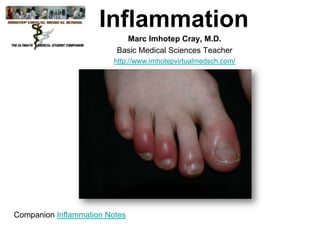
- 1. Inflammation Marc Imhotep Cray, M.D. Basic Medical Sciences Teacher http://www.imhotepvirtualmedsch.com/ Companion Inflammation Notes
- 3. Inflammation Definition EVOLUTION OF “Inflammation is a complex reaction of INFLAMMATION a tissue and its microcirculation to a pathogenic insult. It is characterized • Engulfment/entrapment by the generation of inflammatory mediators and movement of fluid & • Neutralization of irritant • Elimination of injurious leukocytes from the blood into extravascular tissues.” agent 3
- 4. The inflammatory response: • Is fundamentally a protective/defensive response • Persists until the inciting stimulus is removed and the mediators are dissipated or inhibited • Can be potentially harmful: – Anaphylactic shock (peanut allergy) – Systemic inflammatory response syndrome (SIRS) • Is closely intertwined with repair • Therapeutic strategies target critical control points in inflammatory pathways 4
- 5. Acute Inflammation: major components • Vascular changes: – Vasodilation and increased blood flow – Increased vascular permeability • Cellular events: – Leucocyte transmigration – Phagocytosis • Chemical mediators (acute & chronic) 5
- 7. Cardinal Signs (Celsus, 2 AD) • • • • • Redness (rubor) Swelling (tumor) Heat (calor) Pain (dolor) Loss of function (functio laesa) (the fifth cardinal sign added by Virchow) 7
- 8. Inflammation Cardinal Signs Patient with a Methicillin-resistant Staphylococcus aureus wound infection, and classic signs of inflammation. 8
- 9. Inflammation Cardinal Signs X-ray of previous patient showing non-union of fracture. Holes are from orthopedic screws. 9
- 10. Inflammation Cardinal Signs Bone scan of same patient, showing uptake in area of active inflammation. 10
- 11. Acute Inflammation: 1. Vasodilation/increased blood flow 2. Deposition of fibrin and other plasma proteins (exudate) 3. Transmigration and accumulation of neutrophils 11
- 13. Inflammation Stasis and Margination Polymorphs at margin of a vessel in acutely inflamed tissue. 13
- 14. Vascular Leakage: Transudate: (edema) specific gravity <1.015 and protein content low Exudate: (inflammation) specific gravity >1.015 and protein content high 14
- 15. Vascular Leakage (cont.) Rat model: only venules leak and deposit carbon 15
- 16. Leukocyte Extravasation and Phagocytosis • Margination, rolling, activation and adhesion • Transmigration (diapedesis) • Migration toward the site of injury along a chemokine gradient 16
- 17. Adhesion Molecule Modulation • P-selectin is redistributed to the cell surface from the Weibel-Palade bodies due to stimulation by thrombin, histamine, and Platelet Activating Factor (PAF) 17
- 18. Adhesion Molecule Modulation • Induction of E-selectin on endothelium by IL1 and TNF • Increased avidity of binding of integrins (conformational change) 18
- 20. Animation 20
- 21. 21
- 22. 22
- 23. Sequence following acute injury: 23
- 24. Mechanisms of Inflammatory Cell Function • The formation of a ligand-receptor complex leads to activation of a specific phospho-diesterase (phospholipase C) in the plasma membrane (stimulusresponse coupling) • • • Increased phospholipid metabolism Increased intracellular calcium Activation of protein kinases and phosphatases 24
- 25. Biochemical Events in Leucocyte Activation 25
- 26. Chemotaxis • Locomotion along a chemical gradient • Chemoattractants: Exogenous (from bacteria) Endogenous (from mediators) 26
- 27. Chemotaxis • Exogenous mediators, e.g.: – N-formyl methionine terminal amino acids from bacteria – Lipids from destroyed or damaged membranes (including LPS) • Endogenous mediators, e.g.: – Complement proteins (C5a) – Chemokines, particularly IL-8 – Arachidonic acid products (LTB4) 27
- 28. Phagocytosis • Recognition and attachment • Engulfment • Degradation (killing) 28
- 29. Bactericidal activity • Activated oxygen species • • • Superoxide (.O2) - formed via NADPH oxidase Hydrogen peroxide (H2O2) - formed via spontaneous dismutation of superoxide) Hypochlorous acid (HOCl) (Myeloperoxidase) Probably the primary bactericidal agent in neutrophils • Myeloperoxidase converts H2O2 into HOCl. . • Hydroxl radical ( OH) • 29
- 30. Bactericidal activity • Non-oxidative bacterial killing • • • • • • Lysosomal hydrolases (1o and 2o granules) Bactericidal/permeability-increasing protein (1o granules - affects Gram-negatives) Defensins (1o granules) Lactoferrin (2o granules - chelates iron) Lysozyme (1o and 2o granules, lysosomes) Bactericidal proteins of eosinophils 30
- 31. Tissue Injury by Inflammatory Cells "Our arsenals for fighting off bacteria are so powerful, and involve so many different defense mechanisms, that we are more in danger from them than from the invaders. We live in the midst of explosive devices; we are mined." – Lewis Thomas 31
- 32. Tissue Injury by Inflammatory Cells • Activated oxygen species • • • • • Can migrate through intact plasma membranes Initiate lipid peroxidation React with DNA Oxidize sulfhydryl groups of proteins Degrade extracellular matrix components 32
- 33. Tissue Injury by Inflammatory Cells • Lysosomal enzymes • • Since these enzymes are used to degrade microorganisms in lysosomes, obviously they could damage tissue in the extracellular environment Usually protease activity is controlled by a variety of anti-proteases present in plasma ( 1-anti-trypsin, 2-macroglobulin, etc.) 33
- 34. Tissue Injury by Inflammatory Cells • Phagocytic cell adherence • Adherence to basement membranes, other components of the extracellular matrix and other cells by phagocytes enhances the damage caused by reactive oxygen species and lysozyme, because normal inhibitors present in plasma cannot gain access to that space by virtue of the phagocytic cell adherence 34
- 35. Tissue Injury by Inflammatory Cells Abscess, formed via liquifactive necrosis, primarily due to the action of neutrophils. 35
- 36. Tissue Injury by Inflammatory Cells Normal glomerulus, containing 50-100 nuclei. 36
- 37. Tissue Injury by Inflammatory Cells • Post-streptococcal glomerulonephritis, showing a hypercellular glomerulus. • Most of the extra cells are neutrophils, reacting with immune complexes in the glomerular basement membrane. • This is a very bad thing for the glomerulus, and it is usually destroyed and heals with a hyaline scar. 37
- 38. Clinical examples of leukocyte-induced injury 38
- 39. Defects in Phagocytosis • Congenital • Chediak-Higashi Syndrome (autosomal recessive) • • • Job Syndrome (Hyper IgE) Chronic granulomatous disease (x-linked) • • • Defective intracellular transport protein, inability to lyse bacteria No oxidative burst Myeloperoxidase deficiency Acquired • • • • • Iatrogenic immunosuppression (most common) Overwhelming infections Severe trauma or burn Diabetes mellitus Chronic debilitating disease 39
- 40. Summary of Acute Inflammation What You MUST Known 40
- 41. Chemical Mediators of Inflammation • Originate from either plasma or cells • Bind to specific cellular receptors, have direct enzymatic activity or mediate oxidative damage • One mediator can stimulate release of additional mediators (“amplification”) • Effects on multiple cell types, varying effects on different cells • Once activated and released …are short-lived • Have potential to cause harmful effects 41
- 42. Sources of Inflammatory Mediators • Cell-derived: – Proteins sequestered in granules – Membrane phospholipids (via arachidonic acid metabolism) – Vasoactive amines (mast cells and platelets) • Inactive precursors in plasma: – Complement proteins (C3a, C5a) – Coagulation proteins initiated by Hageman factor (FDPs) 42
- 44. Chemical mediators of inflammation • Vasoactive amines: – Histamine – Serotonin • Plasma proteases: – Complement system – Kinin system – Coagulation system • Arachidonic acid metabolites: – Prostaglandins – Leukotrienes – Lipoxins • Platelet activating factor • Cytokines and chemokines • Nitric oxide • Leucocyte lysosomal consituents • Oxygen-derived free radicals • Neuropeptides 44
- 45. 45
- 46. Vasoactive Amines • Histamine: – Source = mast cell granules – Wide distribution around vessels – Main action is vasodilation and increased vascular permeability – Released by trauma, heat or cold, complement, cytokines – G-protein-coupled receptor • Serotonin (5-hydroxytryptamine): – Source = platelet granules – Actions similar to histamine – Released by platelet aggregation (collagen, thrombin, ADP, PAF from mast cells – G-protein-coupled receptor 46
- 47. Plasma Protease Systems • Complement, coagulation, fibrinolytic and kinin systems • All zymogen-based proteolytic cascades with great amplification characteristics • Generate pro-inflammatory by-products • Mutual interactions between systems 47
- 48. Chemical Mediators: proteases • Complement cascade system – Classic and alternative pathways • C3 is the critical control point, and interacts with both pathways • C3a and C5a are known as “anaphylatoxins”, and are capable of releasing histamine from mast cells, along with potent chemotactic abilities (C5a) • Membrane attack complex (MAC) is the active agent of complement lysis and consists of C5-9 48
- 49. Biologic effects of complement fragments • Vascular phenomena: – Increase vascular permeability – Vasodilation • Leucocyte adhesion, chemotaxis: C5a • Phagocytosis: C3b and C3bi – Opsonins 49
- 50. Complement: regulatory mechanisms • Regulation of C3 and C5 convertases: – Decay acceleration and proteolysis • Binding of activated complement components: – C1 inhibitor – CD59 membrane inhibitor of lysis 50
- 51. Chemical Mediators: proteases • Kinin system – Vasoactive peptides called kinins are generated by proteases called kallikrein – Hageman factor is a potent activator of kallikrein – Most important product is bradykinin • Pain + vascular dilation • Coagulation/Fibrinolytic pathway – Both systems are induced by activated factor XIIa (Hageman) – The coagulation and fibrinolytic systems complement and counterbalance each other – Most important molecules are fibrinogen, fibrin, thrombin, plasminogen, and plasmin 51
- 52. Arachidonic Acid Metabolites • Arachidonic acid (AA) is a 20-C polyunsaturated fatty acid released from membrane phospholipids by phospholipase A2 • Mechanical, chemical physical stimuli can activate phospholipase A2 • Metabolites of AA are called eicosanoids, and are generated by cyclooxygenases and lipoxygenases • Cycloxygensases synthesize prostaglandins and thromboxane • Lipoxygenases synthesize leukotrienes and lipoxins • Eicosanoid receptors are G-protein-coupled transmembrane proteins • Eicosanoids mediate all aspects of inflammation 52
- 53. Chemical Mediators of Inflammation • Arachidonic acid metabolites – Synthesized from cell membrane phospholipids though the action of phospho-lipases (inhibited by corticosteroids) – Form leukotrienes via 5-lipoxygenase – Form prostaglandins and thromboxane A2 via cyclo-oxygenase (COX-1 inhibited by aspirin and indomethacin, COX-2 inhibitors now coming on the market) 53
- 54. Inflammatory Action of Eicosanoids 54
- 55. 55
- 56. Chemical Mediators of Inflammation • Platelet activating factor – Numerous effects, including vasodilation and increased vascular permeability, with 100-10,000-fold increased activity, with respect to those actions, when compared to histamine 56
- 57. Chemical Mediators of Inflammation • Cytokines of importance – Interleukin 1- endothelial cell activation – Interleukin 2 (T cell growth factor) – Tumor necrosis factor cachectin) – Tumor necrosis factor (lymphotoxin) – Various colony stimulating factors, named for the stem cell they affect – Chemokines – important in chemotaxis 57
- 58. Cytokines mediating inflammation • Tumor necrosis factor • Interleukin-1 (IL-1) • Chemokines 58
- 59. Inflammatory Cells and their Chemokines 59
- 60. Chemical Mediators of Inflammation • Nitric oxide (NO) – Formed by NO synthase (NOS) • Constitutively expressed in endothelial and neuronal cells. May be increased rapidly by calcium influx. • Inducible NOS – induced in macrophages by TNFor IFN- – Potent vasodilator – Involved in the pathogenesis of septic shock 60
- 61. 61
- 62. Inflammation Neutrophil Granules • • • Primary - contain serine proteases, lysozyme and phospholipase A2 Secondary - similar to primary, but also contain lactoferrin and collagenase Tertiary - present at the leading edge of migrating PMNs, contain gelatinases that are capable of degrading basement membrane 62
- 66. Outcomes of Acute Inflammation • Resolution (the hoped-for result) • Abscess (via liquifactive necrosis) • Scar (sometimes occurs even if pathogen is eliminated) • Persistent inflammation (chronic inflammation) – due to a failure to completely eliminate the pathological insult (injury) 66
- 67. Resolution of acute inflammation • Return to normal vascular permeability • Drainage of edema fluid into lymphatics or pinocytosis into macrophages • Phagocytosis of apoptotic neutrophils and necrotic debris by macrophages • Disposal of macrophages 67
- 68. Chronic Inflammation • • • Inflammation of prolonged duration (weeks, months) Active inflammation, tissue destruction and attempts at repair proceed simultaneously Settings: • • • Persistent infection Prolonged exposure to toxic agent Autoimmunity 68
- 69. Chronic Inflammation • Histologic features – Infiltration by mononuclear cells: macrophages, lymphocytes, and plasma cells – Tissue destruction by ongoing inflammation – Attempts at healing, including angiogenesis, fibroblasts and fibrosis 69
- 70. Chronic Inflammation: Lung • Collection of chronic inflammatory cells • Destruction of parenchyma • Replacement by connective tissue (fibrosis) 70
- 71. Maturation of macrophages From Abbas et al Cellular and Molecular Immunology 1997 71
- 72. Macrophage activation • Via cytokines from immune-activated Tcells • By non-immunologic stimuli: endotoxin, fibronectin, mediators 72
- 74. Chronic Inflammation • Monocyte/Macrophages – Key cell in chronic and granulomatous inflammation – Reproduce locally, at the site of injury – Produce numerous cytokines, which continue to recruit additional cells, including more macrophages – May present antigen to T-cells, producing specific hypersensitivity reactions 74
- 75. Chronic Inflammation Products released from macrophages 75
- 76. Lymphocyte-macrophage interactions • Activated lymphocytes and macrophages influence each other • Release mediators that affect other cells 76
- 77. Chronic Inflammation: other cells • Plasma cells (lymphocytes) • Mast cells • Eosinophils 77
- 78. Granulomatous Inflammation • • • Distinct pattern of chronic inflammation in which the chief reactive cell is an activated macrophage Destruction of tissue is primarily via the action of killer T cells (CD8+), directed by macrophages Hence, the old term for tuberculosis was consumption, for good reason 78
- 80. Granulomatous Inflammation • Tuberculous lung, showing massive destruction by granulomatous inflammation. • This type of response is simply the best the body can do, since the inciting organism cannot be removed. Mycobacteria may live for years, perhaps even a 80 lifetime, within granulomas.
- 81. Granulomatous Inflammation • Characteristic arrangement of cells in a granuloma. • The extracellular matrix is active in orchestrating the precise alignment of cells. • Granulomas often form when indigestible substances are present, such as foreign bodies, or Mycobacteria, which are resistant to oxidative killing. Fibroblasts Lymphocytes Macrophages, Epithelioid Cells, and Giant Cells Caseous Necrosis Non-caseating Granuloma Caseating Granuloma 81
- 82. Granulomatous Inflammation • Granuloma, H&E, showing Langhans giant cells. • These multinucleated cells have macrophage markers on their cell surface, and are apparently the result of fusion of individual macrophages. While useful in diagnosing a granuloma, they are not considered the characteristic cell. • That honor belongs to the epitheliod histiocyte, which also has macrophage cell surface markers. 82
- 83. Granulomatous Inflammation • Granuloma, H&E, showing Langhans giant cells. • These multinucleated cells have macrophage markers on their cell surface, and are apparently the result of fusion of individual macrophages. • While useful in diagnosing a granuloma, they are not considered the characteristic cell. • That honor belongs to the epitheliod histiocyte, which also has macrophage cell surface markers. 83
- 87. Tissue morphology: inflammation exudative peptic ulceration Esophagus Stomach 87
- 88. Systemic Manifestations of Inflammation • • • • Systemic effects can be seen in setting of acute or chronic inflammation Largely mediated by cytokines Fever - clinical hallmark of inflammation • Endogenous pyrogens: IL-1 and TNFLeukocytosis: neutrophils bacterial (pneumonia) lymphocytes viral (infectious mono) eosinophils parasites, allergy • Acute phase reactants - non-specific elevation of many serum proteins - will markedly increase the “sed rate” 88
- 89. Acute phase response • Acute phase proteins: a group of proteins - CRP, mannose-binding protein, SAA, … that modulate inflammation (complement cascade, stimulate chemotaxis of phagocytes) • Inflammatory cytokines (TNF, IL-1, ) stimulate liver cells to sythesize acute phase proteins • Acute phase response provides an early defense before full activation of immune responses 89
- 90. Acute phase reaction • Endocrine/metabolic: C-reactive protein (CRP) Serum amyloid A (SAA) Serum amyloid P (SAP) • Autonomic: blood flow • Physiologic/behavioral (shivering) 90
- 91. Systemic Manifestations of Inflammation • Shock – most common in Gram-negative septicemia (bacteria in the bloodstream), although it can occur with Gram-positive bacteremia • • Lipopolysaccharide (LPS or endotoxin) of Gram-negatives can produce symptoms of shock when injected into animals TNF- can produce a similar syndrome 91
- 92. 92
- 93. Systemic Manifestations of Inflammation • • • Therapy for fever and inflammation targets arachidonic acid metabolism (aspirin) Therapy for shock focuses on fluid resuscitation, and proper selection of antibiotics (more to come later) Experimental therapy for shock using anti-TNF- , and other antibodies directed against cytokines, has, so far, failed to improve survival 93
- 94. THE END THANK YOU Reference: Kumar et al. Robbins and Cotran’s Pathological Basis of Disease 8th edition, Elsevier 2010 Learn more, WebPath • Inflammation: 75 Images Patterns of cellular and tissue injury with inflammation, including acute, chronic, and granulomatous inflammation. • Medical Pathology - What's an Acute Inflammation? (National Institute of Health) Video 94