SlideShare una empresa de Scribd logo
1 de 75
Basic physics of ultrasound SPEAKER: Dr. JAYAPRABHA NARAYAN            1st YEAR PG MODERATOR: Dr. ANAND S H             ASSISTANT PROFESSOR    DEPARTMENT OF RADIODIAGNOSIS
The basis of all diagnostic ultrasound applications are the detection and display of acoustic energy reflected from within the body. In Medical Ultrasound, images representing human organs are formed by transmitting sound waves into the body and receiving back and processing the resultant echoes from the tissues. To accomplish this, medical ultrasound uses a process very similar to an ocean-going vessels “depth sounding” equipment or oceanic survey equipment. All of these systems make use of sound waves and their reflections. Sea
BASIC ACOUSTICS Sound is the result of mechanical energy travelling through matter as a wave   Sound waves are transmitted as a series of alternating pressure waves with high pressure and low pressure pulses. The high pressure areas (compression) are where the particles have been squeezed together; the low pressure areas (rarefaction) are where the particles have been spread apart.  Sound waves cannot travel in vaccum
Audible sound waves range in frequency from 20Hz to 20,000Hz. Ultrasound waves – frequency more than 20 KHz. For medical imaging – 2 to 20 MHz Sound waves – Transverse waves and longitudinal waves 20 MHz Diagnostic    Imaging 1 MHz Ultrasonic  20 KHz Audible sound  20 Hz Infrasonic 0 Hz
Ultrasound waves – propagating through body tissues and fluids - longitudinal waves – producing compression and rarefaction of conducting medium. A zone of compression and an adjacent zone of rarefaction constitute one cycle of an ultrasound wave.
Wavelength :The distance covered by one cycle is the wavelength of the ultrasound wave, λ  The time for the ultrasound wave to pass a given point is Period, T  Frequency: The number of cycles per unit time (cps, or just sec−1) introduced into the medium each second is referred to as the frequency of the wave, expressed in units of Hertz, Kilohertz or Megahertz, where 1Hz equals 1 cps. The maximum height of the wave cycle is the amplitude of the ultrasound wave.
Propagation: in diagnostic ultrasound pressure waves are longitudinal waves and they propagate along the direction of particle movement. The product of the frequency (ν) and the wavelength (λ) is the propagationvelocity of the wave; that is, c = νλ. Propagation depends on the resistance of medium, resistance depends on density and its stiffness or elasticity of the medium .
More stiff/less compressible is the molecules or less dense, more is the velocity in that medium.         i.e. if density is more, velocity will be decresed and vice versa.
On average the propagation velocity of sound in general body tissue is approximately 1540 m/sec
Reflection Current diagnostic ultrasound scanners rely on the detection and display of reflected sound or echoes from interfaces between different tissues in the body.  The fraction of the impinging energy reflected from an interface depends on the difference in acoustic impedance of the media on opposite sides of the interface. The acoustic impedance Z of a medium is the product of the density ρ of the medium and the velocity of ultrasound (c) in the medium: Z = ρc With a large impedance mismatch at an interface, much of the energy of an ultrasound wave is reflected, and only a small amount is transmitted across the interface.
Incident wave Transmitted wave Medium 1 Medium 2 Reflected wave
Specular Reflections, which occur at large change        in impedance producing a large reflection, and also        reducing the continuing wave amplitude. Medium Reflections, which occur with dense tissues        such as muscle. Diffuse Reflections, which occur with soft tissues such         as liver.
Specular reflectors : Diaphragm Wall of urine filled urinary bladder Endometrial stripe Specular reflector                                                   Diffuse reflector
Different types of reflections Scattering
Distance measurement
Refraction  When a propagating ultrasound wave  encounters a Specularinterface at an  oblique angle, it is  Refracted  in the  same way that light is refracted through a lens. The portion of  the wave that is not reflected continues into the second medium, with a change in direction. It is dependent on the velocities of the two medium. If the velocities are equal, There would be no refraction occurred and the beam goes straight into the  second medium.  For the velocities of the different tissues in the human body  are quite close, refraction's can be ignored
Absorption Cause     (a) Removal of energy from the ultrasound beam and     (b) Eventual dissipation of this energy primarily as heat.  Ultrasound is propagated by displacement of molecules of a medium into regions of compression and rarefaction Therefore, the energy of the ultrasound beam is gradually reduced as it passes through the medium.
Attenuation ,[object Object]
 Due to mainly reflection, refraction and absorptionAttenuation of ultrasound wave occurs when it is propagating through the medium.
 Loss of propagating energy will be in the form of heat absorbed by    the tissue, approximately 1 dB/cm/MHz, or caused by wavefront dispersion or wave scattering.
Scattering ,[object Object],reflections from objects even smaller than the wavelength. Many biological interfaces have irregular surfaces, tending to give scatter-like reflection, which is quite   Useful, as it will give at least some echoes even though the beam is not   directly perpendicular to the reflecting interface.    Spherical Scatter-wave
Backscatter Backscatter or Rayleigh scattering occurs with structures smaller than the transmitted wavelength. Reflected energy is very low,  but contributes to the texture of the image.
INSTRUMENTATION
BASIC COMPONENTS TO PERFORM KEY FUNCTIONS A transmitter/pulser – energize the transducer The Transducer A receiver Processor – to detect and amplify the backscattered energy and to manipulate the reflected signals for display A display – that presents ultrasound image or data A method to record or store data or images.
Transducer  A device – generates ultrasound waves for medical imaging. Two functions – Coverts electrical energy provided by transmitter into acoustic pulses directed into the patient.                                also serves as receiver of reflected echoes. Works on Piezoelectric Effect/piezoelectricity.                 it’s the unique property of certain natural or artificial crystals , Piezoelectric crystals – when subjected to electric voltage – crystal changes shape and vibrates with its resonant frequency(realignment and change in dimension) ,  produces sound beam which propagates into tissues( electrical energy converted to mechanical energy) and vice versa.
Piezoelectric Crystals Some naturally piezoelectric occurring materials include Berlinite (structurally identical to quartz), cane sugar, quartz, Rochelle salt, topaz, tourmaline, and dry bone  An example of man-made piezoelectric materials includes barium titanate and lead zirconate titanate (PZT). Most widely used
TRANSDUCER DESIGN A crystal exhibits its greatest response at the resonance frequency The resonance frequency is determined by the thickness of the crystal and propagation velocity in its material Most efficient operation is achieved for a crystal with a thickness equal to half the wavelength of the desired ultrasound  In most of the pulsed mode of operating ultrasound the output sound has frequency both above and below the resonance frequency The range of frequencies are called bandwidth. Generally shorter the pulse length of the transducer more is the bandwidth.  Broad bandwidth helps to reduce speckle by frequency compounding
TRANSDUCER DESIGN contd Spatial pulse length or Pulse length is the number of cycles per pulse In diagnostic ultrasound we need very small pulse length for better axial resolution. So suitable damping materials (backing) are kept behind the transducer material to shorten the pulse length and spacial pulse length by stopping reverberating pulses coming from the interface behind the transducer, thus gives improved axial resolution.                     also reduces ultrasound amplitude and decreases efficiency and sensitivity of the system. Damping materials used – mixture of metal powder(tungsten or aluminum) and a plastic or epoxy
A 1/4λ (quarter-wavelength) thickness matching layer or coating in front of the transducer (generally made up of aluminum powder and epoxy resin) maximizes energy transfer from the transducer to the patient( i.e. increases sound transmission) The filler material enables the transducer assembly to be flat. The elements with its associated case, damping and matching material is called Transducer Assembly Or Probe
Transducer schematic diagram
PULSE – ECHO PRINCIPLE When the crystal is electrically pulsed, it changes shape and vibrates produces sound beam that propagates through tissues and reflected echoes reach the transducer it vibrates and produces electric voltage comparable to returning echo.
The ultrasound pulses produced by the transducer results in a series of wave fronts that form a three dimensional beam of ultrasound.  the features of beam are influenced by constructive and destructive interferences of the pressure waves.
ULTRASOUND BEAMS Ultrasound from a point source creates spherical wave fronts. Ultrasound from a two-dimensional extended source creates planar wave fronts. These sources can be considered to be a collection of point sources, each radiating spherical wave fronts (termed wavelets) into the medium.  In regions where compression zones for one wavelet intersect those of another, a condition of constructive interference is established & the wavelets reinforce each other. Reverse that is cancellation occurs in destructive interference
Interference of the waves
In this figure, the reinforcement and   cancellation of individual wavelets are most noticeable in the region near the source of ultrasound.  They are progressively less dramatic with increasing distance from the ultrasound source The near field where pressure amplitude change is maximum that zone is called “Fresnel zone” Beyond the Fresnel zone, some of the energy escapes along the periphery of the beam to produce a gradual divergence of the ultrasound beam – called Fraunhofer ( or far) zone
For medical applications of ultrasound, beams with little lateral dispersion of energy (i.e., long Fresnel zones) are preferred.  Rules for Transducer Design For a given transducer diameter,    the near-field length increases with increasing frequency;     beam divergence in the far field decreases with increasing frequency For a given transducer frequency     the near-field length increases with increasing transducer diameter;       beam divergence in the far field decreases with increasing transducer diameter.
Focused Transducers A focused ultrasound transducer produces a beam that is narrower at some distance from the transducer face than its dimension at the face of the transducer. In the region where the beam narrows (termed the focal zone of the transducer), the ultrasound intensity may be heightened by 100 times or more compared with the intensity outside of the focal zone.  Because of this increased intensity, a much larger signal will be induced in a transducer from a reflector positioned in the focal zone. The distance between the location for maximum echo in the focal zone and the element responsible for focusing the ultrasound beam is termed the focal length of the transducer An ultrasound beam also may be focused with mirrors and refracting lenses or phasing the linear array electronically
Focused Transducers
Receiver  Returning echoes strike the transducer face (which is on quiescent mode electronically) and they produce minute voltages in the piezoelectric crystals. The receiver detects those signals and amplifies those They also provide compensatory amplification of the weaker signals those are coming from the deeper tissues. This is calledtime gain compensation
Time gain compensation
Time gain compensation We can also suppress increased amplitudes of the near field for better comparable image
Receiver  Another function of the receiver is compression of the wide range of amplitudes returning to the transducer into a range that can be displayed to the user. The range is called dynamic rangeof the transducer. Compression and remapping of the data is required to adapt the dynamic range of backscattered signal intensity to the dynamic range of display
Receiver – dynamic range   The widest dynamic range (60 dB) shown permits the best differentiation of subtle differences in echo intensity and preferred in most imaging application The narrower ranges increase conspicuity of larger echo differences.
ARRAYS(TRANSDUCER TYPES)
Linear Array  The linear array scanners produce sound waves parallel      to each other and produces a rectangular image.  The width of the image and number of scan lines are the same at all tissue levels.  This has the advantage of good near field resolution.  Often used with high frequencies i.E. 7mhz. Can be used for viewing surface texture of the liver.  There disadvantage is artifacts when applied to a      curved part of the body creating air gaps between       skin and transducer.
Sector/Phased array  ( freq 1 to 3MHz in adult, in pediatric sector probes upto 8 MHz) Produces a fan like image that is narrow near the transducer and increase in width with deeper penetration.  It is useful when scanning between the ribs as it fits in the intercostals space. The disadvantage is poor near field resolution
Curved Array Often with frequencies of 2 - 5 MHz (to allow for a range of patients from obese to slender).  It is a compromise of the Linear and Sector scanners. The density of the scan lines decreases with increasing distance from the transducer. Can be difficult to use in curved regions of the body eg. the spleen behind the left costal margin. For abdominal ultrasound curved type scanners are used as the best compromise of two other standard type probes the linear and the sector scanner. 
Annular Array A series of concentric elements nested within one another in a circular piece of piezoelectric crystal to produce an annular array Use of multiple concentric elements enables precise focussing.
Linear & Phased array Linear array-individual elements or groups are fired in sequence resulting parallel US beams Phasic array: produce a sector field of view by firing multiple transducer elements in precise sequence to generate interference of acoustic wave fronts
Transducer Selection For general purpose – convex with 3.5 MHz For obstetric purpose – convex or linear with 3.5 MHz For superficial structures – linear with 5MHz For pediatric or in thin people – 5MHz
COUPLING AGENTS Commonly known as “GEL” Fluid medium needed to provide a link between the transducer and the patient Composition : Carbomer – 10.0 gm              Propylene glycol – 75.0gm(72.4ml) Trolamine – 12.5gm(11.2ml)              EDTA – 0.25gm              Distilled water – upto 500gm or 500ml
IMAGE DISPLAY
A-Mode (amplitude mode) A mode display consists of a horizontal baseline. This baseline represents time and or distance with upward (vertical) deflections (spikes depicting the acoustic interfaces) In the A-mode presentation of ultrasound images, echoes returning from the body are displayed as signals on an oscilloscope.
A-Mode (amplitude mode) A-mode reveals the location of echo-producing structures only in the direction of the ultrasound beam.  A-mode displays are not found on most imaging systems used today.  The concept of A-mode is however, useful in explaining how pixels are obtained from scan lines in B-mode imaging.
B-mode (Brightness Mode) Real-time gray scale B-Mode display A two-dimensional display of ultrasound.  To generate a two dimensional image multiple ultrasound pulses are sent down as a series of successive scan lines, building a 2D representation of echoes arising from the area being scanned. When an ultrasound image js displayed on a black background, signals of greatest intensity appear as white, absence of signal is shown as black(anechoic) and signals of intermediate intensity appear as shades of grey.
B-mode, or brightness mode, ultrasound display.
B-mode images may be displayed as either “static” or “real-time” images.  In static imaging the image is compiled as the sound beam is scanned across the patient, and the image presents a “snapshot” averaged over the time required to sweep the sound beam.
B mode real time imaging In real-time imaging, the image is also built up as the sound beam scans across the patient, but the scanning is performed automatically and quickly, and one image follows another in quick succession.( as many as 30-60 complete images per second) Real-time B-mode images are useful in the display of moving structures such as heart valves.
M-mode Ultrasound (motion mode) Is the motion mode displaying moving structures along a single line in the ultrasound beam A single beam in an ultrasound scan can be used to produce an M-mode picture where movement of a structure such as a heart valve can be depicted in a wavelike manner.   Because of its high sampling frequency (up to 1000 pulses per second) This is useful in assessing rates and motion and is still used extensively in cardiac and fetal cardiac imaging
IMAGE QUALITY Spacial Resolution Axial : higher frequency – low wavelength  higher axial resolution  Lateral resolution: excessive beam width and thickness limit the ability to delineate small features such as tiny cystic changes in atherosclerotic plaque Lateral resolution is controlled by focusing the beam by electronic phasing Elevation or Azimuthal resolution is determined by construction of the transducer and can not be controlled by the user
IMAGE ARTIFACTS
Mirror Image Artifact  • This is where a strong reflector at an angle to the probe causes structures that lie in front and to the side of it to appear as if they lie behind it, just as something viewed through a mirror appears to lie behind it.• e.g. diaphragm
Reverberation artifact When the ultrasound signal reflects repeatedly between highly reflective interfaces that are usually near the transducer Reduced by changing the scan angle and placement of the transducer
Comet tail artifacts • This is the same process as reverberation, but occurs within a very small structure, with smooth highly reflective borders, e.g. metal fragment. • Tiny bright reverberations are seen deep to the structure slowly diminishing is size as if it had a tail
Refraction Artifact This can lead to subtle miss placement of structures and some degradation of image quality when the angle of incidence is particularly acute. Rectus abdominis acting as refracting lens
Range artifact
Side Lobe Artifacts The probe cannot produce a pulse that travels purely in one direction. Pulses also travel off at specific angles. Side lobes may interact with strong reflectors that lie outside the scan plane. These side lobes are relatively weak and so normally do little to degrade the image. Can be reduced by repositioning of the transducer ,or its focal zone or using a different transducer. GB
Acoustic shadowing  Marked reduction in the intensity of ultrasound deep to a strong reflector or attenuator.  ( other cause of loss of image information are improper system gain and TGC setting, poor scanning angles and poor resolution)
Acoustic enhancement Due to high penetration of sound through a structure and then being reflected from the tissue behind it.
Multipath artifact Echoes reflected (e.g. from diaphragm and wall of ovarian cyst) may create complex echo path that delay return of the reflected signal thus showing false deeper/abnormal position of the tissues Ovarian cyst --- false mural nodule
Speckle Artifact Usual grainy appearance of the USG image Due to random interference of the scatterer . Irregular interference pattern. This is generated by more scatterers somewhat randomly distributed. The speckle pattern is thus random too.  Interference pattern. Here is simulated two wave sources or scatterers at the far field (white points).
Speckle Artifact

Más contenido relacionado

La actualidad más candente

ULTRASOUND - ARTIFACTS.pptx
ULTRASOUND - ARTIFACTS.pptxULTRASOUND - ARTIFACTS.pptx
ULTRASOUND - ARTIFACTS.pptxShubhamRai502947
 
Basic Pulse Sequences In MRI
Basic Pulse Sequences In MRIBasic Pulse Sequences In MRI
Basic Pulse Sequences In MRIUpakar Paudel
 
ultrasound transducers and resolution
ultrasound transducers and resolutionultrasound transducers and resolution
ultrasound transducers and resolutionVallabhaneni Bhupal
 
Ultrasound transducer doppler ppt pdf pk
Ultrasound transducer doppler ppt pdf pkUltrasound transducer doppler ppt pdf pk
Ultrasound transducer doppler ppt pdf pkDr pradeep Kumar
 
Doppler Ultrasonography And Advancements in USG
Doppler Ultrasonography And Advancements in USGDoppler Ultrasonography And Advancements in USG
Doppler Ultrasonography And Advancements in USGSudil Paudyal
 
Ultrasound Transducer Types
Ultrasound Transducer TypesUltrasound Transducer Types
Ultrasound Transducer TypesMette Lyng
 
Ultrasound imaging system
Ultrasound imaging system Ultrasound imaging system
Ultrasound imaging system Riadh Al-Haidari
 
Ultrasound artifacts
Ultrasound artifactsUltrasound artifacts
Ultrasound artifactsansaripv
 
Tissue harmonic imaging
Tissue harmonic imaging Tissue harmonic imaging
Tissue harmonic imaging MAMTA PANDA
 
Multi slice ct ppt
Multi slice ct pptMulti slice ct ppt
Multi slice ct pptAnand Rk
 
Basic physics of Ultrasound
Basic physics of UltrasoundBasic physics of Ultrasound
Basic physics of UltrasoundNouman Ahmed
 
Physics of Multidetector CT Scan
Physics of Multidetector CT ScanPhysics of Multidetector CT Scan
Physics of Multidetector CT ScanDr Varun Bansal
 
MRI Coil and Gradient power-point slide pk
MRI Coil and Gradient power-point slide pkMRI Coil and Gradient power-point slide pk
MRI Coil and Gradient power-point slide pkDr pradeep Kumar
 

La actualidad más candente (20)

Ultrasound artifacts
Ultrasound artifactsUltrasound artifacts
Ultrasound artifacts
 
ULTRASOUND - ARTIFACTS.pptx
ULTRASOUND - ARTIFACTS.pptxULTRASOUND - ARTIFACTS.pptx
ULTRASOUND - ARTIFACTS.pptx
 
Basic Pulse Sequences In MRI
Basic Pulse Sequences In MRIBasic Pulse Sequences In MRI
Basic Pulse Sequences In MRI
 
ultrasound transducers and resolution
ultrasound transducers and resolutionultrasound transducers and resolution
ultrasound transducers and resolution
 
Usg artefacts
Usg artefacts Usg artefacts
Usg artefacts
 
Ultrasound transducer doppler ppt pdf pk
Ultrasound transducer doppler ppt pdf pkUltrasound transducer doppler ppt pdf pk
Ultrasound transducer doppler ppt pdf pk
 
Doppler physics ppt
Doppler physics pptDoppler physics ppt
Doppler physics ppt
 
Doppler Physics
Doppler PhysicsDoppler Physics
Doppler Physics
 
Doppler Ultrasonography And Advancements in USG
Doppler Ultrasonography And Advancements in USGDoppler Ultrasonography And Advancements in USG
Doppler Ultrasonography And Advancements in USG
 
Ultrasound Transducer Types
Ultrasound Transducer TypesUltrasound Transducer Types
Ultrasound Transducer Types
 
Ultrasound imaging system
Ultrasound imaging system Ultrasound imaging system
Ultrasound imaging system
 
Mri physics
Mri physicsMri physics
Mri physics
 
Ultrasound artifacts
Ultrasound artifactsUltrasound artifacts
Ultrasound artifacts
 
MRI artifacts
MRI artifactsMRI artifacts
MRI artifacts
 
Usg artifacts
Usg artifactsUsg artifacts
Usg artifacts
 
Tissue harmonic imaging
Tissue harmonic imaging Tissue harmonic imaging
Tissue harmonic imaging
 
Multi slice ct ppt
Multi slice ct pptMulti slice ct ppt
Multi slice ct ppt
 
Basic physics of Ultrasound
Basic physics of UltrasoundBasic physics of Ultrasound
Basic physics of Ultrasound
 
Physics of Multidetector CT Scan
Physics of Multidetector CT ScanPhysics of Multidetector CT Scan
Physics of Multidetector CT Scan
 
MRI Coil and Gradient power-point slide pk
MRI Coil and Gradient power-point slide pkMRI Coil and Gradient power-point slide pk
MRI Coil and Gradient power-point slide pk
 

Destacado

Ultrasound Physics
Ultrasound PhysicsUltrasound Physics
Ultrasound Physicsu.surgery
 
Ultrasound artifacts and contrast enhanced ultrasound
Ultrasound artifacts and contrast enhanced ultrasoundUltrasound artifacts and contrast enhanced ultrasound
Ultrasound artifacts and contrast enhanced ultrasoundArjun Reddy
 
Proposal Writing
Proposal WritingProposal Writing
Proposal WritingProf S
 
Imaging in blunt abdominal trauma
Imaging in blunt abdominal traumaImaging in blunt abdominal trauma
Imaging in blunt abdominal traumaSunil Kumar
 
Hypersonic Sound
Hypersonic SoundHypersonic Sound
Hypersonic SoundEike Maas
 
Ultrsonography Principle and application
Ultrsonography Principle and applicationUltrsonography Principle and application
Ultrsonography Principle and applicationsuniu
 
Biomedical image processing ppt
Biomedical image processing pptBiomedical image processing ppt
Biomedical image processing pptPriyanka Goswami
 
ELECTROENCEPHALOGRAM/ ELECTROENCEPHALOGRAPHY
ELECTROENCEPHALOGRAM/ ELECTROENCEPHALOGRAPHYELECTROENCEPHALOGRAM/ ELECTROENCEPHALOGRAPHY
ELECTROENCEPHALOGRAM/ ELECTROENCEPHALOGRAPHYRoxanneMae Birador
 
General-Purpose Ultrasound
General-Purpose UltrasoundGeneral-Purpose Ultrasound
General-Purpose Ultrasoundmarianahu123
 
EEG Presentation Kassing/Konold
EEG Presentation Kassing/KonoldEEG Presentation Kassing/Konold
EEG Presentation Kassing/KonoldDriphtwood
 
Basic Ecocardiography
Basic EcocardiographyBasic Ecocardiography
Basic Ecocardiographyrahterrazas
 
Emotiv Epoc/EEG/BCI
Emotiv Epoc/EEG/BCIEmotiv Epoc/EEG/BCI
Emotiv Epoc/EEG/BCISuhail Khan
 

Destacado (20)

Ultrasound Physics
Ultrasound PhysicsUltrasound Physics
Ultrasound Physics
 
Ultrasound artifacts and contrast enhanced ultrasound
Ultrasound artifacts and contrast enhanced ultrasoundUltrasound artifacts and contrast enhanced ultrasound
Ultrasound artifacts and contrast enhanced ultrasound
 
Proposal Writing
Proposal WritingProposal Writing
Proposal Writing
 
Imaging in blunt abdominal trauma
Imaging in blunt abdominal traumaImaging in blunt abdominal trauma
Imaging in blunt abdominal trauma
 
Hypersonic Sound
Hypersonic SoundHypersonic Sound
Hypersonic Sound
 
Part 1
Part 1Part 1
Part 1
 
1.5 interference
1.5 interference1.5 interference
1.5 interference
 
Sound physics
Sound physicsSound physics
Sound physics
 
Blunt trauma abdomen
Blunt trauma abdomenBlunt trauma abdomen
Blunt trauma abdomen
 
Superposition theorem
Superposition theoremSuperposition theorem
Superposition theorem
 
Basics Physics of ultrasound
Basics Physics of ultrasoundBasics Physics of ultrasound
Basics Physics of ultrasound
 
Ultrsonography Principle and application
Ultrsonography Principle and applicationUltrsonography Principle and application
Ultrsonography Principle and application
 
Biomedical image processing ppt
Biomedical image processing pptBiomedical image processing ppt
Biomedical image processing ppt
 
ELECTROENCEPHALOGRAM/ ELECTROENCEPHALOGRAPHY
ELECTROENCEPHALOGRAM/ ELECTROENCEPHALOGRAPHYELECTROENCEPHALOGRAM/ ELECTROENCEPHALOGRAPHY
ELECTROENCEPHALOGRAM/ ELECTROENCEPHALOGRAPHY
 
Electroencephalogram
ElectroencephalogramElectroencephalogram
Electroencephalogram
 
General-Purpose Ultrasound
General-Purpose UltrasoundGeneral-Purpose Ultrasound
General-Purpose Ultrasound
 
EEG Presentation Kassing/Konold
EEG Presentation Kassing/KonoldEEG Presentation Kassing/Konold
EEG Presentation Kassing/Konold
 
Basic Ecocardiography
Basic EcocardiographyBasic Ecocardiography
Basic Ecocardiography
 
New Trends in Epilepsy Management
New Trends in Epilepsy ManagementNew Trends in Epilepsy Management
New Trends in Epilepsy Management
 
Emotiv Epoc/EEG/BCI
Emotiv Epoc/EEG/BCIEmotiv Epoc/EEG/BCI
Emotiv Epoc/EEG/BCI
 

Similar a Basic physics of ultrasound.JH

Basic of ultrasound
Basic of ultrasoundBasic of ultrasound
Basic of ultrasoundAsad Moosa
 
Ultrasound physics and image optimization1 (1)
Ultrasound physics and image optimization1 (1)Ultrasound physics and image optimization1 (1)
Ultrasound physics and image optimization1 (1)Prajwith Rai
 
Principles and technology of two dimensional echocardiography (2)
Principles and technology of two dimensional echocardiography (2)Principles and technology of two dimensional echocardiography (2)
Principles and technology of two dimensional echocardiography (2)Kangkan Sharma
 
Physics of ultrasound and echocardiography
Physics of ultrasound and echocardiographyPhysics of ultrasound and echocardiography
Physics of ultrasound and echocardiographyjeetshitole
 
basicsofultrasound-170807183128 (1).pdf
basicsofultrasound-170807183128 (1).pdfbasicsofultrasound-170807183128 (1).pdf
basicsofultrasound-170807183128 (1).pdflittlealphonsa
 
basicsofultrasound-170807183128 (1).pdf
basicsofultrasound-170807183128 (1).pdfbasicsofultrasound-170807183128 (1).pdf
basicsofultrasound-170807183128 (1).pdfMohamedAli690824
 
PHYSICS OF ULTRASOUND AND KNOBOLOGY
PHYSICS OF ULTRASOUND AND KNOBOLOGYPHYSICS OF ULTRASOUND AND KNOBOLOGY
PHYSICS OF ULTRASOUND AND KNOBOLOGYArun Babu
 
Basic Physics Of Transoesophageal Echocardiography For The Workshop2
Basic Physics Of Transoesophageal Echocardiography   For The Workshop2Basic Physics Of Transoesophageal Echocardiography   For The Workshop2
Basic Physics Of Transoesophageal Echocardiography For The Workshop2Anil Ramaiah
 
Ultrasound assignment 3 mark question and answer
Ultrasound assignment 3 mark question and answer Ultrasound assignment 3 mark question and answer
Ultrasound assignment 3 mark question and answer Ganesan Yogananthem
 
Presentation1, basic principle of ultrasound.
Presentation1, basic principle of ultrasound.Presentation1, basic principle of ultrasound.
Presentation1, basic principle of ultrasound.Abdellah Nazeer
 
INTRODUCTION TO ULTRASOUND IMAGING ( ULTRASONGRAPHY ) - ANAND GURMAITA (BSc. ...
INTRODUCTION TO ULTRASOUND IMAGING ( ULTRASONGRAPHY ) - ANAND GURMAITA (BSc. ...INTRODUCTION TO ULTRASOUND IMAGING ( ULTRASONGRAPHY ) - ANAND GURMAITA (BSc. ...
INTRODUCTION TO ULTRASOUND IMAGING ( ULTRASONGRAPHY ) - ANAND GURMAITA (BSc. ...ANAND GURMAITA
 
BASICS OF US & DUPLEX (NEW).ppt
BASICS OF US & DUPLEX (NEW).pptBASICS OF US & DUPLEX (NEW).ppt
BASICS OF US & DUPLEX (NEW).pptmohamedabushanab1
 

Similar a Basic physics of ultrasound.JH (20)

Physics
PhysicsPhysics
Physics
 
Basic of ultrasound
Basic of ultrasoundBasic of ultrasound
Basic of ultrasound
 
Ultrasound physics and image optimization1 (1)
Ultrasound physics and image optimization1 (1)Ultrasound physics and image optimization1 (1)
Ultrasound physics and image optimization1 (1)
 
ULTRASOUND
ULTRASOUND ULTRASOUND
ULTRASOUND
 
Principles and technology of two dimensional echocardiography (2)
Principles and technology of two dimensional echocardiography (2)Principles and technology of two dimensional echocardiography (2)
Principles and technology of two dimensional echocardiography (2)
 
Physics of ultrasound and echocardiography
Physics of ultrasound and echocardiographyPhysics of ultrasound and echocardiography
Physics of ultrasound and echocardiography
 
basicsofultrasound-170807183128 (1).pdf
basicsofultrasound-170807183128 (1).pdfbasicsofultrasound-170807183128 (1).pdf
basicsofultrasound-170807183128 (1).pdf
 
basicsofultrasound-170807183128 (1).pdf
basicsofultrasound-170807183128 (1).pdfbasicsofultrasound-170807183128 (1).pdf
basicsofultrasound-170807183128 (1).pdf
 
Physics of echo i.tammi raju
Physics of echo i.tammi rajuPhysics of echo i.tammi raju
Physics of echo i.tammi raju
 
PHYSICS OF ULTRASOUND AND KNOBOLOGY
PHYSICS OF ULTRASOUND AND KNOBOLOGYPHYSICS OF ULTRASOUND AND KNOBOLOGY
PHYSICS OF ULTRASOUND AND KNOBOLOGY
 
Basic Physics Of Transoesophageal Echocardiography For The Workshop2
Basic Physics Of Transoesophageal Echocardiography   For The Workshop2Basic Physics Of Transoesophageal Echocardiography   For The Workshop2
Basic Physics Of Transoesophageal Echocardiography For The Workshop2
 
Ultrasound assignment 3 mark question and answer
Ultrasound assignment 3 mark question and answer Ultrasound assignment 3 mark question and answer
Ultrasound assignment 3 mark question and answer
 
Paper
PaperPaper
Paper
 
Medical imaging
Medical imagingMedical imaging
Medical imaging
 
Sillu thesis
Sillu thesisSillu thesis
Sillu thesis
 
Presentation1, basic principle of ultrasound.
Presentation1, basic principle of ultrasound.Presentation1, basic principle of ultrasound.
Presentation1, basic principle of ultrasound.
 
USphysics.pptx
USphysics.pptxUSphysics.pptx
USphysics.pptx
 
INTRODUCTION TO ULTRASOUND IMAGING ( ULTRASONGRAPHY ) - ANAND GURMAITA (BSc. ...
INTRODUCTION TO ULTRASOUND IMAGING ( ULTRASONGRAPHY ) - ANAND GURMAITA (BSc. ...INTRODUCTION TO ULTRASOUND IMAGING ( ULTRASONGRAPHY ) - ANAND GURMAITA (BSc. ...
INTRODUCTION TO ULTRASOUND IMAGING ( ULTRASONGRAPHY ) - ANAND GURMAITA (BSc. ...
 
Ocular Ultrasonography
Ocular UltrasonographyOcular Ultrasonography
Ocular Ultrasonography
 
BASICS OF US & DUPLEX (NEW).ppt
BASICS OF US & DUPLEX (NEW).pptBASICS OF US & DUPLEX (NEW).ppt
BASICS OF US & DUPLEX (NEW).ppt
 

Basic physics of ultrasound.JH

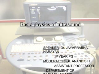
  • 1. Basic physics of ultrasound SPEAKER: Dr. JAYAPRABHA NARAYAN 1st YEAR PG MODERATOR: Dr. ANAND S H ASSISTANT PROFESSOR DEPARTMENT OF RADIODIAGNOSIS
  • 2. The basis of all diagnostic ultrasound applications are the detection and display of acoustic energy reflected from within the body. In Medical Ultrasound, images representing human organs are formed by transmitting sound waves into the body and receiving back and processing the resultant echoes from the tissues. To accomplish this, medical ultrasound uses a process very similar to an ocean-going vessels “depth sounding” equipment or oceanic survey equipment. All of these systems make use of sound waves and their reflections. Sea
  • 3. BASIC ACOUSTICS Sound is the result of mechanical energy travelling through matter as a wave Sound waves are transmitted as a series of alternating pressure waves with high pressure and low pressure pulses. The high pressure areas (compression) are where the particles have been squeezed together; the low pressure areas (rarefaction) are where the particles have been spread apart. Sound waves cannot travel in vaccum
  • 4. Audible sound waves range in frequency from 20Hz to 20,000Hz. Ultrasound waves – frequency more than 20 KHz. For medical imaging – 2 to 20 MHz Sound waves – Transverse waves and longitudinal waves 20 MHz Diagnostic Imaging 1 MHz Ultrasonic 20 KHz Audible sound 20 Hz Infrasonic 0 Hz
  • 5. Ultrasound waves – propagating through body tissues and fluids - longitudinal waves – producing compression and rarefaction of conducting medium. A zone of compression and an adjacent zone of rarefaction constitute one cycle of an ultrasound wave.
  • 6. Wavelength :The distance covered by one cycle is the wavelength of the ultrasound wave, λ The time for the ultrasound wave to pass a given point is Period, T Frequency: The number of cycles per unit time (cps, or just sec−1) introduced into the medium each second is referred to as the frequency of the wave, expressed in units of Hertz, Kilohertz or Megahertz, where 1Hz equals 1 cps. The maximum height of the wave cycle is the amplitude of the ultrasound wave.
  • 7. Propagation: in diagnostic ultrasound pressure waves are longitudinal waves and they propagate along the direction of particle movement. The product of the frequency (ν) and the wavelength (λ) is the propagationvelocity of the wave; that is, c = νλ. Propagation depends on the resistance of medium, resistance depends on density and its stiffness or elasticity of the medium .
  • 8. More stiff/less compressible is the molecules or less dense, more is the velocity in that medium. i.e. if density is more, velocity will be decresed and vice versa.
  • 9. On average the propagation velocity of sound in general body tissue is approximately 1540 m/sec
  • 10. Reflection Current diagnostic ultrasound scanners rely on the detection and display of reflected sound or echoes from interfaces between different tissues in the body. The fraction of the impinging energy reflected from an interface depends on the difference in acoustic impedance of the media on opposite sides of the interface. The acoustic impedance Z of a medium is the product of the density ρ of the medium and the velocity of ultrasound (c) in the medium: Z = ρc With a large impedance mismatch at an interface, much of the energy of an ultrasound wave is reflected, and only a small amount is transmitted across the interface.
  • 11. Incident wave Transmitted wave Medium 1 Medium 2 Reflected wave
  • 12. Specular Reflections, which occur at large change in impedance producing a large reflection, and also reducing the continuing wave amplitude. Medium Reflections, which occur with dense tissues such as muscle. Diffuse Reflections, which occur with soft tissues such as liver.
  • 13. Specular reflectors : Diaphragm Wall of urine filled urinary bladder Endometrial stripe Specular reflector Diffuse reflector
  • 14. Different types of reflections Scattering
  • 16. Refraction When a propagating ultrasound wave encounters a Specularinterface at an oblique angle, it is Refracted in the same way that light is refracted through a lens. The portion of the wave that is not reflected continues into the second medium, with a change in direction. It is dependent on the velocities of the two medium. If the velocities are equal, There would be no refraction occurred and the beam goes straight into the second medium. For the velocities of the different tissues in the human body are quite close, refraction's can be ignored
  • 17. Absorption Cause (a) Removal of energy from the ultrasound beam and (b) Eventual dissipation of this energy primarily as heat. Ultrasound is propagated by displacement of molecules of a medium into regions of compression and rarefaction Therefore, the energy of the ultrasound beam is gradually reduced as it passes through the medium.
  • 18.
  • 19. Due to mainly reflection, refraction and absorptionAttenuation of ultrasound wave occurs when it is propagating through the medium.
  • 20. Loss of propagating energy will be in the form of heat absorbed by the tissue, approximately 1 dB/cm/MHz, or caused by wavefront dispersion or wave scattering.
  • 21.
  • 22. Backscatter Backscatter or Rayleigh scattering occurs with structures smaller than the transmitted wavelength. Reflected energy is very low, but contributes to the texture of the image.
  • 24. BASIC COMPONENTS TO PERFORM KEY FUNCTIONS A transmitter/pulser – energize the transducer The Transducer A receiver Processor – to detect and amplify the backscattered energy and to manipulate the reflected signals for display A display – that presents ultrasound image or data A method to record or store data or images.
  • 25. Transducer A device – generates ultrasound waves for medical imaging. Two functions – Coverts electrical energy provided by transmitter into acoustic pulses directed into the patient. also serves as receiver of reflected echoes. Works on Piezoelectric Effect/piezoelectricity. it’s the unique property of certain natural or artificial crystals , Piezoelectric crystals – when subjected to electric voltage – crystal changes shape and vibrates with its resonant frequency(realignment and change in dimension) , produces sound beam which propagates into tissues( electrical energy converted to mechanical energy) and vice versa.
  • 26. Piezoelectric Crystals Some naturally piezoelectric occurring materials include Berlinite (structurally identical to quartz), cane sugar, quartz, Rochelle salt, topaz, tourmaline, and dry bone An example of man-made piezoelectric materials includes barium titanate and lead zirconate titanate (PZT). Most widely used
  • 27. TRANSDUCER DESIGN A crystal exhibits its greatest response at the resonance frequency The resonance frequency is determined by the thickness of the crystal and propagation velocity in its material Most efficient operation is achieved for a crystal with a thickness equal to half the wavelength of the desired ultrasound In most of the pulsed mode of operating ultrasound the output sound has frequency both above and below the resonance frequency The range of frequencies are called bandwidth. Generally shorter the pulse length of the transducer more is the bandwidth. Broad bandwidth helps to reduce speckle by frequency compounding
  • 28. TRANSDUCER DESIGN contd Spatial pulse length or Pulse length is the number of cycles per pulse In diagnostic ultrasound we need very small pulse length for better axial resolution. So suitable damping materials (backing) are kept behind the transducer material to shorten the pulse length and spacial pulse length by stopping reverberating pulses coming from the interface behind the transducer, thus gives improved axial resolution. also reduces ultrasound amplitude and decreases efficiency and sensitivity of the system. Damping materials used – mixture of metal powder(tungsten or aluminum) and a plastic or epoxy
  • 29. A 1/4λ (quarter-wavelength) thickness matching layer or coating in front of the transducer (generally made up of aluminum powder and epoxy resin) maximizes energy transfer from the transducer to the patient( i.e. increases sound transmission) The filler material enables the transducer assembly to be flat. The elements with its associated case, damping and matching material is called Transducer Assembly Or Probe
  • 31. PULSE – ECHO PRINCIPLE When the crystal is electrically pulsed, it changes shape and vibrates produces sound beam that propagates through tissues and reflected echoes reach the transducer it vibrates and produces electric voltage comparable to returning echo.
  • 32. The ultrasound pulses produced by the transducer results in a series of wave fronts that form a three dimensional beam of ultrasound. the features of beam are influenced by constructive and destructive interferences of the pressure waves.
  • 33. ULTRASOUND BEAMS Ultrasound from a point source creates spherical wave fronts. Ultrasound from a two-dimensional extended source creates planar wave fronts. These sources can be considered to be a collection of point sources, each radiating spherical wave fronts (termed wavelets) into the medium. In regions where compression zones for one wavelet intersect those of another, a condition of constructive interference is established & the wavelets reinforce each other. Reverse that is cancellation occurs in destructive interference
  • 35. In this figure, the reinforcement and cancellation of individual wavelets are most noticeable in the region near the source of ultrasound. They are progressively less dramatic with increasing distance from the ultrasound source The near field where pressure amplitude change is maximum that zone is called “Fresnel zone” Beyond the Fresnel zone, some of the energy escapes along the periphery of the beam to produce a gradual divergence of the ultrasound beam – called Fraunhofer ( or far) zone
  • 36.
  • 37. For medical applications of ultrasound, beams with little lateral dispersion of energy (i.e., long Fresnel zones) are preferred. Rules for Transducer Design For a given transducer diameter, the near-field length increases with increasing frequency; beam divergence in the far field decreases with increasing frequency For a given transducer frequency the near-field length increases with increasing transducer diameter; beam divergence in the far field decreases with increasing transducer diameter.
  • 38. Focused Transducers A focused ultrasound transducer produces a beam that is narrower at some distance from the transducer face than its dimension at the face of the transducer. In the region where the beam narrows (termed the focal zone of the transducer), the ultrasound intensity may be heightened by 100 times or more compared with the intensity outside of the focal zone. Because of this increased intensity, a much larger signal will be induced in a transducer from a reflector positioned in the focal zone. The distance between the location for maximum echo in the focal zone and the element responsible for focusing the ultrasound beam is termed the focal length of the transducer An ultrasound beam also may be focused with mirrors and refracting lenses or phasing the linear array electronically
  • 40. Receiver Returning echoes strike the transducer face (which is on quiescent mode electronically) and they produce minute voltages in the piezoelectric crystals. The receiver detects those signals and amplifies those They also provide compensatory amplification of the weaker signals those are coming from the deeper tissues. This is calledtime gain compensation
  • 42. Time gain compensation We can also suppress increased amplitudes of the near field for better comparable image
  • 43. Receiver Another function of the receiver is compression of the wide range of amplitudes returning to the transducer into a range that can be displayed to the user. The range is called dynamic rangeof the transducer. Compression and remapping of the data is required to adapt the dynamic range of backscattered signal intensity to the dynamic range of display
  • 44. Receiver – dynamic range The widest dynamic range (60 dB) shown permits the best differentiation of subtle differences in echo intensity and preferred in most imaging application The narrower ranges increase conspicuity of larger echo differences.
  • 46. Linear Array The linear array scanners produce sound waves parallel to each other and produces a rectangular image. The width of the image and number of scan lines are the same at all tissue levels. This has the advantage of good near field resolution. Often used with high frequencies i.E. 7mhz. Can be used for viewing surface texture of the liver. There disadvantage is artifacts when applied to a curved part of the body creating air gaps between skin and transducer.
  • 47. Sector/Phased array  ( freq 1 to 3MHz in adult, in pediatric sector probes upto 8 MHz) Produces a fan like image that is narrow near the transducer and increase in width with deeper penetration. It is useful when scanning between the ribs as it fits in the intercostals space. The disadvantage is poor near field resolution
  • 48. Curved Array Often with frequencies of 2 - 5 MHz (to allow for a range of patients from obese to slender). It is a compromise of the Linear and Sector scanners. The density of the scan lines decreases with increasing distance from the transducer. Can be difficult to use in curved regions of the body eg. the spleen behind the left costal margin. For abdominal ultrasound curved type scanners are used as the best compromise of two other standard type probes the linear and the sector scanner. 
  • 49. Annular Array A series of concentric elements nested within one another in a circular piece of piezoelectric crystal to produce an annular array Use of multiple concentric elements enables precise focussing.
  • 50. Linear & Phased array Linear array-individual elements or groups are fired in sequence resulting parallel US beams Phasic array: produce a sector field of view by firing multiple transducer elements in precise sequence to generate interference of acoustic wave fronts
  • 51.
  • 52. Transducer Selection For general purpose – convex with 3.5 MHz For obstetric purpose – convex or linear with 3.5 MHz For superficial structures – linear with 5MHz For pediatric or in thin people – 5MHz
  • 53. COUPLING AGENTS Commonly known as “GEL” Fluid medium needed to provide a link between the transducer and the patient Composition : Carbomer – 10.0 gm Propylene glycol – 75.0gm(72.4ml) Trolamine – 12.5gm(11.2ml) EDTA – 0.25gm Distilled water – upto 500gm or 500ml
  • 55. A-Mode (amplitude mode) A mode display consists of a horizontal baseline. This baseline represents time and or distance with upward (vertical) deflections (spikes depicting the acoustic interfaces) In the A-mode presentation of ultrasound images, echoes returning from the body are displayed as signals on an oscilloscope.
  • 56. A-Mode (amplitude mode) A-mode reveals the location of echo-producing structures only in the direction of the ultrasound beam. A-mode displays are not found on most imaging systems used today. The concept of A-mode is however, useful in explaining how pixels are obtained from scan lines in B-mode imaging.
  • 57. B-mode (Brightness Mode) Real-time gray scale B-Mode display A two-dimensional display of ultrasound. To generate a two dimensional image multiple ultrasound pulses are sent down as a series of successive scan lines, building a 2D representation of echoes arising from the area being scanned. When an ultrasound image js displayed on a black background, signals of greatest intensity appear as white, absence of signal is shown as black(anechoic) and signals of intermediate intensity appear as shades of grey.
  • 58.
  • 59. B-mode, or brightness mode, ultrasound display.
  • 60. B-mode images may be displayed as either “static” or “real-time” images. In static imaging the image is compiled as the sound beam is scanned across the patient, and the image presents a “snapshot” averaged over the time required to sweep the sound beam.
  • 61. B mode real time imaging In real-time imaging, the image is also built up as the sound beam scans across the patient, but the scanning is performed automatically and quickly, and one image follows another in quick succession.( as many as 30-60 complete images per second) Real-time B-mode images are useful in the display of moving structures such as heart valves.
  • 62. M-mode Ultrasound (motion mode) Is the motion mode displaying moving structures along a single line in the ultrasound beam A single beam in an ultrasound scan can be used to produce an M-mode picture where movement of a structure such as a heart valve can be depicted in a wavelike manner. Because of its high sampling frequency (up to 1000 pulses per second) This is useful in assessing rates and motion and is still used extensively in cardiac and fetal cardiac imaging
  • 63. IMAGE QUALITY Spacial Resolution Axial : higher frequency – low wavelength  higher axial resolution Lateral resolution: excessive beam width and thickness limit the ability to delineate small features such as tiny cystic changes in atherosclerotic plaque Lateral resolution is controlled by focusing the beam by electronic phasing Elevation or Azimuthal resolution is determined by construction of the transducer and can not be controlled by the user
  • 65. Mirror Image Artifact • This is where a strong reflector at an angle to the probe causes structures that lie in front and to the side of it to appear as if they lie behind it, just as something viewed through a mirror appears to lie behind it.• e.g. diaphragm
  • 66. Reverberation artifact When the ultrasound signal reflects repeatedly between highly reflective interfaces that are usually near the transducer Reduced by changing the scan angle and placement of the transducer
  • 67. Comet tail artifacts • This is the same process as reverberation, but occurs within a very small structure, with smooth highly reflective borders, e.g. metal fragment. • Tiny bright reverberations are seen deep to the structure slowly diminishing is size as if it had a tail
  • 68. Refraction Artifact This can lead to subtle miss placement of structures and some degradation of image quality when the angle of incidence is particularly acute. Rectus abdominis acting as refracting lens
  • 70. Side Lobe Artifacts The probe cannot produce a pulse that travels purely in one direction. Pulses also travel off at specific angles. Side lobes may interact with strong reflectors that lie outside the scan plane. These side lobes are relatively weak and so normally do little to degrade the image. Can be reduced by repositioning of the transducer ,or its focal zone or using a different transducer. GB
  • 71. Acoustic shadowing Marked reduction in the intensity of ultrasound deep to a strong reflector or attenuator. ( other cause of loss of image information are improper system gain and TGC setting, poor scanning angles and poor resolution)
  • 72. Acoustic enhancement Due to high penetration of sound through a structure and then being reflected from the tissue behind it.
  • 73. Multipath artifact Echoes reflected (e.g. from diaphragm and wall of ovarian cyst) may create complex echo path that delay return of the reflected signal thus showing false deeper/abnormal position of the tissues Ovarian cyst --- false mural nodule
  • 74. Speckle Artifact Usual grainy appearance of the USG image Due to random interference of the scatterer . Irregular interference pattern. This is generated by more scatterers somewhat randomly distributed. The speckle pattern is thus random too.  Interference pattern. Here is simulated two wave sources or scatterers at the far field (white points).
  • 76. Spacial imaging modes Harmonic imaging: to improve spatial resolution and reduction of side lobes Spatial compounding: to reduce speckle using multiple scan angle Spatial compounding
  • 77. “HARDWORK , SINCERITY AND PERSEVERENCE PAVES THE WAY TO SUCCESS” THANK YOU