
Recomendados
Más contenido relacionado
La actualidad más candente
La actualidad más candente (20)
Destacado
Destacado (20)
Similar a Mandible
Similar a Mandible (20)
Último
Último (20)
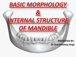
Mandible
- 2. CONTENTS • INTRODUCTION • OSTEOLOGY • ATTACHMENTS AND RELATIONS OF THE MANDIBLE • BLOOD SUPPLY ,NERVE SUPPLY AND LYMPHATIC DRAINAGE • AGE CHANGES • APPLIED ANATOMY • REFERENCES
- 3. INTRODUCTION DEFINITION :- • The mandible(from Latin mandibula, "jawbone") or inferior maxillary bone is the largest, strongest and lowest bone in the face. • It forms the lower jaw and holds the lower teeth in place. • It comprises of BODY,RAMUS, ANGLE,CONDYLAR PROCESS AND CORONOID PROCESS.
- 5. OSTEOLOGY • Mandible is the second bone after clavicle to ossify in the body. • Parts that ossify in cartilage includes: incisive part below the incisor teeth, coronal and condyloid processes • Upper half of ramus above the level of the mandibular foramen • Each half of mandible ossifies from one centre which appears in the 6th week of intrauterine life in mesenchymal shealth of Meckle’s cartilage.
- 6. • Meckel’s cartilage has a close, relationship to the mandibular nerve. • A single ossification centre for each half of the mandible arises in the 6th week of I.U. life in the region of bifurcation of inferior alveolar nerve into mental and incisive branches.As the ossification continues, the meckel’s cartilage become surrounded and invaded by bone. • Ossification stops at the site that will later become the mandibular lingula from where the meckel’s cartilage continues into the middle ear and develops into the auditory ossicles i.e. malleus and incus.
- 8. THE ENDOCHONDRAL OSSIFICATION Endochondral ossification is seen in 3 areas of mandible: 1. The condylar process:- Ossification starts by14th week. 2. The coronoid process:- Ossification starts by about the 10-14 week of IU life. 3. The mental region:- Ossification starts by the 7th month of I.U. life. The two halves of the mandibular body are united by fibrous joint at the symphysis menti which is replaced by the bone within 2nd year.
- 11. THE BODY • Consists of horseshoe-shaped • Has two surfaces and two borders • Surfaces: External or Outer or Lateral Internal or Inner or Medial • Separated by two borders: Upper and Lower
- 12. FEATURES SEEN ON OUTER SURFACE OF THE BODY • Symphysis Menti • Mental Protuberance • Mental Tubercles • Mental foramen • Oblique line • Incisive fossa
- 14. FEATURES ON THE INNER SURFACE OF THE BODY • Genial tubercles • Mylohyoid line • Below the mylohyoid line, surface is slightly hollowed out to form SUBMANDIBULAR FOSSA , which lodges submandibular gland. • Above the mylohyoid line, there is SUBLINGUAL FOSSA , which lodges sublingual gland.
- 16. UPPER BORDER or ALVEOLAR PART • Contains 16 alveoli for the roots of teeth, varying in size and depth, some being multiple. LOWER BORDER OR BASE Near the midline, the base shows an oval depression called as DIGASTRIC FOSSA.
- 17. • Quadrilateral in shape • Consists of two surfaces ,four borders & two processes Two surfaces include:- Lateral Medial • Four border include : Upper Lower Anterior posterior • Two processes include :- Coronoid Condyloid THE RAMUS
- 18. BORDERS • UPPER BORDER: Thin and curved downwards forming the mandibular notch. • LOWER BORDER: Backward continuation of base of mandible. • ANTERIOR BORDER is thin while the POSTERIOR BORDER is thick.
- 19. PROCESSES • CORONOID PROCESS: Flattened triangular upward projection from the anterosuperior part of the ramus. • CONDYLOID PROCESS: Strong upward projection from posterosuperior part of the ramus.
- 20. ON THE LATERAL SURFACE: 1. From The Oblique line : buccinator and In front of this origin: depressor labii inferioris and depressor anguli oris below the mental foramen 2. Incisive fossa: gives origin to MENTALIS and mental slips of ORBICULARIS ORIS. 3. Whole of lateral surface of ramus except posterosuperior part provides insertion to MASSETER. 4.Posterosuperior part : covered by PAROTID GLAND. ATTACHMENTS AND RELATIONS OF THE MANDIBLE
- 21. 5. Lateral surface of the neck provides insertion to the LATERAL LIGAMENT OF TMJ. 6. Parts of both the inner and outer surfaces just below the alveolar margins are covered by mucous membrane of the mouth. 7. PLATYSMA is inserted into the lower border. 8. The deep cervical fascia ( investing layer) is attached to the whole length of the lower border.
- 23. 1. Digastric fossa: arises ANTERIOR BELLY OF DIGASTRIC 2. Genial tubercles: arises GENIOGLOSSUS and GENIOHYOID. 3. Mylohyoid line : arises MYLOHYOID MUSCLE. 4. From an area above the posterior end of mylohyoid line: arises SUPERIOR CONSTRICTOR OF PHARYNX. 5. PTERYGOMANDIBULAR RAPHE: Attached immediately behind the third molar tooth in continuation with the origin of superior constrictor . ON THE MEDIAL SURFACE
- 24. 7.Below and behind the mylohyoid groove: insertion of MEDIAL PTERYGOID muscle . 8.At the apex of coronoid process : TEMPORALIS is inserted ;extend downwards on ant. Border of ramus. 9.Into the pterygoid fovea: insertion of LATERAL PTERYGOID. 10.Sphenomandibular ligament : is attached to the lingula.
- 26. FORAMINA AND RELATIONS TO NERVES AND VESSELS 1. MENTAL FORAMEN: Transmits the mental nerve and vessels. 2. MANDIBULAR FORAMEN: Inferior alveolar nerve and vessels enter the mandibular canal via this foramen. 3. Mylohyoid nerve and vessels lie in the mylohyoid groove.
- 27. 4. The lingual nerve is related to the medial surface of the ramus in front of the mylohyoid groove. 5. The area above and behind the mandibular foramen is related to the INFERIOR ALVEOLAR NERVE and VESSELS ; and MAXILLARY ARTERY respectively. 6. The masseteric nerve and vessels pass through the mandibular notch. 7. The auricotemporal nerve is related to the medial side of the neck of the mandible.
- 28. BLOOD SUPPLY OF THE MANDIBLE 1. Central blood supply via THE INFERIOR ALVEOLAR ARTERY except the coronoid process , which is supplied by temporalis muscle vessels. 2. Peripheral blood supply via the PERIOSTEUM.. periosteal supply ,which generally runs parallel to cortical surfaces of bone, giving off nutrient vessels those penetrate cortical bone and anastomose with the branches of inferior alveolar artery.
- 29. NERVE SUPPLY OF MANDIBLE • It is basically derived from mandibular branch of trigeminal nerve. 1. The long buccal nerve: The anterior division of the mandibular nerve. It supplies mucosa opposite the last three mandibular molars on their buccal aspect. 2. The inferior alveolar nerve: The posterior division of the mandibular nerve. It supplies all lower jaw teeth, lower lip, buccal mucosa from the incisors to the premolar & the skin over the chin. 3. The lingual nerve: The posterior division of the mandibular nerve. It gives sensory supply to the anterior 2/3rd of tongue, the mucosa on the lingual aspect of the lower teeth & the floor of mouth.
- 30. LYMPHATIC DRAINAGE • Most of the mandible & lower teeth drain into the submandibular group of lymph nodes . • Except a small wedge in the symphysis region & the lower incisors which drain into the submental group of lymph nodes. • From the submental group the lymph drains to the submandibular group of nodes. • Most of the submandibular nodes ultimately drain to the jugulo-omohyoid group of deep cervical lymph nodes. • Few extremely posterior submandibular nodes drain to jugulo-digastric group of deep cervical lymph nodes.
- 31. AT BIRTH The body of the bone is a mere shell, containing the sockets of the two incisor, the canine, and the two deciduous molar teeth, imperfectly partitioned off from one another. The mandibular canal is of large size, and runs near the lower border of the bone; the mental foramen opens beneath the socket of the first deciduous molar tooth. The angle is obtuse (175°), and the condyloid portion is nearly in line with the body. The coronoid process is of comparatively large size, and projects above the level of the condyle AGE CHANGES IN THE MANDIBLE
- 33. CHILDHOOD • The two segments of the bone become joined at the symphysis, from below upward, in the first year; but a trace of separation may be visible in the beginning of the second year, near the alveolar margin. • The body becomes elongated in its whole length, but more especially behind the mental foramen, to provide space for the three additional teeth developed in this part. • The depth of the body increases owing to increased growth of the alveolar part, to afford room for the roots of the teeth. • The angle becomes less obtuse, owing to the separation of the jaws by the teeth; about the fourth year it is 140°.
- 35. 1. After the eruption of permanent teeth the mental foramen lies mid-way between the upper & lower borders of the bone. 2. Growth of the rami takes place posteriorly & vertically by the process of remodeling. Posterior growth accommodates the eruption of permanent molars & reduces the angle of mandible to almost 110º-115º. Vertical growth allows the condylar process to lie higher than the coronoid process. IN ADULTS
- 37. 1. Teeth fall out and the alveolar border is absorbed so that the height of the body is markedly reduced. 2. The mental foramen and the mandibular canal are close to the alveolar border. 3. The angle again becomes obtuse about 140 degrees because the ramus is oblique. IN OLD AGE
- 39. APPLIED ANATOMY OF MANDIBLE 1. Parasymphysis region lateral to the mental prominence is a naturally weak area susceptible for parasymphyseal fracture. This is because of the presence of incisive fossa and mental foramen. 2. The body of the mandible is considerably thicker than the ramus and the junction between these two portions constitutes a line of structural weakness. 3. Strength of the lower jaw varies with the presence or absence of teeth. The presence of impacted lower third molars or excessive long roots of canines make the area more vulnerable for fracture.
- 40. 4. With the advancing age, the loss of teeth and resorption of alveolar bone leads to a decrease in the vertical height of the mandible, making it prone to fracture. 5. The slender neck of the mandibular condyles renders it particularly liable to fracture as a result of direct violence applied to the chin. This acts as a safety mechanism , as a fracture of neck of the condyle prevents injury to the middle cranial fossa. Direct blow to the chin region can lead towards fracture of one or both condyles. Sideways blow can bring about fracture of the opposite condylar neck along with the parasymphysis fracture at the same side of the blow.
- 41. 6. It is possible to split the ramus of the mandible in sagittal plane bilaterally thereby correcting micrognathia this procedure is called sagittal split osteotomy. 7. Osteomyelitis of the mandible is more commoner than the maxilla as the maxilla has rich blood supply.
- 42. REFERENCES • B.D CHAURASIA’S HUMAN ANATOMY – 6TH EDITION • TEXTBOOK OF ANATOMY BY INDERBIR SINGH- 5TH EDITION • GRAY’S ANATOMY – 2ND EDITION • SICHER AND DuBRUL’S ORAL ANATOMY – 8TH EDITION • TEXTBOOK OF ORAL AND MAXILLOFACIAL SURGERY , NEELIMA ANIL MALIK- 2ND EDITION • INTERNET