
Recomendados
Más contenido relacionado
La actualidad más candente
La actualidad más candente (20)
Similar a Oogenesis
Similar a Oogenesis (20)
Último
Último (20)
Oogenesis
- 2. Oogenesis is the period of growth differentiation and maturation occurring in the female gonads or ovaries during which the egg or ovum acquires its developmental potential Ovum is a unique cell which has all the properties to develop into a new individual when segregated from the organism VON BAER in 1827,discovered the mammalian ovum and studied embryonic development of many animals
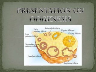
- 3. • In development at the time of gastrulation a small group of cells are "put aside" to later form oocytes and spermatozoa. This population of cells is described as the primordial germ cells (PGCs). •These cells also migrate initially into the posterior endoderm that forms the hindgut and from there into the genital ridge that will be the site of the developing gonad ,from this time there occurs a multiplication phase leading to the formation of oogonia which is diploid in number. •The following cycle depicts different phases of Oogenesis during meiotic events, it also shows follicular development of an ovary.
- 10. The primordial germ cell migrate from the endoderm. Multiply to form oogonia initiating the multiplication phase. Thus leading to the formation of primary oocytes.
- 11. This is a differentiation phase where primary and secondary oocytes formation takes place. It includes various sub-stages namely:- > Previtellogenesis and > Vitellogenesis Previtellogenesis further includes stages involved before formation of the yolk where as vitellogenesis refers to the stages involved in formation of yolk.
- 12. GROWTH PHASE OF PRIMARY OOCYTES Magnitude of growth Rate of oocytes growth Mode of growth of oocytes
- 13. OOCYTE grows to enormous proportions, normally to become the largest cell of the animal body. Most of the PGC’s are approximately 10 microns in diameter, which is the size of an average body cell. In case of mammals, some species may reach a diameter of 200 micron, in frog the increase is up to 2000 microns where as in birds the diameter of the ovum is as large as 40,000 microns. This growth is not so much in active cytoplasm as compared to an increase in the reserves such as yolk.
- 14. Rate of growth of primary oocytes may be slow or higher for example in case of drosophila it needs only 3 days for oocytes growth, whereas the mouse needs 16 days and the frog needs 3 years. In case of humans its around 27 days.
- 15. During growth phase of primary oocyte qualitative and quantitative changes take place both in the nucleus and cytoplasm. It includes:- 1. growth of nuclear substances. 2. formation of mitochondrial clouds 3. formation of cortical granules
- 16. In oocytes not surrounded by nurse cells, oocyte nuclei swells and chromosomes begin to elongate, such chromosomes are called lampbrush chromosomes whereas in oocyte surrounded by the nurse cells the lamp brush chromosomes are absent and the nuclei of the nurse cell enlarges, become metabolically active and synthesises the gene products which are stored in oocyte cytoplasm.
- 17. LAMPBRUSH CHROMOSOMES • Homologous and bivalent • Paternal and maternal chromosomes are held together by chiasmata(sites of crossing over) • Long axis of each chromosome is made up of two chromatids, with tight coils called chromomeres, and the regions which are uncoiled are called lampbrush loops. • This shape enhances the production of mRNA, which produce proteins of storage value
- 18. Gene Amplification • One set of gene is replicated selectively in rDNA(ribosomal DNA) of NOR(nucleolar organiser) of oocytes in nucleolus, such that large number of ribosomes are manufactured. • Eg:- Xenopus eggs are very large cells(1.3 mm in diameter) that accumulates large number of ribosomes(10^12)
- 19. Large number of mitochondria accumulates in the oocyte, more than required for respiratory metabolism. In young oocytes mitochondria is present at the periphery, but as it grows it is dispersed in the cytoplasm. During growth, mitochondria segregates and forms “mitochondrial clouds”. Mitochondria arises either from autonomous replication of the circular DNA or from nurse cells, migrating down the nutritive chords into oocyte cytoplasm
- 20. membrane bound spherical bodies of diameter from 0.8 micron to 2 micron. formed from cisternae of Golgi apparatus, initially they lie inside the cup-shaped space formed by Golgi membrane then move to the periphery and get arranged in layer close to the ectoplasm. contain mucopolysaccharides required for the synthesis of fertilization membrane during fertilization.
- 21. synthesis and deposition of yolk. Yolk - major nutritional reserve of the oocyte - required for the nourishment of embryo - composed of proteins, phospholipids and natural fats. types of yolk - protein yolk(present in the form of granules or yolk platelets) - fatty yolk(present in the form of fat droplets i.e. lipoproteins or lipochondria)
- 22. Protein yolk:-Yolk that has more of protein than lipids. Fatty yolk:- yolk that has more of fat contents than proteins Granular yolk:- yolk in forms of fine granules, evenly distributed in cytoplasm of oocyte Yolk platelets:- yolk in form of large granules, oval in shape, flattened in one plane -two main proteinaceous substances:- -phosphitin -lipovitellin -Phosphitin-highly phosphorylated protein -Lipovitellin-protein with large molecule and bound lipids -2 mol of phosphitin+ 1 mol of lipovitellin = 1phospholipoprotein/ vitellogenin
- 23. 1. Yolk is morphologically designated as the molecule that takes part in the nourishment of an embryo. 2. Yolk is made up of phospholipoprotein called vitellogenin. 3. Vitellogenin is of crystalized form and is made of 2 molecules of phosphotin + 1 molecule of lipovitellin. 4. Formation of yolk invertebrates occurs in liver under the influence of estrogen. 5. In this case the vitellogenin is present in dephosphorylated form and is soluble by the action of protein kinase and ATP get phosphorylated and is stored in the insoluble form in the ovary. 6. In fishes and amphibians it is present in solid form where as its present in liquid or semisolid form in reptiles and birds.
- 25. 1. MICROLECITHAL OR OLIGOLECITHAL EGGS – These are small-sized eggs containing very small amount of yolk. These type of eggs are found in Hydra and sea urchin. 2. MESOLECITHAL EGGS- These contains moderate amount of yolk and are found in annelid worms, molluscs and amphibians. 3. MEGALECITHAL, MACROLECITHAL OR POLYLECITAL EGGS- These eggs contain enormous amount of yolk and are found in insects, reptiles and birds.
- 27. 1. HOMOLECITHAL OR ISOLECITHAL-In this the amount of yolk is so little that it is found scattered almost uniformly throughout the egg cytoplasm. 2. TELOLECITHAL EGGS- These types of eggs have a polarized distribution of yolk in the ooplasm and are found in the mesolecitthal and macrolecithal eggs. 3. CENTROLECITHAL- In some hydrozoa and insects, the yolk is concentrated in the center and the active cytoplasm forms a thin peripheral layer around the yolk
- 28. 1. What do you mean by oogenesis,depict using a flowchart. 2. Explain follicular development of ovary? 3. What are PGC’s? 4. Name various sub stages of meiotic phase? 5. What are the factors involved in the growth phase of primary oocyte in previtellogenesis? 6. Define the following 1. Lamp brush chromosomes 2. Mitochondrial clouds 3. Cortical granules 4. Lipochondria 5. Vitellogenin 7. What is composition of yolk? 8. What is the phenomenon involved in yolk formation? 9. Write a short note on egg types based on :- 1. Amount of yolk 2. Distribution of yolk QUESTIONAIRE
- 29. In the absence of lampbrush chromosomes which cells surround the oocyte. Sperm mother cells PGCs Nurse cells Oogonia In which phase primary oocytes get arrested Metaphase I Anaphase I Telophase I Prophase I • Primary oocytes leads to formation of how many secondary oocytes. 4 3 2 1