Episiotomy
•
628 recomendaciones•283,904 vistas

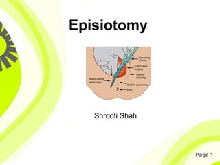
A surgically planned incision on the perineum and the posterior vaginal wall during the second stage of labour is called episiotomy.

Recomendados
Más contenido relacionado
La actualidad más candente
La actualidad más candente (20)
Destacado
Destacado (20)
Similar a Episiotomy
Similar a Episiotomy (20)
Más de Shrooti Shah
Más de Shrooti Shah (10)
Último
Último (20)
Episiotomy
- 2. Page 2 Definition • A surgically planned incision on the perineum and the posterior vaginal wall during the second stage of labour is called episiotomy.
- 3. Page 3 Types of perineal tear
- 4. Page 4 Objectives • To enlarge the vaginal introitus so as to facilitate easy and safe delivery of the fetus- spontaneous or manipulative. • To minimise overstretching and rupture of the perineal muscles and fascia; to reduce the stress and strain on the fetal head.
- 5. Page 5 Indications • In elastic rigid perineum • Anticipating perineal tear: Big baby, face to pubis delivery, Breech delivery, Shoulder dystocia • Operative delivery: Forceps delivery, Ventouse delivery • Previous perineal surgery: Pelvic floor repair, Perineal reconstructive surgery
- 6. Page 6 Indications Common indications are: 1. Threatened perineal injury in primigravidae 2. Rigid perineum 3. Forceps, breech, occipito-posterior or face delivey
- 7. Page 7 Timing of the episiotomy Bulging thinned perineum during contraction just prior to crowning is the ideal time.
- 8. Page 8 Advantages • Maternal: – A clear and controlled incision is easy to repair and heals better than a lacerated wound that might occur otherwise. – Reduction in the duration of second stage – Reduction of trauma to the pelvic floor muscles • Fetal: – It minimises intracranial injuries specially in premature babies or after coming head of breech
- 9. Page 9 Types • Medio-lateral • Median • Lateral • T-shaped
- 10. Page 10 Relatives merit of median and mediolateral episiotomy Median • The muscles are not cut. • Blood loss is least. • Repair is easy • Post operative comfort is maximum • Healing is superior • Wound disruption is rare • Dyspareunia is rare Medio-lateral • Relatively safety from rectal involvement from extension. • If necessary, the incision can be extennded
- 11. Page 11 Relatives demerits of median and mediolateral episiotomy Median • Extension, if occurs, may involve the rectum. • Not suitable for manipulative delivery or in malpresentation Medio-lateral • Apposition of the tissues is not so good • Blood loss is little more • Post operative discomfort is more. • Relative increased incidence of wound disruption • Dyspareunia is comparatively more
- 12. Page 12 Structure cut are: • Posterior vaginal wall • Superior and deep transverse perineal muscles, bulbospongiosus and part of levator ani. • Fascia covering those muscles • Transverse perineal branches of pudendal vessels and nerves • Subcutaneous tissue and skin
- 13. Page 13 Steps of episiotomy • Provide emotional support and encouragement. • Use local infiltration with lignocaine. • Make sure there are no known allergies to lignocaine or related drugs. • Infiltrate beneath the vaginal mucosa, beneath the skin of the perineum and deeply into the perineal muscle. • Note: Aspirate (pull back on the plunger) to be sure that no vessel has been penetrated
- 14. Page 14 Steps of episiotomy
- 15. Page 15 Steps of episiotomy • Wait 2 minutes and then pinch the incision site with forceps. • Wait to perform episiotomy until: - the perineum is thinned out; and - 3–4 cm of the baby’s head is visible during a contraction.
- 16. Page 16 Steps of episiotomy • Wearing high-level disinfected gloves, place two fingers between the baby’s head and the perineum. • Use scissors to cut the perineum about 3– 4 cm in the mediolateral direction
- 17. Page 17 Steps of episiotomy • Use scissors to cut 2–3 cm up the middle of the posterior vagina. • Control the baby’s head and shoulders as they deliver. • Carefully examine for extensions and other tears and repair
- 18. Page 18 Repair of episiotomy • Apply antiseptic solution to the area around the episiotomy. • If the episiotomy is extended through the anal sphincter or rectalmucosa, manage as third or fourth degree tears, respectively • Close the vaginal mucosa using continuous 1-0 suture
- 19. Page 19 Repair of episiotomy • Start the repair about 1 cm above the apex (top) of the episiotomy. Continue the suture to the level of the vaginal opening. • At the opening of the vagina, bring together the cut edges of the vaginal opening • - Bring the needle under the vaginal opening and out through the incision and tie.
- 20. Page 20 Repair of episiotomy • Close the perineal muscle using interrupted 1-0 sutures • Close the skin using interrupted (or subcuticular) 1-0 sutures
- 21. Page 21 Post operative care • Dressing • Comfort • Ambulation • Removal of stitches
- 22. Page 22 Complications • Immediate • Extension of the incision to involve the rectum • Vulval haematoma • Infection • Wound dehiscence • Injury to anal sphincter causing incontinence of flatus or faeces • Rectovaginal fistula (Rarely) • Necrotising fascitis
- 23. Page 23 Complications • Remote • Dyspareunia • Chance of perineal lacerations • Scar endometriosis (rare)