Laparoscopic Cholecystectomy
•
155 recomendaciones•27,311 vistas

Laparoscopic Cholecystectomy

Recomendados
Recomendados
Más contenido relacionado
La actualidad más candente
La actualidad más candente (20)
Similar a Laparoscopic Cholecystectomy
Similar a Laparoscopic Cholecystectomy (20)
Más de Dr. Shouptik Basu
Más de Dr. Shouptik Basu (8)
Último
Último (20)
Laparoscopic Cholecystectomy
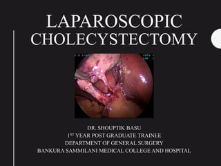
- 1. LAPAROSCOPIC CHOLECYSTECTOMY DR. SHOUPTIK BASU 1ST YEAR POST GRADUATE TRAINEE DEPARTMENT OF GENERAL SURGERY BANKURA SAMMILANI MEDICAL COLLEGE AND HOSPITAL
- 2. Objectives • Development and Dissemination of Lap Chole. • Basic Instruments • Standard Laparoscopic Cholecystectomy • Avoiding Bile Duct Injuries • Exit Strategies for a Difficult gall bladder • Variations in Standard Laparoscopic Cholecystectomy • The Future : Recent Advances
- 3. Prof. (Dr.) Med Erich Mühe – [1985] A Surgeon ahead of his time ReynoldsW.The First Laparoscopic Cholecystectomy. JSLS : Journal of theSociety of Laparoendoscopic Surgeons. 2001;5(1):89-94. Galloscope
- 4. SAGES AND EAES recognize his work in 1999 1999 Annual Karl Storz Lecture in NewTechnology, which was given Friday, March 26, 1999, in San Antonio,Texas. Dr Mühe's lecture was titled “The First Laparoscopic Cholecystectomy: Overcoming the Roadblocks on the Road to the Future.” Rejection in the GSS, 1986 Mühe received the GSS Anniversary Award for his pioneering work in endoscopic surgery. In receiving this award, his laparoscopic cholecystectomy was described by Franz Gall, president of the GSS, as one of the greatest original achievements of German medicine in recent history.
- 5. DEVELOPMENT OF Laparoscopic Cholecystectomy THE FRENCH REVOLUTION Moret, Gynecologist First Lap Cholecystectomy 1987 Dubois and Perrisat , Surgeon performed LC and Cholecystostomy and removal of stones , 1988 THE US REVOLUTION “The French Technique” Bill Saye (Gyne) and Barry Mc Kernan (Surg) 1st Lap Chole in US , 1988 Eddy Joe Reddick Douglas Olsen
- 6. The first Indian Lap Chole ? “I soon realised the value of diagnostic laparoscopy in a surgical unit in a developing country and tried to pass on my enthusiasm to all my colleagues. Surgeons in large cities viewed my passion with indifference if not scorn. To my gratification surgeons in small towns, in the course of innumerable workshops, were very receptive, specially so since they lacked other diagnostic facilities and very many of them had laparoscopy equipment as part of the family planning programme. From these somewhat primitive beginnings, laparoscopy and its logical sequel laparoscopic surgery has grown in the country in a phenomenal way. The first laparoscopic cholecystectomy in India was performed in 1990 at the JJ Hospital, Mumbai, followed a few months later in Pune by Dr. Jyotsna Kulkarni. ” Dr.Tehemton Erach Udwadia Honoured with Padma Bhushan
- 7. INDICATIONS FOR Lap Chole • Asymptomatic Cholelithiasis No indication except Immunosuppression Porcelain Gall Bladder with Gallstones-Risk of CA • Symptomatic Cholelithiasis Biliary colic and Cholecystitis (Acute/Chronic) • Complicated Cholelithasis Gall Stone pancreatitis close to discharge Choledocolithiasis with Cholangitis after the Cholangitis is resolved (pre- Op ERCP is a prerequisite) Cholelithiasis Conditions unrelated to Gall bladder Disease • Acute Acalculous cholecystitis • Biliary Diskinesia (dec EF on HIDA scan) • Polyps, Cholesterosis and Adenomyomatosis (Size > 1cm)
- 8. Contra-INDICATIONS FOR Lap Chole Absolute Relative • Major upper abdominal surgery, • History of ascites, • Coagulopathy. • Extremely limited
- 9. Status of Lap Chole Today… More than 50 techniques have been described in Literature These are modifications by Surgeons to improve Post operative outcomes or cosmesis Mainly Reduction in port size/number.
- 10. Armamentarium INSUFFULATOR CAMERAAND RECORDING UNIT LIGHT SOURCE & CABLE MONITOR TROCARS DISPOSABLE TROCARS TELESCOPE REDUCER VERESS NEEDLE
- 11. Commonly used Hand Instruments PALMER GRIP LIGA CLIP APPLICATOR ASPIRATION NEEDLE MARYLAND TRAUMATIC GRASPER ATRAUMATIC GRASPER FINE SCISSORS (METZENBAUM) MONOPOLAR L - HOOK STONE EXTRACTOR ALLIGATOR / GB EXTRACTOR SUCTION-IRRIGATION CANNULA
- 12. TECHNIQUES STANDARD LC MODIFIED LC REDUCED PORT SIZE REDUCED PORT NUMBER SSLC/TUSS HYBRID NOTES
- 13. STANDARD Lap Cholecystectomy • Most Commonly done • Four Ports (10-10-5-5) • Operating Table must allow Fluoroscopy (Optional) • American (Surgeon on the Left) / French position (Surgeon in- between patient’s legs) • Under General Anaesthesia, patient supine arms abducted 90 deg. (Some use arms tucked in) • Ryles tube decompression may be required EpigastricTrocar through the Falciform Lig MidAxillary trocar inserted towards theGB 3 working ports Triangulating onto the GB
- 14. Hepatocystic triangle dissection • Patient is placed in Reverse Trendelenburg position • Dissect the omental adhesions to the Gall bladder • A locking grasper holds the fundus and retracts it superiorly and towards the right shoulder • Another grasper holds the infundibulum and retracts it laterally, to the right. This separates the cystic duct from the Common Bile Duct. • Dissect the Hepato-cystic triangle with short bursts of Electro cautery or blunt graspers. • Identify the node of Calot in this location and gently separate it from the gallbladder. [Don’t go below it…] adhesiolysis Post. dissection Ant. dissection
- 15. • After the anterior and posterior dissection are complete and a window is created, and the cystic duct and artery are skeletonized. It is advisable to dissect the superior peritoneal fold to reveal the cystic plate. Hence the Critical view of safety is attained.
- 16. • Apply clips to the cystic duct and artery and divide them. • Intra operative Cholangiogram can be done any time prior to dividing the duct for aberrant anatomy and CBD stones
- 17. • Gall bladder is removed from the liver bed by using L-hook electrocautery • Before specimen extraction. • Check the liver bed, and Cystic structures for haemostasis. • Drain is optional (HRPOM)
- 18. • A severely contaminated specimen or CA GB specimen is best extracted in an Endobag (or - Gloves as endobag) • Umbilical or Epigastric post may be used for extraction. • Ports are closed with Port Vicryl
- 19. • Haemorrhage • Bile Leak • Bile Duct Injury • Failure to recognize Aberrant Anatomy Complications of Laparoscopic Cholecystectomy
- 20. The SAGES Safe Cholecystectomy Program Strategies for Minimizing Bile Duct Injuries: Adopting a Universal Culture of Safety in Cholecystectomy (COSIC) Why ? • Bile duct injury rates have increased (3 per 1,000 procedures) • Life altering complications leading to significant morbidity and cost. • Because bile duct injuries are relatively infrequent, definitive studies comparing methods to minimize these complications will likely never be performed. AIM: Secure Identification of Structures
- 21. Why do Bile Duct Injuries Occur…? • Incorrect interpretation of anatomy Incorrect Traction Blind Haemostasis • Accidental Mechanical – Scissors Energy Driven
- 23. 6 strategies 1. Use the Critical View of Safety Method (CVS) 2. Intra-op time out before cutting/clipping 3. Understand potential aberrant anatomy 4. Liberal use of Cholangiogram 5. Recognize a risky dissection- HALT !!! – Finish safely 6. Get help from an experienced Surgeon • The hepatocystic triangle is cleared of fat and fibrous tissue. • The lower one third of the gallbladder is separated from the liver to expose the cystic plate. • Two and only two structures should be seen entering the gallbladder.
- 24. RATIONALE….Secure Identification Open Cholecystectomy Lap. Cholecystectomy
- 25. What’s wrong with the Infundibular Technique…?
- 26. Aberrant Ducts ? May God Help you !!! • After dividing the artery and peritoneal attachments. • GB- Duct junction is mobilized to give an Elephant head appearance. • Visually ensures no aberrant duct is entering the Cystic duct. • Don’t Clip the duct until this is achieved. • Surest way to identify aberrant ducts. ComprehensiveTextbook of Laparoscopic Surgery , by IAGES 2 Ed
- 27. Orientation: Hepato-cystic Triangle • Area between cystic duct and Gall Bladder & the Liver and the Hepatic Duct. • Most Bile Duct andVascular Injuries occur HERE !!! Variation duringTraction & Dissection
- 28. Calot’s triangle Hepato-cystic triangle Based on the location of the CysticArtery CysticArtery is neither consistently present or Anatomically Precise… Preffered NOMENCLATURE V/S PLEASE FORGET MY TRIANGLE I KNOWYOU CAN’T !!!
- 29. Orienting various Anatomic Landmarks I. FALCIFORM LIGAMENT • Lies between Segments 3 and 4 • The CHD is between the mid-plane of the liver between Segments 4 and 5
- 30. Rouviere’s sulcus usually “points” to the neck of the gallbladder (the narrow part that then tapers into the cystic duct) and can then be used as a reference point to facilitate identification and dissection in Calot’s triangle. The Right Portal pedicle enters the liver here. Identifiable in most healthy livers (Absent 20%). Hugh et al reported that fewer common bile duct injuries occur during laparoscopic cholecystectomy if dissection begins ventral / anterior to Rouviere’s sulcus. Scar Open Closed II. The importance of the Rouviere’s sulcus … [Stay anterior to it]
- 31. III. EPICHOLEDOCHAL PLEXUS Produces a pattern of vessels on the outside of the common duct.Thus differentiates this from the cystic duct by its external appearance.
- 32. IV. DUODENUM Relationship of the duct to the Duodenum… Any duct that goes directly behind the Duodenum is the Common Bile duct.
- 33. Intraoperative Cholangiogram (IOC) • Delineate the biliary anatomy and to evaluate the common bile duct for filling defects, obstruction (pathologic or iatrogenic), or contrast extravasation indicative of injury. • Routine IOC vs Selective IOC debatable. SAGES says it diminishes Bile Duct Injury.
- 34. Indocyanine Green (Fluorescence Cholangiography) • Sends fluorescent signal detected by an advanced Laparoscope • Excreted through Bile as it concentrates there • Better visualization of Ductal Anatomy • Near Infra-red light is used
- 35. • fundus was punctured • 50 % methylene blue diluted by saline solution was injected into the gall bladder for coloration of biliary tree. “Conclusion: We believe that the incidence of bile duct injury related to anatomic misidentification can be decreased or even totally suppressed by intraoperative injection of methylene blue into the gall bladder fundus and visualisation of the gall bladder, cystic duct and ductus choledochus.” Other Dyes : Methylene Blue
- 36. Light Cholangiography (LCP)… Cold Light source through the Duodenum may help “In group I cold light was used to illuminate the common bile duct by leading an optical fiber into the common duct with a duodenoscope at the time of LC.The light coming from the fiber in the CBD could clearly illuminate the location of CBD and hepatic duct establishing its location relative to the cystic duct.This method was compared with the dye injection technique using methelenum coeruleum.” CONCLUSION: LCP is the only technique that can clearly and directly show the location of the extrahepatic biliary system and may be useful in selecting cases of uncertain anatomy in the prevention of bile duct injury.
- 37. Trouble shooting (Intra Operative) • Spilled stones • Slipped Clips • Heavy Stone load
- 38. Exit Strategies for a Difficult Gallbladder I. Difficult Fundus Retraction – Aspiration of bile from the gallbladder, with a long laparoscopic needle – large stone or contracted gallbladder, the lateral-most trocar can be upsized to a 10 mm dilating trocar, and a large claw forceps can be used to hold the gallbladder II. Inflamed and Indurated Calot’s Triangle / Acute Cholecystitis – gentle dissection by an experienced surgeon – Increase the number of ports (4 to 5) if required. – suction irrigator can be used bluntly to delineate the structures. – Use electrocautery judiciously (prevents injury) – intraoperative cholangiogram – If the gallbladder is severely contracted and adherent to the liver bed, it would be wise to leave the posterior wall of the GB in place.
- 39. III . Fundus First Approach – indicated when the triangle of Calot cannot be easily visualized (dense inflammation) – port placement is similar – assistant’s job is to grab the gallbladder just as it meets the uppermost portion of the liver bed – the surgeon grabs next to the assistant’s placement on the gallbladder side and uses Electro-cautery to dissect the peritoneum around the Gall Bladder – Cautery is then used to carefully separate the gallbladder away from the liver bed – The gallbladder is circumferentially dissected, with the surgeon – Blunt dissection, as well as using a suction irrigation, may be necessary – As the gallbladder is approached inferiorly, the first structure that should be identified is the cystic artery -> It should be clipped – The dissection then continues along the infundibulum of the gallbladder until no structure, other than the cystic duct, remains. – Once the cystic duct is completely skeletonized and the cystic artery taken, the cystic duct can be traversed with clips – Gall bladder removed in a standard fashion
- 40. Reduced Port Size LC • Epigastric port can be changed to 5mm -> Specimen Retrieved through Umbilicus but a Camera change to 5mm-> 5mm Clip applicator is used and Endoloop ligation of the Cystic Duct.-> Bipolar to cut the Cystic Artery • Epigastric Port Remains as 10mm and Umbilicus is 5mm -> Specimen extracted via Epigastrium and normal Clip applicators are used. • Two ports may be converted to 3mm then the configuration becomes 10-5-3-3. Here 3mm instruments are used • Minilaparoscopic Cholecystectomy one 10 mm port and rest all 5 mm one 5 mm and other <5 mm ports one 10 mm port, one 5 mm and rest 2 to 3 mm ports • Micro-Laparoscopic Cholecystectomy 10 mm and rest all 2-3 mm ports have been used
- 41. Reduced Number LC • Initial attempts were made by surgeons to reduce the port numbers from 4 to 3 for performing standard LC. (10-10-5 3 port Modified LC) – It’s Fallacy: vision achieved is quite different no major difference Some Surgeon’s claim there is less pain, faster recovery and less scars however this view is based on personal experiences of surgeons. Best for – Short Gall bladder and a Floppy Liver • 3 ports with Sutured FundalTraction (5-10-5 SF Modified LC) – 18 G needle to take a figure of 8 bite on the fundus for traction. Standard Clip applicator can be applied – Demerits: Minor bile leak from the fundal bite Epigastric 10mm port causes more pain comparatively No cosmetic advantage if a 10 mm umbilical port is truncated to 5mm
- 42. • One can also use micro laparoscopic instruments, i.e. 3 mm or 2 mm instruments for performing reduced port LC -> here intracorporeal ligation of cystic duct is advisable because of the paucity of good 3mm clip applicators. • 2 port LC with 2 SutureTractions (10-10-SF-SH) or (5-10-SF-SH) – Two traction sutures are on the Fundus (SF) and on the Hartmann’s pouch (SH) – So how does this work ? For anterior as well as posterior dissection of the Calot’s triangle, a doublesuture swinging traction of the Hartmann’s can be employed with one suture extracted from the epigastric port and one from the right lumbar port.Thus with traction on the right lumbar suture, anterior dissection of Calot’s triangle is possible, while with an epigastric suture traction posterior dissection is possible. – Fallacy- Quality ofTraction is not like instrument traction Can be combined with Micro-Laparoscopy but one port must be 10mm for GB extraction
- 43. Single Site Laparoscopic Cholecystectomy (SSLC) / Trans-umbilical Single Site Surgery (TUSS) • SSLC means when all ports are placed at single site; here it is practically in or around umbilicus. • First Reported by Piskun • Single skin and sheath incision or single skin and separate sheath incisions or separate skin and sheath incisions but at the same site. • Port devices such as SILS port (Covidien),Tri port or Quad port (Olympus) or X cone(Storz) through the single incision. • larger skin incision (at least 20 mm) then that needs in standard LC. (size should increase if it’s a thick Gallbladder with multiple stones) • Risk of Incisional Hernias
- 44. Bhandarkar, D., Mittal, G., Shah, R., Katara, A., & Udwadia, T. E. (2011). Single-incision laparoscopic cholecystectomy: How I do it? Journal of Minimal Access Surgery, 7(1), 17–23. • Standard instruments / Multiple curved instruments are used • No more than 3 instruments around the umbilicus as its difficult to handle them • Additional 3mm / 5mm trocars can be used in case of difficulty • Sutures may be used for traction • Hybrid procedures combine the principles of SILS and Multiport methods
- 45. Another French Revolution Langenbuch:1867 Moret :1987 B. Dallemange, J Marescaux,S. Perretta-2007 Trans-Gastric Cholecystectomy
- 46. NOTES No Laparoscope… A flexible endoscope Hybrid: uses a 5mm port through the umbilicus and the endoscope through the vagina Trans-gastric and trans colonic approaches First Reported by: R.Zorron and B. Dellmange and S. Perratta Techniques are in infancy One Reported case of Rectal Injury during th vaginal puncture
- 47. The Future…Hybrid • Combination of a flexible transumbilical double-channel endoscope and a 3-mm rigid transcutaneous trocar placed in the left hypochondrium for liver retraction • Instruments used through the two working channels of the endoscope were either a grasping forceps or snare for grasping and pulling and a hot-biopsy forceps for cold and hot preparation and dissection. Endoclips were used for cystic duct and artery closure. • So far, our endoscope-based transumbilical cholecystectomy technique has not yielded satisfactory results in humans.
- 48. Will Robots take over… • The future direction lies in the development of robotic surgery. • Transcontinental robot-assisted remote surgery (telesurgery) has been reported. • No clinical trials available for verifying the advantages of robotic over conventional surgery • Offers the advantage of surgeon comfort, elimination of surgeon tremor and improved imaging and increased degrees of freedom of the operative instruments • More time consuming.
- 49. • One of the major drawbacks of laparoscopic surgery has been due to the carbon dioxide pneumoperitoneum. • Carbon dioxide pneumoperitoneum can have severe physiological disturbances. • It is especially useful when operating on critical patients with a cardiorespiratory problem, who would benefit most from laparoscopic surgery due to the reduced trauma and advantages for recovery. • Mechanical lifting of the abdomen is based on the traction and subsequent elevation of the abdominal wall. • Trocars may not be required. Laparolift System. Gasless Laparoscopic Cholecystectomy Mühe's Modifications: Gasless Technique, LC through Trocar Sleeve, and “Open Laparoscope”
- 50. • Absorbable clips have been used, but have not been found to be advantageous • Pre-tied loop or an Endo-GIA stapler can be used for a dilated cystic duct • Harmonic scalpel has been used as the sole instrument for dividing the cystic duct and artery (‘clipless laparoscopic cholecystectomy’) • A combined method of endoscopic sphincterotomy with common bile duct stone extraction and laparoscopic cholecystectomy under general anesthesia, for a single-session treatment of patients with gallstones with simultaneous CBD stones is described, - the so called “rendez-vous” technique. Miscellaneous
- 51. Controversies of Lap Chole : Should I practice Open Chole in 2018 … ? Which is better – open or laparoscopic cholecystectomy? There can be no doubt that with laparoscopic cholecystectomy, the pain felt by the patient is less, overall morbidity is less, recovery is faster, hospital stay is reduced, cosmesis is better and return to work is earlier. As more and more experience is gained, the contraindications to the procedure have shrunk, so that the only absolute contraindications to laparoscopic cholecystectomy are the same as those for open cholecystectomy. There was an initial increased incidence of iatrogenic complications, especially bile duct injury, but even this is gradually coming down.
- 52. To conclude • Laparoscopic Cholecystectomy is the Gold standard Surgery for the Gall bladder. • Considering the many techniques , a surgeon makes the decision to choose the technique based on his competency. • It’s important to Recognize complications early. • Always consult a Senior Hepato-biliary surgeon when in doubt or when in trouble. • Recent Advancements in guidelines, instruments and techniques make surgery easier and decrease the complications.