
Recommended
More Related Content
What's hot
What's hot (20)
Similar to Musculo skeletal system
Similar to Musculo skeletal system (20)
More from Saugat Chapagain
More from Saugat Chapagain (14)
Recently uploaded
Recently uploaded (20)
Musculo skeletal system
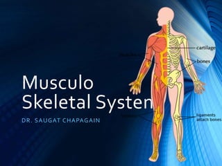
- 1. Musculo Skeletal System DR. SAUGAT CHAPAGAIN
- 2. Course of content • Fractures • Arthritis • Osteomyelitis • Osteoporosis • Leprosy • Gout • Muscular dystrophy • Myasthenia gravis
- 3. Normal anatomy • Two components 1. cortical/ compact bone 2. Trabecular / cancellous bone • Histology 1. Osteoblasts 2. Osteocytes 3. Osteoclasts 4. Osteoid matrix
- 4. Fracture
- 5. Introduction • Structural break in continuity of the bone • Complete/ incomplete break in continuity of the cortex of the bone Causes: • Traumatic (in normal bones) or pathological (in diseased bones) • Green stick (in immaturity) • <fall/ accident/ tumors/ immaturity/ drugs>
- 6. Classification • Based on etiology • Traumatic • Fatigue/ stress • Pathological • Clinical basis • Closed • Open • Based on involvement of joint • Extra capsular / articular • Intracapsular/ articular • Based on number of fragments • Simple • Comminuted • Involvement of vital structures • Simple • Complicated • Pattern • Linear • Transverse • Oblique • Spiral • Comminuted • Compacted • Compression • Greenstick • Impacted
- 7. Pathophysiology • Injury/ stress periosteum and blood vessels in cortex, marrow and surrounding soft tissue disturbed hematoma formation replaced by granulation tissue • Pathophysiology is d/t inflammatory response • Osteoblasts produce callus (osteoid) • Osteoclasts remodelling and resorption • Osteoblasts mature into osteocytes
- 8. Clinical Features • Pain • Deformity, swelling, tenderness • Muscular spasm • Broken skin with bone protruding outside • Limited range of motion • Ecchymosis, DNVS altered/ intact • Crepitus/ clicking sound on movement
- 9. Investigations • Imaging • X ray • CT scan • MRI
- 10. Management • First Aid measures • Airway/ Breathing/ Circulation • Emergency management • Splinting the limb (immobilization) • Analgesic, TT prophylaxis • RICE (rest, Ice packing, Compression/cast, elevation) • In case of blood loss, • Compress site of bleeding (the bleeding vessel) • Fluid resuscitation to prevent haemodynamic shock • Surgical management • Closed reduction • K-wire fixation • Open reduction • Rods/ plates and screws
- 11. osteomyelitis
- 12. Introduction • Myelo – marrow • Infection/inflammation of bone + marrow • Usu. By pyogenic organisms • Pathologically significant types: • Pyogenic osteomyelitis • Tuberculous osteomyelitis
- 13. Classification Based on origin: • Primary • Secondary Based on cause/ duration: • Acute (7 days) – Staph., Streptococcus, Pneumococcus, Salmonella • Sub acute – over 21 days • Chronic – discharging sinus e.g. tuberculous
- 14. ETIOLOGY Age group Most common organisms Newborns (< 4 months) S. aureus, Enterobacter spp.,Gp.A beta hemolytic Streptococcus Children (4 months – 4 years) S. aureus, Gp.A beta hemolytic Streptococcus, Haemophilus influenzae, Enterobacter spp. Children/ adolescents (4 yrs+) S. aureus, Gp.A beta hemolytic Streptococcus, H. influenzae, Enterobacter spp. Adult (18+) S. aureus, occasionally Enterobacter or Streptococcus spp.
- 15. Pathogenesis • Pathogen grows in a hematoma or in weakened area or site of infection travels through blood to metaphysea end of marrow cavity pus formed pressure built spread along marrow cavity (leads to periosteitis) • Infection may form subperiosteal abscess or drain via sinuses. • Combination of suppuration and impaired blood supply erosion, infarction and necrosis of cortex (sequestrum). • New bone formed beneath periosteum over infected bone (involucrum)
- 16. Pathogenesis (cntd….) • Acute osteomyelitis may be contained to a localized area and sealed by fibrous tissues and granulation tissue BRODIE’s Abscess • After involucrum formation; resolution may occur or complications may occur • Basic pattern: • Suppuration ischemic necrosis healing by fibrosis/ bony repair
- 17. Complications • Septicemia • Acute bacterial arthritis • Pathological fractures • Progression to chronic osteomyelitis • Development of carcinoma • Secondary amyloidosis • Vertebral osteomyelitis may cause: • Vertebral collapse • Paravertebral abscess • Epidural abscess • Cord compression • Neurological deficits
- 18. Clinical features • Pain • History of or current infection source • Restricted movement • Local rise of temperature • Tenderness over affected area • Swelling and redness • In infants, continuous crying, fever, malaise, ill and toxic look. • Discharging pus from sinus
- 19. Investigations • Blood –TC, DC, ESR, Hb% WBC and ESR elevated • Blood C/S • Imaging – X ray, CT scan, MRI • Bone scan
- 20. Treatment • Immobilization, traction, bed rest • Supportive measures (analgesic, fluid support) • I & D, C/S of pus. • Acute osteomyelitis • Systemic antibiotic, intracavitary instillation via closed system, continuous irrigation with low intermittent suction • Limited irrigation • Packed wet antibiotic soaked dressing • Chronic osteomyelitis (poor prognosis) • Surgery for removal of dead bone and abscess • Hyperbaric O2 • Skin, bone and muscle grafts
- 22. Introduction • Group of congenital primary muscular diseases • Progressive, symmetrical wasting of skeletal muscles without neural or sensory deficits. • Major forms: • Duchenne’s • Becker’s • Myotonic • Facio- scapulo-humeral • Limb girdle • occulopharyngeal
- 23. Common to all forms of muscular dystrophies are muscle fiber necrosis, regenerative activity, replacement by interstitial fibrosis and adipose tissue.
- 24. Pathophysiology • Metabolic changes that causes the muscles to die are present from foetal life itself. • Abnormally permeable cell membrane leakage of a variety of muscle enzymes (esp. creatinine kinase). • Phagocytosis of muscle cells by inflammatory cells may cause scarring and loss of muscle function. • Skeletal muscles replaced by fat and connective tissues deformed bones progressive immobility. • Fibrosis of cardiac and smooth muscles.
- 25. Clinical features • Weakness on site of dystrophy • Enlarged firm calf muscles, toe walking and lumbar lordosis • Difficulty in climbing stairs, inability to raise arms above head • Winging of scapula, abnormal movement
- 26. Investigations • Electromyography • Muscle biopsy combination of muscle cell degeneration as well as regeneration • Genetic analysis
- 27. Treatment Supportive care only • Breathing exercise • Awareness of early signs / symptoms • Orthopedic appliances, physiotherapy • Surgery to correct deformity (contractures) • Dietary regulation - Low calorie, high protein, high fiber diet • Genetic counseling
- 28. Gout / Podagra
- 29. Introduction Disorder of purine metabolism with one or more of the following: 1. Increased sr. uric acid concentration (hyperuricemia) 2. Recurrent attacks of characteristic type of acute arthritis (with monosodium urate monohydrate <tophi> crystals seen inWBCs of synovial fluid) 3. Aggregated deposits of tophi in and around the joints 4. Renal diseases 5. Uric acid nephrolithisasis.
- 30. Types 1. Primary gout (metabolic) • Cause : • genetic defect in purine metabolism, idiopathic. 2. Secondary gout (usu. Renal origin) • 90% cases d/t reduced renal excretion • Causes: • Obesity, DM, HTN, Sickle cell anaemiaa • Drugs (hydrochlorthiazide, pyrazinamide)
- 31. Pathophysiology • Altered purine metabolism/ decreased renal excretion of uric acid supersaturation in blood tophi-accumulations of urate salts in connective tissues • Crystals triggers inflammation PMNs begin to ingest crystals lysosomal secretions tissue damage • Crystals deposit in joints, tendons and in surrounding tissues • Acute gouty arthritis chronic tophaceous arthritis tophi in soft tissues renal lesions
- 32. CLINICAL FEATURES • Joint pain, redness, swelling • Tophi in great toe, ankle, ear pinna, etc. • Elevated skin temperature • HTN INVESTIGATIONS • CBC – leucocytosis • ESR – elevated • Serum uric acid – elevated • X ray of affected joint – punched out lesions with calcifications • Aspiration of synovial fluid – urate crystals
- 33. Treatment • Acute gout: • Immobilization and protection of joint • Increased fluid intake • Colchicines – inhibit phagocytosis of crystals • NSAID for pain and inflammation (indomethacin) • Chronic gout: • Allopurinol – suppress uric acid formation • Colchicines – prevent acute attacks • Uricosurics (probenecid/ sulfinpyrazone) – promote UA excretion and inhibit accumulation • Avoid alcohol and high protein diet
- 35. Arthritis
- 36. Types: • Osteoarthritis • Rheumatoid arthritis • Suppurative arthritis • Tuberculous arthritis • Gouty arthritis
- 37. Rheumatoid Arthritis • Chronic, multisystem disease • Symetrical, destructive, deforming poly arthritis of small and large synovial joints with associated systemic disturbances. • 80% cases are seropositive for RF (also elevated in hepatitis, cirrhosis, sarcoidosis and leprosy) • Other lab findings: • Normocytic normochromic RBC • Elevated ESR • Leucocytosis • Hypergamma globulinaemia
- 38. Etiopathogenesis Occurs in immunologically predisposed individuals to the effect of microbial agents acting as triggers. • Immunological derangements • Trigger events: • Infectious agents – mycoplasma, EBV, CMV, rubella. • Male: female = 3:1 • Common between 3rd and 5th decades
- 39. Antigenic exposure CD4+T cells activated Cytokine secretion activate endothelial cells, B lymphocytes and macrophages IgM against IgG (anti-IgG)/ Rheumatoid factor damage to synovium, small blood vessel and collagen. Inflammatory cell collection stimulated macrophages cytokines pannus formation Damage & destruction of bone and cartilage fibrosis and ankylosis JOINT DEFORMITIES
- 40. Diagnosis criteria 1. Morning stiffness (>1 hr) 2. Arthritis of >3 joint areas 3. Arthritis of hand joints 4. Symmetrical arthritis 5. Rheumatoid nodules 6. Rheumatoid factor 7. Radiological changes Duration > 6 weeks Diagnosis – if 4 or more criteria (+)
- 42. Clinical features • Onset is gradual • Joint pain, stiffness and symmetrical swelling of a number of peripheral joints • Initially – pain only on movement of joints • Later – pain at rest & early morning stiffness • First affects small joints of fingers and toes wrist, elbows, shoulder, knees and ankle, subtalar and mid tarsal joints. • PIP swelling spindle appearance of fingers • MTP swelling broad forefoot
- 43. Investigations • Blood picture – anaemia, ESR elevated • Serology – RA factor (+) • X ray of affected joint – periartcular osteoporosis, loss of articular space, erosions, subluxation and ankylosis • Synovial fluid analysis/ arthroscopy
- 45. Microscopic appearance Articular lesions: • Diffuse synovitis with pannus formation • Foci of fibrinoid necrosis and fibrin deposition • Intense inflammatory infiltration Extra articular lesions: • Non specific inflammatory changes seen • Rheumatoid nodules in SC tissues over pressure points (elbows, occiput and sacrum)
- 46. Variants • Juvenile RA • <16 yrs of age with fever and predominant involvement of ankle and knee. • Felty’s syndrome • Polyarticular RA + splenomegaly/ hypersplenism • Ankylosing spondylitis • Rheumatoid involvement of spine, esp SI joints in young males with HLA B27 association
- 47. Treatment • General supportive care – rest, nutrition, physiotherapy • Local measures – splints, intra articular steroid injection • NSAIDS • IMMUNOMODULATION – azathioprine, cyclophospamide, cyclosporine. • Medical synovectomy • Surgical correction and rehabilitation
- 52. Introduction • Myasthenia – muscular weakness • Gravis – serious • Neuromuscular disorder of autoimmune origin in which AchReceptors in motor end plates are damaged. • Adult women: adult men = 3:2 (15-50 yrs) • Descending polyneuropathy • In late cases, muscle fatigue and respiratory paralysis death • c/f aggravate on use and relieved on rest
- 55. Investigations • Tensilon test : • Injection of endrophonium or neostigmine temporarily improves muscle function within 30-60 secs to 30 mins. • Sr. Ach receptor antibody titer – increased • EMG – electromyelography
- 56. Treatment • Oral anticholiesterase drugs – neostigmine/ pyridostigmine • Immunosuppresant therapy – corticosteroids, azathioprine, cyclosporine and cyclophospamide. • Thymectomy, IV-IgG, plasmapheresis
- 57. Osteoporosis
- 58. Introduction • Clinical syndrome involving multiple bones • Quantitative reduction of otherwise normal bone tissue mass • Results in fragile skeleton which is a/w increased risk of fractures and consequent pain n deformity. • Common in elderly and post menopausal women. • Radiologically evident after >30% of bone mass has been lost • Sr. calcium, phosphorus, ALP usu. Normal
- 59. Pathogenesis
- 60. Primary osteoporosis Cause: osteopenia without underlying disease or medication • Idiopathic – in young and juveniles • Involutinoal – postmenopausal women, ageing individuals Risk factors • Genetics – more in whites and asians • Sex – females> males • Reduced physical activities (old age) • Deficiency of sex hormones – oestrogen (post menopausal), androgen (in male) • Combined deficiency of calcitonin and oestrogen • Hyperparathyroidism • Deficiency ofVit D
- 61. Secondary osteoporosis • Immobilization • Chronic anaemia • Acromegaly • Hepatic diseases • Hyperparathyroidism • Hypogonadism • Thyrotoxicosis • Starvation • Effect of medications – hypercortisonism, adminsitration of anticonvulsants, large dose of heparin.
- 62. IT’S OVER………. IT’S FINALLY OVER……
Editor's Notes
- PANNUS – damaged joint tissues with vascularization of cartilage Ankylosis - abnormal stiffening and immobility of a joint due to fusion of the bones.