
Recomendados
Más contenido relacionado
La actualidad más candente
La actualidad más candente (20)
Similar a Lymphoma
Similar a Lymphoma (20)
Más de Saugat Chapagain
Más de Saugat Chapagain (14)
Último
Último (20)
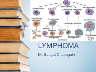
Lymphoma
- 3. Conceptual classification • Lukemia – based on cell type – Acute/ chronic – Myeloid/ lymphoid • Lymphoma – malignant tumor of lymphoreticular tissues: – Hodgkin’s lymphoma/ disease – Non Hodgkin’s lymphoma
- 4. Categories of haematopoeitic & lymphoid neoplasia 1. Myeloid neoplasms – Of myeloid cell lineage – RBC, platelets, granulocytes and monocytes 2. Lymphoid neoplams: – Of lymphoid cell lineage – Lukemias and lymphomas of B,T or NK origin 3. Histiocytic neoplasms – Proliferation of histiocytes – E.g. langerhans cell histicytosis
- 5. Etiology • Heridity – identical twins, family history, genetic disorders • Infections – HTLV-I, EBV, HCV, HIV, etc. • Environmental factors – radiation, carcionogen, etc. • Association with disease of immunity
- 6. Pathogenesis • Genetic damage to single clone of target cells – Proliferation of transformed clone • Chromosomal translocations – Lukemias have translocation of Philadelphia (Ph) chromosome in 70-90% cases with CML (long arm of chromosome 22 to chromosome 9) • Maturition defect – In acute lukemia – AML – maturition defect beyond the myeloblast/ promyelocytic level – ALL – lymphoblast level
- 7. Pathogenesis (Cntd…) • Myelosuppression – Accumulation of leukemic cells suppression of normal hematopoeitic stem cells • Organ infiltration – Cells proliferate primarily in the bone marrow infiltrate other sites • Cytokines – Reed sternberg cells secrete cytokines reactive inflammation (in HL/ HD)
- 8. Lab investigations 1. Peripheral Blood smear – RBC – WBC – Platelet 2. Bone marrow examination – Cellularity – Myeloid cells – Erythropoiesis – Megakaryocytes – Cytogenetics 3. Cytochemistry
- 10. Introduction • Lymphoid cells constitute immune system of the body • Differentiation and maturation occurs in lymphoid tissues • Neoplasm of cells leukemia • Neoplasm of tissues lymphoma • ALL, CLL, NHL, HD
- 11. WHO classification (1999) 1. Hodgkin’s disease 2. Precursor (immature) B- cell malignancies 3. Peripheral (mature) B-cell malignancies 4. Precursor (immature) T-cell malignancies 5. Peripheral (mature) T-cell malignancies 2 & 4 – Blastic type; e.g. ALL 3 & 5 – CLL and other lymphomas
- 12. General comments • Incidence – – NHL (62%); HD (8%); plasma cell disorders (15%); CLL (9%, most common lymphoid leukemia) & ALL (4%) • Diagnosis – – Lymph node biopsy – Blood picture – Bone marrow Biopsy • Staging – Ann Arbor • Ancillary studies – CT scan, PET scan & gallium scan • Immune abnormalities
- 14. Introduction • Primarily arises within LN • Extranodal secondaries • Peak incidence – between 15-35 and after 50s • Male> female • Proliferation of lymphocytes, histiocytes, eosinophils • Diagonistic feature presence of (Dorothy) Reed Sternberg cells
- 15. Reed Sternberg Cell • Diagonistic of Hodgkin’s lymphoma/ disease • Classic RS cell – – Large, with bilobed nucleus (mirror image) / miltilobed sometimes. – Each nuclear lobe contains eosinophilic granular halo (owl eye appearance) • Lacunar type – – Smaller with pericellular space (lacuna) d/t cell shrinkage – Usu. In Nodulo sclerotic HD • Polyploid (popcorn) type – – Larger size with popcorn like lobulated nucleus • Pleomorphic – – Pleomorphic and atypical nuclei.
- 16. Classification of HD Rye classification (1966) 1. Lymphocyte predominance type 2. Nodular sclerosis type 3. Mixed cellularity type 4. Lymphocyte depletion type WHO classification – Nodular lymphocyte predominant HD – Classic HD (includes all 4 types in rye classification)
- 17. Pathophysiology • Proliferation of tumor with only a small proportion of cells are malignant • Mostly normal lymphocytes • RS cells – multinucleated giant cell mutations of T- lymphocytes • Infiltration leads to LN necrosis and fibrosis
- 18. Clinical Features • Painless swelling in one of lymph nodes (usu. Cervical region) with a h/o URTI • Persistent fever, night sweats, fatigue, weight loss • Malaise, pruritis, extreme pain, nerve irritation • Absence of pulse d/t rapid enlargement of the LN • Neck vein engorgement • Enlargement of reteroperitoneal nodes, spleen and liver
- 19. Investigations
- 20. Treatment • Chemotherapy and/or radiation therapy • Antiemetics/ antidiarrhoeal drugs, sedatives • Bone marrow transplant • Blood transfusion • Immunotherapy
- 23. Pathophysiology • Pathophysiologically similar to HD • No RS cells • Most common lymphoma (62%) • Mature/ immature B/T cell malignancies
- 24. Clinical features • Enlarged tonsils and adenoids • Painless rubbery enlargement of lymphatic tissue, usu. Cervical or supraclavicular nodes • In children – cervical nodes affected first with dyspnoea and coughing • An advancing disease • s/s specific to involved structure • Systemic c/c – fatigue, malaise, weight loss, fever, etc
- 25. Investigations • Blood count – anaemia • Uric acid level – elevated/ normal • Sr. calcium – elevated • Imaging – bone & CXR, lymphangiography, liver and spleen scans, CT abdomen f/s/o metastasis • LN biopsy – cell type revealed • Biopsy of tonsil, bone marrow, liver or spleen malignant cells
- 26. Treatment • Radiation – esp in early and localized lesions • Total nodal irradiation • Chemotherapy • Bone marrow transplant • Blood transfusion (supportive)