SlideShare una empresa de Scribd logo
1 de 3
Descargar para leer sin conexión
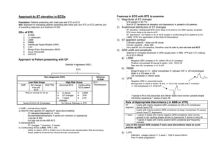
Approach to ST elevation in ECGs                                                                  Features in ECG with STE to examine
                                                                                                  1) Magnitude of ST changes
Population: Patients presenting with chest pain and STE on ECG                                       -   STE greater in MI PTs
Aim: Approach to managing patients presenting with chest pain and STE on ECG with the aim            -   Sum of ST deviations (ie elevation and depression) is greated in MI patients
of expediting diagnosis and treatment of AMI                                                      2) Anatomical distribution of ST changes
                                                                                                     -   ST elevation: widespread STE more likely to be due to non-AMI causes, localised
DDx of STE:                                                                                              STE more likely to be due to aCS
     1.     STEMI                                                                                    -   ST depression: not helpful in dx of ACS except in confounding ECG patterns (LVH,
     2.     LV aneurysm                                                                                  LBBB, VPR) thru application of the Rule of Discordance
     3.     LBBB                                                                                  3) ST segment contour
     4.     RBBB                                                                                     -   Concave upwards – likely non-MI cause
     5.     Ventricular Paced Rhythm (VPR)                                                           -   Convex upwards – likely AMI
     6.     LVH                                                                                      -   Very specific but not sensitive, therefore used to rule in, but not rule out ACS
     7.     Benign Early Repolarisation (BER)                                                     4) QRS width and amplitude
     8.     Acute Pericarditis                                                                       -   widened or increased amplitude of QRS usually seen in BBB, VPR and LVH, making
     9.     NSIVCD                                                                                       dx of ACS difficult
                                                                                                     a) LBBB
Approach to Patient presenting with CP                                                                   -    Negative QRS complex in V1 (either QS or rS complex)
                                                                                                         -    Positive monophasic R waves in leads I, aVL, V5 & V6
                                                    Stabilise if necessary (ABC)                         -    May see QS complexes in III & aVF
                                                               ECG                                   b) RBBB
                                                                                                         -    Broad R wave in V1 – either monophasic R, biphasic rSR’ or qR morphologies
                                                                                                         -    Wide S or RS wave in V6
                                                                                                         -    QS complexes in inferior leads
                             Non-diagnostic ECG                                       Obvious
                                                                                       STEMI         c) VPR
                                                                                                         -    Negative QRS in precordial leads
             Low Risk Group                         High Risk Group                PCI /
                                                                                                         -    Monophasic R wave in leads I & aVL +/- V5 & V6, usually a/w T inversion
  NSR           No change        NSSTTW       Abnormal ECG      Confounding        Thrombolysis
                                                                                                         -    +/- QS complexes in II, III & AVF
                 from old                                       ECG pattern
                   ECG
          Rely on clinical hx for dx      •      Q waves         •    LBBB
                                          •      ST changes      •    VPR                                -    T waves in Rt to mid precordial and inferior leads have convex upwards shape
                                          •      T wave          •    LVH                                     mimicking hyperacute T waves of early MI
                                              changes
     Serial ECG & CE if indicated               Dx Clinical Pathway in STE                               Rule of Appropriate Discordance ( in BBB or VPR)
                                                                                                         ST         Leads with mainly negative QRS complexes (ie QS or rS complexes)
1) NSR – normal sinus rhythm                                                                             segment    should have STE
2) NSSTW (Non-specific ST segment/T wave abnormalities)                                                             Leads with mainly positive QRS complexes (ie large monophasic R waves)
     -   ST elevation/depression of <1mm                                                                            should have ST depression
     -   Blunted/flattened/biphasic T waves w/o inversion or hyperacuity                                 T wave     T waves in leads with mainly negative QRS complexes have convex
     -   Low risk of AMI                                                                                            upward or tall vaulting shapes similar to hyperacute T waves in early MI
     -   but high risk of non-AMI ACS                                                                               T waves in leads with mainly positive QRS complexes are frequently
3) Abnormal ECG                                                                                                     inverted
     -   ST changes, T inversion, Q waves                                                                Loss of this normal QRS complex-T wave axes discordance imply an acute
4) Confounding ECGs (LBBB, VPR, LVH)                                                                     process eg AMI
     -   ability to detect ACS is limited due to the abnormal repolarisation that accompany
         these patterns of abnormal intraventricular conductions                                     d) LVH
                                                                                                         -    Definition: voltage criteria V1 S wave + V5/6 R wave ≥35mm
                                                                                                         -    Poor R wave progression
-    V1 & V2: loss of septal R wave in Rt to mid precordial leads, usually resulting in
             QS pattern (ie mostly negative); usually a/w concave pattern STE and prominent
             T waves
        -    Leads I, aVL, V5 & V6: ST depression with downsloping ST segment; prominent
             R waves; assymetrical (gradual downsloping initial limb with abrupt return to
             baseline), biphasic or inverted T waves

Diagnostic Clinical Pathway in STE
Overview
   1)   Identify chest pain patients with STE
   2)   For STE in LVH, BBB & VPR (ie confounding ECG patterns)
        -     Use highly specific criterias to rule in AMI
        -     If negative, use serial ECG to f/u the patient for dynamic changes suggestive of
              AMI
   3) For STE in uncomplicated ECGs
        -     Look for specific features to rule in AMI
        -     If negative, use serial ECG to f/u the patient for dynamic changes suggestive of
              AMI
   Use of specific criterias aims to diagnose AMI quickly based on a single ECG so as to
   allow for rapid diagnosis and institution of thrombolysis/PCI.


                                                                                                                                         Digitally signed by DR WANA HLA SHWE
                                                                                                                                         DN: cn=DR WANA HLA SHWE, c=MY, o=UCSI
                                                                                                                                         University, School of Medicine, KT-Campus, Terengganu,
                                                                                                                                         ou=Internal Medicine Group, email=wunna.
                                                                                                                                         hlashwe@gmail.com
                                                                                                                                         Reason: This document is for UCSI University, School of
                                                                                                                                         Medicine students.
                                                                                                                                         Date: 2009.03.05 08:55:25 +08'00'


                                                                                                  Based on Clinical Decision-making in Adult Chest Pain Patients with
                                                                                                  Electrocardiographic ST-segment Elevation: STEMI vs Non-AMI Causes of ST-
                                                                                                  Segment Abnormality. William J Brady, Andrew Homer. Emergency Medicine
CP patient
                                                                        - Perform ECG


                                                                        STE
                                                                        ≥ 1mm in ≥2 contiguous leads

                                                                                                             a) VPR          Sgarbossa Criteria – to rule in AMI in VPR
Aim: Predict AMI in ECGs with confounding patterns (ie LVH, BBB, VPR)


                                                                        1) Widened QRS                                       (any one of 3)


                                                                                                       Yes
                                                                                                                               STE ≥5mm with negative QRS
                                                                        complex?                                                                                                              Likely MI – initiate Rx
                                                                                                                               STE ≥1mm with positive QRS                        Positive
                                                                                                                               ST depression ≥1mm with negative QRS              Negative     Serial ECG to look for dynamic changes
                                                                               No                            b) LBBB         Sgarbossa Criteria – to rule in AMI in LBBB                         ≥0.2mm change in STE in single lead OR       Positive      Likely MI
                                                                                                                             (any one of 3)                                                      ≥0.1mm change in STE in 2 leads              Negative      No ECG
                                                                                                                               STE ≥1mm with positive QRS                                                                                                   evidence of
                                                                                                                               ST depression ≥1mm in V1,2 or 3                                                                                              MI
                                                                                                                               STE ≥5mm with negative QRS
                                                                                                             Sgarbossa Criteria is based on principle of appropriate discordance to look for abnormal ST segment changes
                                                                                                               It has low sensitivity & specificity, hence it is used to rule in PTs with MI in VPR/LBBB so as to allow for early initiation of Rx
                                                                                                               Does not rule out MI in VPR/LBBB, hence the need for serial ECG monitoring in PTs who do not meet Sgarbossa criteria. Aim is to look for dynamic changes
                                                                                                               that indicates ischaemic induced changes of an evolving AMI, as ST & T wave abnormalities are assumed to be temporally static in VPR/LBBB


                                                                        2) Large amplitude                   LVH                             STE                   Concave up           Serial ECG q4hr to look for dynamic changes
                                                                                                                                                                                          0.05mm change in STE or depression
                                                                                                       Yes


                                                                        QRS                                  SV1 + RV 5/6 ≥ 35mm             morphology                                   Q wave devt
                                                                                                                                             High specificity                                                                                 Positive      Likely MI
                                                                                                                                                                                          T inversion in ≥2 anatomically continuous           Negative      No ECG
                                                                                                                                             but low sensitivity
                                                                               No                                                                                                         leads                                                             evidence of
                                                                                                                                             - used to rule in
                                                                                                                                                                                          Loss of ST segment concavity                                      MI
                                                                                                                                             AMI, not to rule
                                                                                                                                                                   Convex up or         Suspicious for MI
                                                                                                                                             out AMI
                                                                                                                                                                   obliquely flat


                                                                                                                                                                      ST segment morphology             Convex upwards or obliquely flat
                                                                        3) STE in                            Exclude AMI                                                                                - specific but not sensitive, hence
                                                                                                       Yes


                                                                                                                                                                                                        used to rule in AMI                       Suspicious for AMI
                                                                        uncomplicated ECGs
Aim: Predict AMI in uncomplicated ECGs


                                                                                                                                                                      Reciprocal ST depression          Positive
                                                                                                                                                                      (in absence of LVH, BBB,          - specific but not sensitive, hence
                                                                                                                                                                      VPR)                              used to rule in AMI

                                                                                                             Exclude Benign Early Repolarisation                           J point notching present                                              Suspicious for AMI


                                                                                                             Exclude Acute Pericarditis                                    PR depression OR                                                      Suspicious for AMI
                                                                                                                                                                           Ratio of ST height at J point in V6 to T wave
                                                                                                                                                                           apex height in V6 ≥0.25


                                                                                                             Exclude AMI                                                   Ratio of sum off T wave apex heights in V1-4                          Suspicious for AMI
                                                                                                                                                                           to sum of QRS apex heights in V1-4 >0.22


                                                                                                             Serial ECG                                                                                    Positive         Likely MI
                                                                                                               ≥0.05mm change in ST elevation or depression                                                Negative         No ECG
                                                                                                               Q wave devt                                                                                                  evidence of MI
                                                                                                               T inversion in ≥2 anatomically continuous leads
                                                                                                               Loss of ST segment concavity

Más contenido relacionado

Destacado

Normal labour, first stage by Yin Moe
Normal labour, first stage by Yin MoeNormal labour, first stage by Yin Moe
Normal labour, first stage by Yin MoeDr. Rubz
 
Diabetes mellitus summary
Diabetes mellitus summaryDiabetes mellitus summary
Diabetes mellitus summaryDr. Rubz
 
Hyperthyroidism summary
Hyperthyroidism summaryHyperthyroidism summary
Hyperthyroidism summaryDr. Rubz
 
Nephrotic syndrome summary
Nephrotic syndrome summaryNephrotic syndrome summary
Nephrotic syndrome summaryDr. Rubz
 
Regional Overview in HIV by Steve Kraus
Regional Overview in HIV by Steve KrausRegional Overview in HIV by Steve Kraus
Regional Overview in HIV by Steve KrausDr. Rubz
 
Hypertension summary
Hypertension summaryHypertension summary
Hypertension summaryDr. Rubz
 
Bohomolets 3rd year Surgery Tumors
Bohomolets 3rd year Surgery TumorsBohomolets 3rd year Surgery Tumors
Bohomolets 3rd year Surgery TumorsDr. Rubz
 
Shock summary
Shock summaryShock summary
Shock summaryDr. Rubz
 
Game Changer by Dr Shaari Ngadiman
Game Changer by Dr Shaari NgadimanGame Changer by Dr Shaari Ngadiman
Game Changer by Dr Shaari NgadimanDr. Rubz
 
Acromegaly Summary
Acromegaly SummaryAcromegaly Summary
Acromegaly SummaryDr. Rubz
 
Meningitis by Prof Khin
Meningitis by Prof KhinMeningitis by Prof Khin
Meningitis by Prof KhinDr. Rubz
 
Peadiatric pneumonia by Teo Yan
Peadiatric pneumonia by  Teo YanPeadiatric pneumonia by  Teo Yan
Peadiatric pneumonia by Teo YanDr. Rubz
 
Atrial fibrillation management summary
Atrial fibrillation management summaryAtrial fibrillation management summary
Atrial fibrillation management summaryDr. Rubz
 
Testicular torsion by Dr Teo
Testicular torsion by Dr TeoTesticular torsion by Dr Teo
Testicular torsion by Dr TeoDr. Rubz
 
Other scrotal swelling by Dr. Teo
Other scrotal swelling by Dr. TeoOther scrotal swelling by Dr. Teo
Other scrotal swelling by Dr. TeoDr. Rubz
 
Legislative and Ethical Framework of Organ Donation and Transplantation in Ma...
Legislative and Ethical Framework of Organ Donation and Transplantation in Ma...Legislative and Ethical Framework of Organ Donation and Transplantation in Ma...
Legislative and Ethical Framework of Organ Donation and Transplantation in Ma...Dr Hirman Ismail
 
Gout summary
Gout summaryGout summary
Gout summaryDr. Rubz
 
Ventral hernia by Dr Teo
Ventral hernia by Dr TeoVentral hernia by Dr Teo
Ventral hernia by Dr TeoDr. Rubz
 
Psychosexual disorders
Psychosexual disorders Psychosexual disorders
Psychosexual disorders Dr. Rubz
 

Destacado (20)

Normal labour, first stage by Yin Moe
Normal labour, first stage by Yin MoeNormal labour, first stage by Yin Moe
Normal labour, first stage by Yin Moe
 
Diabetes mellitus summary
Diabetes mellitus summaryDiabetes mellitus summary
Diabetes mellitus summary
 
Hyperthyroidism summary
Hyperthyroidism summaryHyperthyroidism summary
Hyperthyroidism summary
 
Long case 17.5.14
Long case 17.5.14Long case 17.5.14
Long case 17.5.14
 
Nephrotic syndrome summary
Nephrotic syndrome summaryNephrotic syndrome summary
Nephrotic syndrome summary
 
Regional Overview in HIV by Steve Kraus
Regional Overview in HIV by Steve KrausRegional Overview in HIV by Steve Kraus
Regional Overview in HIV by Steve Kraus
 
Hypertension summary
Hypertension summaryHypertension summary
Hypertension summary
 
Bohomolets 3rd year Surgery Tumors
Bohomolets 3rd year Surgery TumorsBohomolets 3rd year Surgery Tumors
Bohomolets 3rd year Surgery Tumors
 
Shock summary
Shock summaryShock summary
Shock summary
 
Game Changer by Dr Shaari Ngadiman
Game Changer by Dr Shaari NgadimanGame Changer by Dr Shaari Ngadiman
Game Changer by Dr Shaari Ngadiman
 
Acromegaly Summary
Acromegaly SummaryAcromegaly Summary
Acromegaly Summary
 
Meningitis by Prof Khin
Meningitis by Prof KhinMeningitis by Prof Khin
Meningitis by Prof Khin
 
Peadiatric pneumonia by Teo Yan
Peadiatric pneumonia by  Teo YanPeadiatric pneumonia by  Teo Yan
Peadiatric pneumonia by Teo Yan
 
Atrial fibrillation management summary
Atrial fibrillation management summaryAtrial fibrillation management summary
Atrial fibrillation management summary
 
Testicular torsion by Dr Teo
Testicular torsion by Dr TeoTesticular torsion by Dr Teo
Testicular torsion by Dr Teo
 
Other scrotal swelling by Dr. Teo
Other scrotal swelling by Dr. TeoOther scrotal swelling by Dr. Teo
Other scrotal swelling by Dr. Teo
 
Legislative and Ethical Framework of Organ Donation and Transplantation in Ma...
Legislative and Ethical Framework of Organ Donation and Transplantation in Ma...Legislative and Ethical Framework of Organ Donation and Transplantation in Ma...
Legislative and Ethical Framework of Organ Donation and Transplantation in Ma...
 
Gout summary
Gout summaryGout summary
Gout summary
 
Ventral hernia by Dr Teo
Ventral hernia by Dr TeoVentral hernia by Dr Teo
Ventral hernia by Dr Teo
 
Psychosexual disorders
Psychosexual disorders Psychosexual disorders
Psychosexual disorders
 

Similar a Approach to ST elevation in ECG sumary

Broad complex tachycardia
Broad complex tachycardiaBroad complex tachycardia
Broad complex tachycardiaJeya Balaji
 
Bundle branch blocks by Dr Sujith Chadala
Bundle branch blocks by Dr Sujith ChadalaBundle branch blocks by Dr Sujith Chadala
Bundle branch blocks by Dr Sujith ChadalaDr Sujith Chadala
 
Cardiology 2.1. ECG or EKG - by Dr. Farjad Ikram
Cardiology 2.1. ECG or EKG - by Dr. Farjad IkramCardiology 2.1. ECG or EKG - by Dr. Farjad Ikram
Cardiology 2.1. ECG or EKG - by Dr. Farjad IkramFarjad Ikram
 
Vt in normal and abnormal hearts my ppt copy
Vt in normal and abnormal hearts my ppt   copyVt in normal and abnormal hearts my ppt   copy
Vt in normal and abnormal hearts my ppt copyRahul Chalwade
 
A Guide TO ECG Interpretation
A Guide TO ECG InterpretationA Guide TO ECG Interpretation
A Guide TO ECG Interpretationmeducationdotnet
 
Beginners Guide for ECG Interpretation
Beginners Guide for ECG InterpretationBeginners Guide for ECG Interpretation
Beginners Guide for ECG Interpretationmeducationdotnet
 
Myocardial infarction (MI) ecg localisation
Myocardial infarction (MI) ecg localisationMyocardial infarction (MI) ecg localisation
Myocardial infarction (MI) ecg localisationMalleswara rao Dangeti
 
3rd part ECG Basics QRS complex Dr Salah Mabrouk Khallaf
3rd part ECG Basics  QRS complex Dr Salah Mabrouk Khallaf3rd part ECG Basics  QRS complex Dr Salah Mabrouk Khallaf
3rd part ECG Basics QRS complex Dr Salah Mabrouk KhallafDr Salah Mabrouk Khallaf
 
Wide complex tachycardia drneeraj
Wide complex tachycardia drneerajWide complex tachycardia drneeraj
Wide complex tachycardia drneerajDrNeeraj Nirala
 
GRAPHICS-WIDE COMPLEX TACHYCARDIAS- APPROACH.pptx
GRAPHICS-WIDE COMPLEX TACHYCARDIAS- APPROACH.pptxGRAPHICS-WIDE COMPLEX TACHYCARDIAS- APPROACH.pptx
GRAPHICS-WIDE COMPLEX TACHYCARDIAS- APPROACH.pptxAditya Madhavpeddi
 

Similar a Approach to ST elevation in ECG sumary (20)

WIDE QRS TACHYCARDIA
WIDE  QRS TACHYCARDIAWIDE  QRS TACHYCARDIA
WIDE QRS TACHYCARDIA
 
Broad complex tachycardia
Broad complex tachycardiaBroad complex tachycardia
Broad complex tachycardia
 
All info about ecg
All info about ecgAll info about ecg
All info about ecg
 
Ecg
EcgEcg
Ecg
 
ECG Interpretation
ECG InterpretationECG Interpretation
ECG Interpretation
 
E c g interpretation
E c g interpretationE c g interpretation
E c g interpretation
 
Bundle branch blocks by Dr Sujith Chadala
Bundle branch blocks by Dr Sujith ChadalaBundle branch blocks by Dr Sujith Chadala
Bundle branch blocks by Dr Sujith Chadala
 
ECG: Wolff-Parkinson-White syndrome
ECG: Wolff-Parkinson-White syndromeECG: Wolff-Parkinson-White syndrome
ECG: Wolff-Parkinson-White syndrome
 
Ecg made easy
Ecg made easyEcg made easy
Ecg made easy
 
Cardiology 2.1. ECG or EKG - by Dr. Farjad Ikram
Cardiology 2.1. ECG or EKG - by Dr. Farjad IkramCardiology 2.1. ECG or EKG - by Dr. Farjad Ikram
Cardiology 2.1. ECG or EKG - by Dr. Farjad Ikram
 
LBBB
LBBBLBBB
LBBB
 
ECG: Wide Complex Tachycardia
ECG: Wide Complex TachycardiaECG: Wide Complex Tachycardia
ECG: Wide Complex Tachycardia
 
Vt in normal and abnormal hearts my ppt copy
Vt in normal and abnormal hearts my ppt   copyVt in normal and abnormal hearts my ppt   copy
Vt in normal and abnormal hearts my ppt copy
 
A Guide TO ECG Interpretation
A Guide TO ECG InterpretationA Guide TO ECG Interpretation
A Guide TO ECG Interpretation
 
Beginners Guide for ECG Interpretation
Beginners Guide for ECG InterpretationBeginners Guide for ECG Interpretation
Beginners Guide for ECG Interpretation
 
Myocardial infarction (MI) ecg localisation
Myocardial infarction (MI) ecg localisationMyocardial infarction (MI) ecg localisation
Myocardial infarction (MI) ecg localisation
 
3rd part ECG Basics QRS complex Dr Salah Mabrouk Khallaf
3rd part ECG Basics  QRS complex Dr Salah Mabrouk Khallaf3rd part ECG Basics  QRS complex Dr Salah Mabrouk Khallaf
3rd part ECG Basics QRS complex Dr Salah Mabrouk Khallaf
 
SD BRUGADA ECG 2014.pdf
SD BRUGADA ECG 2014.pdfSD BRUGADA ECG 2014.pdf
SD BRUGADA ECG 2014.pdf
 
Wide complex tachycardia drneeraj
Wide complex tachycardia drneerajWide complex tachycardia drneeraj
Wide complex tachycardia drneeraj
 
GRAPHICS-WIDE COMPLEX TACHYCARDIAS- APPROACH.pptx
GRAPHICS-WIDE COMPLEX TACHYCARDIAS- APPROACH.pptxGRAPHICS-WIDE COMPLEX TACHYCARDIAS- APPROACH.pptx
GRAPHICS-WIDE COMPLEX TACHYCARDIAS- APPROACH.pptx
 

Más de Dr. Rubz

HIV discrimination among health providers in Malaysia by Dr Rubz
HIV discrimination among health providers in Malaysia by Dr RubzHIV discrimination among health providers in Malaysia by Dr Rubz
HIV discrimination among health providers in Malaysia by Dr RubzDr. Rubz
 
Pre and post HIV counseling (VCT)
Pre and post HIV counseling (VCT)Pre and post HIV counseling (VCT)
Pre and post HIV counseling (VCT)Dr. Rubz
 
Testicular cancer for public awareness by Dr Rubz
Testicular cancer for public awareness by Dr RubzTesticular cancer for public awareness by Dr Rubz
Testicular cancer for public awareness by Dr RubzDr. Rubz
 
Prostate cancer for public awareness by DR RUBZ
Prostate cancer for public awareness by DR RUBZProstate cancer for public awareness by DR RUBZ
Prostate cancer for public awareness by DR RUBZDr. Rubz
 
Breast Cancer for public awareness by Dr Rubz
Breast Cancer for public awareness by Dr  RubzBreast Cancer for public awareness by Dr  Rubz
Breast Cancer for public awareness by Dr RubzDr. Rubz
 
Sex work presentation 9.18.13a
Sex work presentation 9.18.13aSex work presentation 9.18.13a
Sex work presentation 9.18.13aDr. Rubz
 
Hernia by Dr. Rubzzz
Hernia by Dr. RubzzzHernia by Dr. Rubzzz
Hernia by Dr. RubzzzDr. Rubz
 
Benign breast disease by Dr. Kong
Benign breast disease by Dr. KongBenign breast disease by Dr. Kong
Benign breast disease by Dr. KongDr. Rubz
 
Breast CA by Dr. Celine Tey
Breast CA by Dr. Celine TeyBreast CA by Dr. Celine Tey
Breast CA by Dr. Celine TeyDr. Rubz
 
Uk malaria treatment guideline
Uk malaria treatment guidelineUk malaria treatment guideline
Uk malaria treatment guidelineDr. Rubz
 
Tuberculosis summary
Tuberculosis summaryTuberculosis summary
Tuberculosis summaryDr. Rubz
 
Short case approach to acromegaly summary
Short case approach to acromegaly summaryShort case approach to acromegaly summary
Short case approach to acromegaly summaryDr. Rubz
 
Short case approach to cushing summary
Short case approach to cushing summaryShort case approach to cushing summary
Short case approach to cushing summaryDr. Rubz
 
Short case approach to parkinson's dz summary
Short case approach to parkinson's dz summaryShort case approach to parkinson's dz summary
Short case approach to parkinson's dz summaryDr. Rubz
 
Short case approach to speech analysis summary
Short case approach to speech analysis summaryShort case approach to speech analysis summary
Short case approach to speech analysis summaryDr. Rubz
 
Stroke ( concise long case approach ) summary
Stroke ( concise long case approach ) summaryStroke ( concise long case approach ) summary
Stroke ( concise long case approach ) summaryDr. Rubz
 
Thrombocytopenia summary
Thrombocytopenia summaryThrombocytopenia summary
Thrombocytopenia summaryDr. Rubz
 
Scleroderma long case summary
Scleroderma long case  summaryScleroderma long case  summary
Scleroderma long case summaryDr. Rubz
 
Rheumatoid arthritis summary
Rheumatoid arthritis summaryRheumatoid arthritis summary
Rheumatoid arthritis summaryDr. Rubz
 
Pyrexia of unknown summary
Pyrexia of unknown summaryPyrexia of unknown summary
Pyrexia of unknown summaryDr. Rubz
 

Más de Dr. Rubz (20)

HIV discrimination among health providers in Malaysia by Dr Rubz
HIV discrimination among health providers in Malaysia by Dr RubzHIV discrimination among health providers in Malaysia by Dr Rubz
HIV discrimination among health providers in Malaysia by Dr Rubz
 
Pre and post HIV counseling (VCT)
Pre and post HIV counseling (VCT)Pre and post HIV counseling (VCT)
Pre and post HIV counseling (VCT)
 
Testicular cancer for public awareness by Dr Rubz
Testicular cancer for public awareness by Dr RubzTesticular cancer for public awareness by Dr Rubz
Testicular cancer for public awareness by Dr Rubz
 
Prostate cancer for public awareness by DR RUBZ
Prostate cancer for public awareness by DR RUBZProstate cancer for public awareness by DR RUBZ
Prostate cancer for public awareness by DR RUBZ
 
Breast Cancer for public awareness by Dr Rubz
Breast Cancer for public awareness by Dr  RubzBreast Cancer for public awareness by Dr  Rubz
Breast Cancer for public awareness by Dr Rubz
 
Sex work presentation 9.18.13a
Sex work presentation 9.18.13aSex work presentation 9.18.13a
Sex work presentation 9.18.13a
 
Hernia by Dr. Rubzzz
Hernia by Dr. RubzzzHernia by Dr. Rubzzz
Hernia by Dr. Rubzzz
 
Benign breast disease by Dr. Kong
Benign breast disease by Dr. KongBenign breast disease by Dr. Kong
Benign breast disease by Dr. Kong
 
Breast CA by Dr. Celine Tey
Breast CA by Dr. Celine TeyBreast CA by Dr. Celine Tey
Breast CA by Dr. Celine Tey
 
Uk malaria treatment guideline
Uk malaria treatment guidelineUk malaria treatment guideline
Uk malaria treatment guideline
 
Tuberculosis summary
Tuberculosis summaryTuberculosis summary
Tuberculosis summary
 
Short case approach to acromegaly summary
Short case approach to acromegaly summaryShort case approach to acromegaly summary
Short case approach to acromegaly summary
 
Short case approach to cushing summary
Short case approach to cushing summaryShort case approach to cushing summary
Short case approach to cushing summary
 
Short case approach to parkinson's dz summary
Short case approach to parkinson's dz summaryShort case approach to parkinson's dz summary
Short case approach to parkinson's dz summary
 
Short case approach to speech analysis summary
Short case approach to speech analysis summaryShort case approach to speech analysis summary
Short case approach to speech analysis summary
 
Stroke ( concise long case approach ) summary
Stroke ( concise long case approach ) summaryStroke ( concise long case approach ) summary
Stroke ( concise long case approach ) summary
 
Thrombocytopenia summary
Thrombocytopenia summaryThrombocytopenia summary
Thrombocytopenia summary
 
Scleroderma long case summary
Scleroderma long case  summaryScleroderma long case  summary
Scleroderma long case summary
 
Rheumatoid arthritis summary
Rheumatoid arthritis summaryRheumatoid arthritis summary
Rheumatoid arthritis summary
 
Pyrexia of unknown summary
Pyrexia of unknown summaryPyrexia of unknown summary
Pyrexia of unknown summary
 

Approach to ST elevation in ECG sumary

  • 1. Approach to ST elevation in ECGs Features in ECG with STE to examine 1) Magnitude of ST changes Population: Patients presenting with chest pain and STE on ECG - STE greater in MI PTs Aim: Approach to managing patients presenting with chest pain and STE on ECG with the aim - Sum of ST deviations (ie elevation and depression) is greated in MI patients of expediting diagnosis and treatment of AMI 2) Anatomical distribution of ST changes - ST elevation: widespread STE more likely to be due to non-AMI causes, localised DDx of STE: STE more likely to be due to aCS 1. STEMI - ST depression: not helpful in dx of ACS except in confounding ECG patterns (LVH, 2. LV aneurysm LBBB, VPR) thru application of the Rule of Discordance 3. LBBB 3) ST segment contour 4. RBBB - Concave upwards – likely non-MI cause 5. Ventricular Paced Rhythm (VPR) - Convex upwards – likely AMI 6. LVH - Very specific but not sensitive, therefore used to rule in, but not rule out ACS 7. Benign Early Repolarisation (BER) 4) QRS width and amplitude 8. Acute Pericarditis - widened or increased amplitude of QRS usually seen in BBB, VPR and LVH, making 9. NSIVCD dx of ACS difficult a) LBBB Approach to Patient presenting with CP - Negative QRS complex in V1 (either QS or rS complex) - Positive monophasic R waves in leads I, aVL, V5 & V6 Stabilise if necessary (ABC) - May see QS complexes in III & aVF ECG b) RBBB - Broad R wave in V1 – either monophasic R, biphasic rSR’ or qR morphologies - Wide S or RS wave in V6 - QS complexes in inferior leads Non-diagnostic ECG Obvious STEMI c) VPR - Negative QRS in precordial leads Low Risk Group High Risk Group PCI / - Monophasic R wave in leads I & aVL +/- V5 & V6, usually a/w T inversion NSR No change NSSTTW Abnormal ECG Confounding Thrombolysis - +/- QS complexes in II, III & AVF from old ECG pattern ECG Rely on clinical hx for dx • Q waves • LBBB • ST changes • VPR - T waves in Rt to mid precordial and inferior leads have convex upwards shape • T wave • LVH mimicking hyperacute T waves of early MI changes Serial ECG & CE if indicated Dx Clinical Pathway in STE Rule of Appropriate Discordance ( in BBB or VPR) ST Leads with mainly negative QRS complexes (ie QS or rS complexes) 1) NSR – normal sinus rhythm segment should have STE 2) NSSTW (Non-specific ST segment/T wave abnormalities) Leads with mainly positive QRS complexes (ie large monophasic R waves) - ST elevation/depression of <1mm should have ST depression - Blunted/flattened/biphasic T waves w/o inversion or hyperacuity T wave T waves in leads with mainly negative QRS complexes have convex - Low risk of AMI upward or tall vaulting shapes similar to hyperacute T waves in early MI - but high risk of non-AMI ACS T waves in leads with mainly positive QRS complexes are frequently 3) Abnormal ECG inverted - ST changes, T inversion, Q waves Loss of this normal QRS complex-T wave axes discordance imply an acute 4) Confounding ECGs (LBBB, VPR, LVH) process eg AMI - ability to detect ACS is limited due to the abnormal repolarisation that accompany these patterns of abnormal intraventricular conductions d) LVH - Definition: voltage criteria V1 S wave + V5/6 R wave ≥35mm - Poor R wave progression
  • 2. - V1 & V2: loss of septal R wave in Rt to mid precordial leads, usually resulting in QS pattern (ie mostly negative); usually a/w concave pattern STE and prominent T waves - Leads I, aVL, V5 & V6: ST depression with downsloping ST segment; prominent R waves; assymetrical (gradual downsloping initial limb with abrupt return to baseline), biphasic or inverted T waves Diagnostic Clinical Pathway in STE Overview 1) Identify chest pain patients with STE 2) For STE in LVH, BBB & VPR (ie confounding ECG patterns) - Use highly specific criterias to rule in AMI - If negative, use serial ECG to f/u the patient for dynamic changes suggestive of AMI 3) For STE in uncomplicated ECGs - Look for specific features to rule in AMI - If negative, use serial ECG to f/u the patient for dynamic changes suggestive of AMI Use of specific criterias aims to diagnose AMI quickly based on a single ECG so as to allow for rapid diagnosis and institution of thrombolysis/PCI. Digitally signed by DR WANA HLA SHWE DN: cn=DR WANA HLA SHWE, c=MY, o=UCSI University, School of Medicine, KT-Campus, Terengganu, ou=Internal Medicine Group, email=wunna. hlashwe@gmail.com Reason: This document is for UCSI University, School of Medicine students. Date: 2009.03.05 08:55:25 +08'00' Based on Clinical Decision-making in Adult Chest Pain Patients with Electrocardiographic ST-segment Elevation: STEMI vs Non-AMI Causes of ST- Segment Abnormality. William J Brady, Andrew Homer. Emergency Medicine
  • 3. CP patient - Perform ECG STE ≥ 1mm in ≥2 contiguous leads a) VPR Sgarbossa Criteria – to rule in AMI in VPR Aim: Predict AMI in ECGs with confounding patterns (ie LVH, BBB, VPR) 1) Widened QRS (any one of 3) Yes STE ≥5mm with negative QRS complex? Likely MI – initiate Rx STE ≥1mm with positive QRS Positive ST depression ≥1mm with negative QRS Negative Serial ECG to look for dynamic changes No b) LBBB Sgarbossa Criteria – to rule in AMI in LBBB ≥0.2mm change in STE in single lead OR Positive Likely MI (any one of 3) ≥0.1mm change in STE in 2 leads Negative No ECG STE ≥1mm with positive QRS evidence of ST depression ≥1mm in V1,2 or 3 MI STE ≥5mm with negative QRS Sgarbossa Criteria is based on principle of appropriate discordance to look for abnormal ST segment changes It has low sensitivity & specificity, hence it is used to rule in PTs with MI in VPR/LBBB so as to allow for early initiation of Rx Does not rule out MI in VPR/LBBB, hence the need for serial ECG monitoring in PTs who do not meet Sgarbossa criteria. Aim is to look for dynamic changes that indicates ischaemic induced changes of an evolving AMI, as ST & T wave abnormalities are assumed to be temporally static in VPR/LBBB 2) Large amplitude LVH STE Concave up Serial ECG q4hr to look for dynamic changes 0.05mm change in STE or depression Yes QRS SV1 + RV 5/6 ≥ 35mm morphology Q wave devt High specificity Positive Likely MI T inversion in ≥2 anatomically continuous Negative No ECG but low sensitivity No leads evidence of - used to rule in Loss of ST segment concavity MI AMI, not to rule Convex up or Suspicious for MI out AMI obliquely flat ST segment morphology Convex upwards or obliquely flat 3) STE in Exclude AMI - specific but not sensitive, hence Yes used to rule in AMI Suspicious for AMI uncomplicated ECGs Aim: Predict AMI in uncomplicated ECGs Reciprocal ST depression Positive (in absence of LVH, BBB, - specific but not sensitive, hence VPR) used to rule in AMI Exclude Benign Early Repolarisation J point notching present Suspicious for AMI Exclude Acute Pericarditis PR depression OR Suspicious for AMI Ratio of ST height at J point in V6 to T wave apex height in V6 ≥0.25 Exclude AMI Ratio of sum off T wave apex heights in V1-4 Suspicious for AMI to sum of QRS apex heights in V1-4 >0.22 Serial ECG Positive Likely MI ≥0.05mm change in ST elevation or depression Negative No ECG Q wave devt evidence of MI T inversion in ≥2 anatomically continuous leads Loss of ST segment concavity