Alloimmune factors in recurrent pregnancy loss
•Descargar como PPT, PDF•
47 recomendaciones•7,112 vistas

This was a presentation at FOGSI workshp on RPL at Chandrapur on alloimmune factors Dr Rajesh Gajbhiye

Recomendados
Más contenido relacionado
La actualidad más candente
La actualidad más candente (20)
Destacado
Destacado (20)
Similar a Alloimmune factors in recurrent pregnancy loss
Similar a Alloimmune factors in recurrent pregnancy loss (20)
Más de Rajesh Gajbhiye
Más de Rajesh Gajbhiye (12)
Último
Último (20)
Alloimmune factors in recurrent pregnancy loss
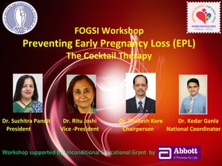
- 1. FOGSI Workshop Preventing Early Pregnancy Loss (EPL) The Cocktail Therapy Dr. Suchitra Pandit Dr. Ritu Joshi Dr. Shailesh Kore Dr. Kedar Ganla President Vice -President Chairperson National Coordinator Workshop supported by Unconditional Educational Grant by
- 2. Alloimmune Background in Early Pregnancy Loss (EPL) Dr Rajesh Gajbhiye Nagpur
- 3. Introduction • About 50% of pregnancies are lost after conception • About 15 % are lost after clinically detectable pregnancies • Causes are not known in more than 50% of cases of RPL • Such large unexplained causes has fuelled interest in immunological causes.
- 4. Pregnancy is a semi-graft 50% of the antigens are foreign
- 5. Immunomodulation during pregnancy Embryo / Fetal antigens produce two types of antibodies: • T Helper I Cell response • T Helper II Cell response
- 6. Immune reaction during pregnancy Fetus with Paternal antigens T helper 1 cell response Abortion of The Fetus T helper 2 cell response Protection of The Fetus
- 7. Embryo / Fetus T helper 1 cell response activated Tumor Necrosis Factor ά Interleukin2 Natural killer Cells Lymphokine Activated Killer Cells Abortion of Fetus Cascade Reaction
- 8. What are Cytokines ? Secreted molecules that regulate the intensity and duration of the immune response by exerting a variety of effects on lymphocytes and other immune cells Cytokines are the messengers of the Immune System (Th1 : TNFα, Il-2, Th2 : IL-4, IL-10) just as Hormones are the messengers of the Endocrine System
- 9. Cytokines and TNF- b Th-1 cytokines trigger thrombotic / inflammatory processes at the maternal uteroplacental blood vessels by activation of vascular endothelial cell procoagulant. Th-2 cytokine inhibit Th-1 induced tissue destruction by monocytes. TNF-b is supposed to suppress the growth of trophoblasts by inducing apoptotic changes in these cells.
- 10. Role of Interleukin in the Luteal Phase Luteal phase of young healthy women is associated with decline in Interleukin 2 levels Hormone Metab. Res. 2001: 33; 348-53. Luteal phase in healthy non pregnant women Decrease in IL-2 blood levels. Decrease in intracellular IL-2 containing lymphocytes. Seen as a start of immune suppression necessary for embryo nidation. Could also be the cause of premenstrual infections seen in young women.
- 11. Embryo protective Immunomodulation - How is this brought about? Normal Pregnancy Progesterone(P) Receptor Activation Progesterone Induced Blocking Factor(PIBF) Blocks Cascade Reaction, Shift to Th type 2 Embryo Protective Immunomodulation Protection of Embryo / Fetus
- 12. Embryo Protective Immunomodulation – What is it? 3 Positive responses T helper 2 cell response NK Activity Asymmetric Antibodies No binding with Antigen No activation of Complement Cascade Protection of Fetus Protective Cytokines IL 3 IL 4 IL 5 IL 6 IL 10 IL 13 Raghupathy et al., (2000): Cytokine production by maternal lymphocytes during normal human pregnancy and in unexplained recurrent spontaneous abortion. Hum. Reprod. 15(3); 713-18.
- 13. Progesterone-induced Blocking Factor (PIBF) Link between the Endocrine and Immune System Progesterone PIBF Th2 Normally Progressing Pregnancy Progesterone PIBF Th1 Miscarriage Ru 486 Progesterone PIBF +anti-PIBF Th1 Miscarriage Szekeres - Bartho J et al. Int Immunopharm 2001; 1:1037-1048.
- 14. Increased Th1 Cytokine Response in Women with RSA Losses & with Multiple Implantation Failures after IVF Comparison of Th1/Th2 cytokine producing CD3+/CD8– (T Control, n = 21 RSA, n = 26 IVF failure, no Hx SA, n = 14 helper) cell ratios ** p < 0.01 * p < 0.05 ** * Kwak -Kim JY et al. Hum Reprod. 2003 Apr;18(4):767-73. 70 60 50 40 30 20 10 0 * ** * IFNγ / IL-4 IFNγ / IL-10 TNFα / IL-4 TNFα / IL-10
- 15. 160 140 120 100 80 60 40 20 7-19 20-29 30-37 38-41 Weeks of gestation PIBF concentration (ng/ml) Normal pregnancy Miscarriage / Preterm labour <41 L PIBF Concentrations in Normal and High Risk Pregnancies * p<0.05 Polgar B et al. Biol Reprod 2004; 71:1699-1705. * * *
- 16. P-receptors in Pregnancy Lymphocytes Activation γ/δ PR+ P P P P PIBF + Normally Progressing Pregnancy Th2 / Th1 + Szekeres-Bartho J et al. Int Immunopharm 2001; 1:10371-1048. γ/δ Natural Killer Cell Activity
- 17. Decidual NK cells • Decidual NK Cells appear to be mainly involved in alloimmune abortion. • Under influence of Th1 cytokines they damage the trophoblasts. • Patients who abort have increased NK cell activity and NK cells of CD3,CD 56,CD 16 types.
- 19. Immunomodulation during pregnancy Molecules like dydrogesterone inhibit the cytokines through PIBF, thereby enhancing chances of successful pregnancy. (Rajraghupati – Abstract World Congress. Hong Kong, Dec 2-5, 2001). No other progesterone has so far been experimented on this aspect of immunomodulation
- 20. IMMUNOLOGIC FACTORS Autoimmune Alloimmune (directed to self) (directed to foreign tissues/cells) -Systemic Lupus Erythmatosus An abnormalmaternal -Antiphospholipid Syndrome immune response to fetal or placental antigen.
- 21. Autoimmune • Systemic Lupus Erythmatosus (SLE) -Risk for loss is 20%,mostly in 2nd and 3rd trimester of pregnancy and associated with antiphospholipid antibodies. • Antiphospholipid syndrome (APA) – 5 - 15 % of womenwith RPL may have APA APA likely induce microthrombi at placentation site. Altered vascularity affects developing embryo, induces abortion
- 22. Antiphospholipid syndrome – An Autoimmune disorder having specific clinical & lab criteria. --Sapporo criteria Diagnosis requires at least one of each. CLINICAL 1) Thrombolic events-arterial,venous,small vessel 2)Pregnancy loss- ≥3 losses at <10wks gestation, fetal death after 10wks,premature birth at <34wks associated with severe preeclampsia or placental insufficiency. LABORATORY 1) Lupus Anticoagulant 2) Anticardiolipin antibodies(IgG or IgM) Any lab test results must be observed on at least 2 separate occasions 6 wks apart.
- 23. • Recent metaanalysis shows that the combination of Aspirin + Heparin is better than Aspirin alone in achieving live births in women with recurrent pregnancy loss and antiphospholipid antibodies Mak A et al, Rheumatology (Oxford) 2010 23 Aspirin alone v/s Aspirin + Heparin
- 24. • There is controversy as to whether LMW Heparin is effective in preventing recurrent pregnancy loss • Consider costs, convenience and compliance before initiating therapy • Therapy should be started when fetal cardiac activity is demonstrated and continued throughout pregnancy and postpartum • Heparin in prophylactic doses needs to be stopped for about 24 hours around the time of labor and delivery 24
- 25. • Heparin in prophylactic doses NEED not be monitored and does not require monitoring by coagulation parameters • Standard doses – Unfractionated heparin – 5000 units sc bd – Enoxaparin – 40 mg sc daily or in two doses 25
- 26. • Meta analysis • Heparin with Aspirin imroved live birth • 25-75% • In 20-30% loss ,inspite of therapy • Alt treatment glucocoticids or IVIG • IVIG is no more effective than aspirin and Heparin in pts of APS
- 27. Alloimmune mechanism Normally pregnancy(foreign tissue graft) is tolerated by the maternal immune system through formation of antigen blocking antibodies. Felt that in couples that share similar types of HLA, there is inadequate formation of blocking antibodies in the maternal environment. Therefore the maternal immune system mounts an immune response to the implanting pregnancy and a spontaneous abortion occurs.
- 28. Alloimmune mechanism Although previous studies have concluded that there was a higher degree of HLA sharing in couples with recurrent abortion, multiple recent studies have not confirmed this. Multiple investigators have attempted to modulate the immune response using 1) paternal WBC immunization 2) IV Immunoglobulin 3) donor seminal plasma vaginal suppositories
- 29. ALLOIMMUNITY DIAGNOSIS HLA Typing-HLA sharing-Insufficient antigenic stimulus • Antipaternal antibodies- absence maternal unreponsiveness • Husband’s lymphocytes + wife’s serum to find antibodies None of the above test Diagnostic
- 30. • In practice study of Peripheral blood NK cells is used. • NK cell markers -Immunotherapy
- 31. Immunologic Factors -Treatment • Immunostimulating Therapies-Leukocyte Immunization • Immunosuppressive Therapies
- 32. Immunostimulating Therapies- Leukocyte Immunization – stimulation of the maternal immune system using alloantigens on either paternal or pooled donor leukocytes – a number of reports support possible mechanism for potential therapeutic value – however, there is no credible clinical or laboratory method to identify a specific individual who may benefit from such therapy – leukocyte immunization also poses significant risk to both the mother and her fetus • graft-versus-host disease, severe intrauterine growth retardation, and autoimmune and isoimmune complications
- 33. TREATMENT Acive immunisation-Transfusion of husband’s lymphocytes Pure suspension of husband’s lymphocytes [ 300ml of blood = 10ml of suspension ] Inject 5ml IV, 1 ml subcu and 1ml intradermal
- 34. • Effective presentation of paternally derived antigen and regulate maternal response through suppression of NK cell activity • Increases Th2 type immune response • Cochrane review 2006 doesnot support this therapy. • ASRM it has higher side effects and marginal benefits
- 35. Intravenous immunoglobulin Passive immuisation • Mechanism – Blockage of allogenic cytotoxic reactions – Suppresses NK cell activity • disadvantage – expensive, invasive, and time-consuming, requiring multiple intravenous infusions over the course of pregnancy • side effects – nausea, headache, myalgias, hypotension, anaphylaxis
- 36. • 0.5 g/Kg body weight IVIG started at 5 wks • Followed by 0.35g/kg every 3 weeks until 24 weeks. • Only therapeutic solution increased activity of NK Cells • When used as homogenous group not effective in various meta-analysis but in properly selected pts beneficial.
- 37. FUTURE • Cytokines GM-CSF9Granulocyte macrophage colony stimulating Factor) • TGF-b
- 38. • In addition to immunotherapy hormonal support (Progesterone and HCG) has been used to improve the live birth rate in recurrent abortion by modulating balance between Th1 and Th2 cytokines.
- 39. Progesterone • Mechanism – inhibits Th1 immunity – shift from Th1-to Th2 type responses • administered – intramuscularly – intravaginally • may increase local, intrauterine concentration • averting any adverse systemic side effects
- 40. Progesterone favours the development of human T helper cells producing Th2-type cytokines and promotes IL – 4 production. Piccinni MP, Gindizi MG et.al , J. Immunal 1995; 155 : 128-133 Progesterone inhibits in vitro embryotoxic Th1 cytokine production in trophoblast in women with recurrent pregnancy loss Choi BC, Polgar K et. Al Hum. Reprod 2000; 15 (supp 1) 46- 59 Hormonal Immunomodulation Progesterone
- 41. Hormonal Immunomodulation Modulation of cytokine production by dydrogesterone in lymphocytes from women with recurrent miscarriage. Dydrogesterone inhibits the production of the Th1 cytokines IFN- γ and TNF–α from lymphocytes and up-regulates the production of the Th2 cytokines IL-4 & IL-6 inducing Th1 to Th2 cytokine shift. Raj Raghupathy et. al BJOG 112; 1 – 6 2005
- 42. Progestogens in Implantation in ART Cycles • LPD & implantation failure results from abnormal serum E2:P ratio or abnormal E2:P receptor ratios • Mitigate the deleterious effects of hyper estrogenism on endometrial development
- 43. Effective Luteal Phase Means: • Effective secretory endometrial preparation “Firm” implantation • Effective endometrial immunomodulation to prevent embryonic rejection
- 44. Effective Luteal phase Both these immunophysiological functions are carried out primarily by Progesterone backed up by estrogen. Estrogen induces nuclear progesterone receptors Progesterone then acting through its own receptor produces a mediator protein known as progesterone induced blocking factor (PIBF).
- 45. Luteal phase defect (LPD) • LPD is the failure of the uterine lining to be in the right phase at the right time • May be due to inadequate progesterone production by corpus luteum • Or inadequate response of endometrium to the normally circulating level of progesterone.
- 46. Incidence of LPD: 26.5% in infertile women (Sahmay et al; Fert. Menopausal Stud. 1995;40 (6) : 316-321). 45% in patient suffering from recurrent miscarriage (Daya et al; Am.J. Obst. Gyn. 1988;158 : 225- 232).
- 47. Cause of LPD in Non ART cycles Poor follicular phase inadequate Luteal phase. Premature LH Stimulation of immature granulose cells. Abnormal patterns of LH secretion in the Attenuated “LH” & Luteal phase “FSH” ovulatory surge Decreased endometrial nuclear progesterone receptors Can also be found with normal levels of progesterone
- 48. Causes of LPD in ART cycles • CC INDUCTION Antiesterogenic to endometrium Reduced endometrial progesterone receptor production • ART Long downregulation resulting in LPD (Smitz, Devroey 1988) Use of Antagonist Supraphysiological level of E2 – Leutolytic Multiple puncture of follicles – damage to granulosa cells.
- 49. • Diagnosis • Sreum progesterone level less than 10ng/ml in midluteal phase. • 3 estimation between day 5-8 should be 30ng/ml. • Out of phase endometrium – Histological lags 2days behind menstrual datesNot used now a days
- 50. • USG Doppler • CL high resistence flow • Ovarian RI is similar in both ovaries Newer tests PIBF measurements
- 51. Treatment for LPD Progesterone supplementation given in suspected cases or empirically Prevents 33% miscarriages Reduces 28% to 6% along with follicular maturing drugs • HCG supplementation-majority have PCOS and LPD
- 52. Progestogens for Luteal phase support in ART cycles • Needs to be given daily basis either oral, vaginal, IM routes • Neutralize the negative effects of hyper estrogenism on endometrial development • Immunosuppressive effect facilitating implantation (Immunomodulation)
- 53. HCG for Luteal phase support in ART cycles • Frequent dosing intervals of 3 - 4 days • Increases incidence of OHSS • HCG increases Luteal E2 to undesirable levels, upsetting E2 : P ratio
- 54. • HCG is good for luteal support • Natural micronised progesterone is drug of choice for LPD • Vaginal route is preferred over others.
- 55. • Chochrane review –IVIG is ineffective • In poor prognosis patients in whom other treatments have failed and in properly selected patients IVIG has been found to improve birth rate. • If immunotherapy fails and embryo is karyotypically normal then Surrogacy is advised.
- 56. • Many questions to be answered • Many stages of maternal immune response remain unclarified • Available diagnostic methods can only provide indirect marker • Results must be interpreted with caution • Appropriate immnointervention be admisnistered.
- 57. Acknowledgement Thanks to YOU - For being wonderful audience EPL Team - who conceptualized and created this program - Team Lead by • Dr . Suchitra N. Pandit - President FOGSI • Dr. Ritu Joshi - Vice President FOGSI • Dr. Shailesh Kore - Chairperson Genetic & Fetal Medicine - Committee, FOGSI • Contributors - Dr . Nozer Sheriar, Dr. Kedar Ganla, Dr. Ameet Patki, Dr. Sarita Bhalerao, Dr. Parikshit Tank, Dr. Bhaskar Pal, Dr. Bharti Dhorepatil, Dr. Atul Ganatra, Dr. Kalyani Ingle Dr. Ameya Purandare, Dr. Kundan Ingle, and all the Doctor Speakers. For supporting the program with unconditional educational grant For organizing and managing the program across the country
- 58. Thankyou
- 59. Outline • Basic facts • Levels • Treatment of low progesterone levels • Role of progesterone in pregnancy • Role of progesterone in ART • Conclusion
- 60. Basic facts: Progesterone support in pregnancy has been in use for nearly 60 years, dating back to the 1940s. Its initial use was in patients who had habitual spontaneous abortion caused by Luteal phase deficiency (LPD).This is due to a failure of the function of the corpus luteum in the production of progesterone ,which is indispensable during the first seven weeks of pregnancy.
- 61. Symmetric Antibodies Exact alignment between binding surfaces of the paternal antigens and maternal antibodies causes activation of the complement cascade Abortion of the fetus Von Wolff et al., (2000): Regulated expression of cytokines in human endometrium throughout the menstrual cycle: dysregulation in habitual abortion. Mol. Hum. Reprod. 6; 626-34.
- 62. Basic facts: Surgical removal of the corpus luteum during this period of time results in pregnancy loss and progesterone replacement can help maintain the pregnancy. There is evidence of support in the concept that progesterone given in early pregnancy may be useful in some women with recurrent miscarriage and that the measurement of serum progesterone levels in early pregnancy can be an adjunctive marker for the further assessment of pathologic pregnancies.
- 63. Levels Progesterone levels are based on her menstrual cycle and in the stage of pregnancy. Before Pregnancy Prior to ovulation: Progesterone levels tend to be < 2 ng/ml After Ovulation: > 5 ng/ml In Pregnancy Progesterone levels rise with pregnancy. This can often indicate the health of a pregnancy. First Trimester: 9-47 ng/ml Second Trimester: 17-147 ng/ml Third Trimester: 55-200 ng/ml
- 64. Progesterone in the Luteal Phase and Pregnancy Progesterone HCG FSH LH 2 4 6 8 10 12 14 16 18 20 Days 4 6 8 10 12 14 Weeks Menses Ovulation Implantation Speroff L et al. Clinical Gyn Endo and Infert 1999; 6: 235.
- 65. Progesterone levels in Pregnancy
- 66. Maintenance of Early Pregnancy Syncytiotrophoblast Cytotrophoblast Yolk sac Amnioti c cavity Chorionic cavity hCG Lacunar network Progesterone Regulation of prostaglandin production VDGF-VEGF receptors Angiogenesis Progesterone Blood vessels Endometrial gland Decidualized endometrial stroma Chorion Maternal blood sinusoid Facilitation of immune tolerance Suppressor macrophage Corpus luteum of ovary Decreased complement activity Altered antigen presenttaion (HLA-G) Regulation of leukocyte traffic by cytokines and chemokines Indoleamine 2,3-dioxygenase Norwitz et al., N Engl J Med. 2001; 345(19):1400-8.
- 67. Classification of Progestogens Schindler AE et al. Maturitas 2003; 46SI:S7-S16. Natural Found in nature Synthetic Structurally related to progesterone or testosterone 17a- hydroxyprogesterone 19-nortestosterone •Medroxyprogesteron e acetate (MPA) • Megestrol acetate • Cyproterone acetate GONANES • Norgestrel •Norgestimate •Gestodene 19-norprogesterone • Trimegestone ESTRANES • Norethisterone = Norethindrone acetate (NETA) • Allylestrenol • Progesterone SSTTEERROOIIDDSS Retro progesterone Dydrogesterone
- 68. Biological Activities of the Progestogens Progestogen Progestogenic Estrogenic Androgenic Glucocorticoid Dydrogesterone + − − − Progesterone + − − + Cyproterone + − − + acetate MPA + − ± + Norethisterone + + + − Schindler AE et al. Maturitas 2003; 46SI:S7-S16.
- 69. Progesterone and Fetal Brain development • Brain is sensitive to progesterone during critical periods of development and maturation • Dramatic sex differences in progesterone receptor (PR) expression, in which males express higher levels of PR than females in specific regions, suggest PR may play an important role in the sexual differentiation of brain and behavior. Wagner CK, Endocrinology 2008 June;149(6):2743-9
- 70. Re Genesis – Pilot Study (Phase I) 2002-04 315 ICSI and 94 recipients on donor oocyte program were included in the trial. 89 patients were at risk of OHSS (E2 > 2000 pg/ml) All were matched for Demographic factors including age, social status, infertility factors and years of infertility. All patients underwent long term down regulation with GnRHa for a minimum of 10 days. Controlled ovarian stimulation with Gonadotropins Recipients on the donor oocyte program were started on Tab. Progynova (estradiol valerate) in an increasing dose from day of stimulation of the Donor.
- 71. Re Genesis – Pilot Study (Phase I) 2002-04 Patients on any other protocol were excluded from the study. All patients received 600 mg of micronized progesterone (utrogestan) vaginally from day of oocyte retrieval. In addition, patients were randomized to either receive Dydrogesterone 20 mg or placebo daily from the day of embryo transfer till serum b hCG which if more than 50 mIU/ ml, the treatment was continued. A USG was done to confirm a viable pregnancy after 3 weeks. Only intrauterine viable pregnancies were considered as positive for pregnancy.
- 72. Results: I ICSI Patients: 315 Treatment with Dydrogesterone 112 (35.5%) Treatment with no Dydrogesterone 203 (64.4%) Pregnancy Rate With Dydrogesterone 37 (33.03%) With no Dydrogesterone 48 (23.64%) P value NS
- 73. Results: II Patients at risk of OHSS : 89 Treatment with Dydrogesterone 57 (64.04%) Treatment with no 32 (35.95%) Dydrogesterone Pregnancy Rate With Dydrogesterone 21 (36.84%) With no Dydrogesterone 09 (28.12%) P value NS
- 74. Results: III Recipient on Donor Oocyte Program : 94 Treatment with Dydrogesterone 49 (52.12%) Treatment with no Dydrogesterone 45 (47.87%) Pregnancy Rate With Dydrogesterone 21 (42.85%) With no Dydrogesterone 07 (15.55%) P < 0.001 Chi. Square test
- 75. Re Genesis – (Phase II) 2004-06 450 ICSI and 120 recipients on donor oocyte program were included in the trial. 105 patients were at risk of OHSS (E2 > 2000 pg/ml) All were matched for Demographic factors including age, social status, infertility factors and years of infertility. All patients underwent long term down regulation with GnRHa for a minimum of 10 days. Controlled ovarian stimulation with Gonadotropins Recipients on the donor oocyte program were started on Tab. Progynova (estradiol valerate) in an increasing dose from day of stimulation of the Donor.
- 76. Re Genesis – (Phase II) Patients on any other protocol were excluded from the study. Patients were randomized to either receive 600 mg daily of micronized progesterone (utrogestan) vaginally or Dydrogesterone 30 mg daily from day of oocyte retrieval. Serum b hCG was tested 14 days from embryo transfer and if more than 50 mIU/ ml, the treatment was continued. A USG was done to confirm a viable pregnancy after 3 weeks. Only intrauterine viable pregnancies were considered as positive for pregnancy. 2004- 05
- 77. Results - I ICSI : 450 Patients Treatment with Dydrogesterone 248 pts [55.1%] Treatment with Micronised 202 pts [44.9%] Progesterone Pregnancy Rate With Dydrogesterone 97 [*39.1%] With Micronised 54 [26.7%] • By Chi Square Test * P<0.01 Significant
- 78. Results - II At risk of OHSS – 105 Patients Treatment with Dydrogesterone 60 [57.13%] Treatment with Micronised 45 [42.8%] Progesterone Pregnancy Rate With Dydrogesterone 25 [*41.6%] With Micronised Progesterone 16 [35.5%] • By Chi Square Test * P<0.01 Significant
- 79. Results - III Donor oocyte program – 120 Patients Treatment with Dydrogesterone 58 [48.3%] Treatment with Micronised 62 [51.6%] Progesterone Pregnancy Rate With Dydrogesterone 28 [*48.2%] With Micronised Progesterone 21 [33.8%] • By Chi Square Test * P<0.001 Significant
- 80. Paper presented at … • Five National conferences in India • Teaching Universities in China Egypt and Vietnam This paper is published in Gynecological Endocrinology, October 2007; 2(S1): 68-72
- 81. Functions of progesterone Converts the endometrium to its secretory shape to prepare uterus for implantation. Affects the vaginal epithelium and cervical mucus , making it thick to un-penetrable sperm. During implantation & pregnancy ,progesterone appears to decrease the maternal immune response to allow for acceptance of pregnancy. Decreases contractility of uterine smooth muscle. Inhibits lactation during pregnancy. Fall in the levels following delivery is one of the triggers for milk production. Drop in progesterone is possibly one step that facilitates the onset of labor.
- 83. Treatment for low progesterone levels Low progesterone levels are associated with increased risk of miscarriage in the first trimester. There are many forms of treatment or supplementation that help to increase and maintain the production of progesterone. The treatment options listed below are generally followed for the first trimester of pregnancy. Dosages vary based on specific need or deficiency. Intra-muscular progesterone injections- self administered for the first trimester of pregnancy Oral progesterone capsules (standard or sustained release) progesterone vaginal pressaries HCG- Human chorionic gonadotropin
- 84. Routes of administration • Intramuscular (painful, low patient compliance) • Vaginal (very effective, available as pessaries, gel, effervescent tablets) • Oral (Micronised progesterone not effective due to the hepatic first bypass )
- 85. Implantation Shedding of the zona pellucida Orientation Apposition Attachment Adhesion
- 86. Role of Progestins in the Treatment of Threatened and Habitual Abortion Spontaneous abortion occurs in approximately 15% of all clinically recognized pregnancies but up to 45% after 3 consecutive miscarriages Speroff L et al. Clinical Gyn Endo and Infert 1999; 6:1043-1044.
- 87. Progestogens for preventing miscarriage No evidence for routine use However in women with recurrent miscarriages of 3 or more , statistically significant decrease rate in miscarriage compared to placebo or no treatment. No statistical difference between route of administration (oral, IM, vaginal) Cochrane Database Reviews 2008 Issue 2 Haas DM, Ramsay PS
- 88. Adverse effects Cramps, abdominal pain, skeletal pain, headaches Diarrhoea, nausea, vomiting Breast enlargement, joint pains Thirst, increased appetite, drowsiness, excessive urination at night. Mood swings, emotional instability, abnormal crying, insomnia, sleep disorders. Plays an important role in the signaling of insulin release & pancreatic function, affect susceptibility to diabetes. Women with high levels of progesterone during pregnancy are likely to develop abnormalities
- 89. Immunomodulation during pregnancy If T helper I cell response is activated it produces harmful cytokines and fetus is rejected.
- 90. Interferon's and progesterone for establishment and maintenance of pregnancy, interactions among novel cell signaling pathways Interferon's (IFNs) Type I and /or Type II are important in establishing uterine receptitivity to implantation in mammals. Uterine receptivity to implantation is progesterone dependent. Hence IFNs and progesterone activate complimentary cell signaling pathways to modulate expression of genes for attachment of trophectoderm to uterine luminal and superficial glandular epithelia . Repmed Biol 2008 Nov:8 (3);179-211
- 91. Luteal Phase Support in Assisted Reproduction Cycles (Cochrane Review) • Selection Criteria for Review : 59 randomized controlled trials of luteal phase support after ART treatment • Objectives /Conclusions 1) Does luteal phase support after assisted reproduction increases the pregnancy rate? - Yes (2) What is the optimal hormone for luteal phase support? - hCG does not provide better results than progesterone and is associated with a greater risk of OHSS when used with GnRHa (3) What is the optimal route of progesterone administration? - This has not yet been established Daya S, Gunby J. Cochrane Review 2004; Issue 3.
- 92. • Natural miracle known as immunological paradox of pregnancy.
- 93. APAS • Treatment 1. Low Molecular weight Heparin – 3000 IU subcut twice a day – Expensive treatment 1. Un-fractionated Heparin is better option 2. Low dose Aspirin 3. Steroids? Mainly for anti nuclear antibodies – 10 – 20 mg prednisolone / day
- 94. Immunosuppressive Therapies – To antiphospholipid antibodies and to inappropriate cellular immunity toward the implanting fetus • intravenous immunoglobulin • progesterone
Notas del editor
- Pregnancy is a semi-graft – 50% of the antigens are foreign. In some pregnancies, the mother recognizes these foreign paternal antigens and sets up an immune rejection reaction to them, which causes abortion. In habitual abortion, this reaction is repeated in every subsequent pregnancy. In healthy pregnancy, however, the fetus is not compromised by an allogenic immune response. In these pregnancies, a cross talk between the pre-embryo and the mother-to-be starts early after fertilization, and results in a protective immune response in the mother.
- Cytokines are secreted molecules that regulate the intensity and duration of the immune response by exerting a variety of effects on lymphocytes and other immune cells. Cytokines are the messengers of the Immune System just as Hormones are the messengers of the Endocrine System
- Sterility is of utmost importance during collection of all samples. Tissue culture techniques are used in the karyotyping and FISH procedures. If a culture fails due to contamination, a repeat sample will have to be given at extra cost.
- Szekeres-Bartho J, Barakonyi A, Par G, Polgar B, Palkovics T, Szereday L. Progesterone as an immunomodulatory molecule. Int Immunopharmacol. 2001 Jun;1(6):1037-48. Increased progesterone sensitivity of pregnancy lymphocytes is due to activation-induced appearance of progesterone binding sites in the lymphocytes. Following recognition of fetally derived antigens gamma/delta TCR+ cells develop progesterone receptors. Progesterone binding results in the synthesis of a mediator protein named the progesterone-induced blocking factor (PIBF). PIBF by acting on the phospholipase A2 enzyme interferes with arachidonic acid metabolism, induces a Th2 biased immune response, and by controlling NK activity exerts an anti-abortive effect.
- Kwak-Kim JY, Chung-Bang HS, Ng SC, Ntrivalas EI, Mangubat CP, Beaman KD, Beer AE, Gilman-Sachs A. Increased T helper 1 cytokine responses by circulating T cells are present in women with recurrent pregnancy losses and in infertile women with multiple implantation failures after IVF. Hum Reprod. 2003 Apr;18(4):767-73. BACKGROUND: We aimed to study T-helper 1 (Th1) and Th2 intracellular cytokine expression in peripheral blood lymphocytes of women with recurrent spontaneous abortions (RSA) or infertility with multiple implantation failures after IVF cycles. METHODS: Twenty-six women with three or more RSA and 23 with two or more IVF failures (14 with no history of spontaneous abortion (SAB) and nine with more than one SAB) comprised the two study groups. Twenty-one non-pregnant healthy multiparous women served as controls. Proportions (%) of lymphocytes containing IFN-gamma, TNF-alpha, IL-4 and IL-10 and the Th1/Th2 ratios of IFN-gamma/IL-4, IFN-gamma/IL-10, TNF-alpha/IL-4 and TNF-alpha/IL-10 in CD3+, CD3+/CD8- (T helper) and CD3+/CD8+ (T suppressor) cells were measured by 4-colour flow cytometry. RESULTS: RSA women demonstrated significantly higher Th1/Th2 ratios of IFN-gamma/IL-4 (P &lt; 0.01), TNF-alpha/IL-4 and TNF-alpha/IL-10 (P &lt; 0.05 each) in CD3+/CD8- T helper cells than those of controls. The proportion of TNF-alpha producing CD3+/CD8- cells (P &lt; 0.05), and the Th1/Th2 ratios of TNF-alpha/IL-4 (P &lt; 0.05) and TNF-alpha/IL-10 (P &lt; 0.005) in CD3+/CD8- cells were significantly higher in women with multiple IVF failures without SAB as compared with those of controls. CONCLUSIONS: The prevalence of dominant Th1 immune responses in peripheral blood lymphocytes may reflect the systemic contribution of Th1 cytokines to RSA or multiple implantation failures in IVF cycles. Chart derived from figure 3 and table 4 in article
- Polgar B, Nagy E, Miko E, Varga P, Szekeres-Bartho J. Urinary progesterone-induced blocking factor concentration is related to pregnancy outcome.Biol Reprod. 2004 Nov;71(5):1699-705. Peripheral lymphocytes from healthy pregnant women secrete a mediator protein named the progesterone-induced blocking factor (PIBF) that exerts an immunomodulatory function and contributes to the maintenance of pregnancy in mice. The gene coding for PIBF mRNA has been cloned and sequenced, and now the recombinant human protein is available. The aim of this study was to develop an ELISA test for determining PIBF concentrations in biological samples of pregnant women. We determined urinary PIBF concentrations of 86 healthy nonpregnant individuals and from almost 500 pregnant women by ELISA. During normal pregnancy, the concentration of PIBF continuously increased until the 37th gestational week and was followed by a sharp decrease after the 41st week of gestation. In pathological pregnancies, urinary PIBF levels failed to increase. The onset of labor was predictable on the basis of this test, whether it was term or preterm delivery. In urine of patients with preeclampsia, PIBF concentrations were significantly lower than in normal pregnancy and showed a correlation with the number of symptoms presented. These data, in line with previous in vivo findings, suggest that PIBF production is a characteristic feature of normal pregnancy, and determination of PIBF concentration in urine might be of use for the diagnosis of threatened premature pregnancy termination.
- The P-receptors on the pregnancy lymphocytes, CD56+ cells and PBMC, are not regulated by steroids but are rather up-regulated by immune mechanisms. After trophoblastic invasion into the endometrium, the pregnancy lymphocytes are activated by exposure to fetal antigens. This leads to an up-regulation of P-receptors. Once the P-receptors are activated by progesterone, PIBF is produced. This leads to at Th2 dominance and decrease the NKcells which gives an anti-abortive effect. Szekeres-Bartho J, Barakonyi A, Par G, Polgar B, Palkovics T, Szereday L. Progesterone as an immunomodulatory molecule. Int Immunopharmacol. 2001 Jun;1(6):1037-48. Increased progesterone sensitivity of pregnancy lymphocytes is due to activation-induced appearance of progesterone binding sites in the lymphocytes. Following recognition of fetally derived antigens gamma/delta TCR+ cells develop progesterone receptors. Progesterone binding results in the synthesis of a mediator protein named the progesterone-induced blocking factor (PIBF). PIBF by acting on the phospholipase A2 enzyme interferes with arachidonic acid metabolism, induces a Th2 biased immune response, and by controlling NK activity exerts an anti-abortive effect.
- Sterility is of utmost importance during collection of all samples. Tissue culture techniques are used in the karyotyping and FISH procedures. If a culture fails due to contamination, a repeat sample will have to be given at extra cost.
- Sterility is of utmost importance during collection of all samples. Tissue culture techniques are used in the karyotyping and FISH procedures. If a culture fails due to contamination, a repeat sample will have to be given at extra cost.
- To achieve a healthy secretory phase of the endometrium in the second half of the menstrual cycle, adequate secretion of progesterone is required. Thereafter, once pregnancy is achieved, the progesterone levels steadily increase. The hormonal interplay between estrogen and progesterone is controlled by secretions from the hypothalamus and the pituitary gland, as well as by negative feedback from estrogen and progesterone themselves.
- This slide shows the network necessary to allow trophoblast invasion and thus, the maintenance of early pregnancy. It is a very complicated situation
- Schindler AE, Campagnoli C, Druckmann R, Huber J, Pasqualini JR, Schweppe KW, Thijssen JH. Classification and pharmacology of progestins. Maturitas 2003;46 Suppl 1:S7-S16. Besides the natural progestin, progesterone, there are different classes of progestins, such as retroprogestogens (e.g. dydrogesterone), progesterone derivatives (e.g. medrogestone), 17-hydroxyprogesterone derivatives (e.g. chlormadinone acetate, cyproterone acetate, medroxyprogesterone acetate, megestrol acetate), 19-norprogesterone derivatives (e.g. nomegestrol, promegestone, trimegestone, nesterone), 19-nortestosterone derivatives (e.g. norethisterone, lynestrenol, levonorgestrel, desogestrel, gestodene, norgestimate, dienogest) and spironolactone derivatives (e.g. drospirenone). Some of the synthetic progestins are prodrugs, which need to be metabolized to become active compounds. Besides the progestogenic effect, which is common to all progestins, there is a wide range of biological effects that differ between the various progestins and must be taken into account when treatment is being considered. The different progestogenic, estrogenic, androgenic and glucocorticoid effects for 5 different progestins are shown in the table.
- Sterility is of utmost importance during collection of all samples. Tissue culture techniques are used in the karyotyping and FISH procedures. If a culture fails due to contamination, a repeat sample will have to be given at extra cost.
- Spontaneous abortion is one of the most devastating and painful experiences for couples expecting a child. When abortion occurs repeatedly, the effect can be extremely traumatic for both partners. Approximately 15% of all recognized pregnancies between 4-20 weeks of gestation (pregnancy) will undergo clinically recognized spontaneous miscarriages. In itself this figure is already high, but the risk of a spontaneous abortion can be up to 45% in women with a history of 3 consecutive miscarriages with the same partner.
- Daya S, Gunby J Luteal phase support in assisted reproduction cycles (Cochrane Review). In: The Cochrane Library, Issue 3, 2004. Chichester, UK: John Wiley & Sons, Ltd. Background: The aspiration of the granulosa cells that surround the oocyte and the use of gonadotropin releasing hormone agonists (GnRHa) during assisted reproduction technology (ART) treatment can interfere with the production, during the luteal phase, of progesterone, which is necessary for successful implantation of the embryo. Providing hormonal supplementation during the luteal phase with either progesterone itself, or human chorionic gonadotropin (hCG), which stimulates progesterone production, may improve implantation and, thus, pregnancy rates. Objectives: To determine (1) if luteal phase support after assisted reproduction increases the pregnancy rate, (2) the optimal hormone for luteal phase support, i.e. hCG, progesterone, or a combination of both, and (3) the optimal route of progesterone administration. Reviewers&apos; conclusions Luteal phase support with hCG or progesterone after assisted reproduction results in an increased pregnancy rate. hCG does not provide better results than progesterone, and is associated with a greater risk of OHSS when used with GnRHa. The optimal route of progesterone administration has not yet been established.