

Recomendados
Más contenido relacionado
La actualidad más candente
La actualidad más candente (20)
Destacado
Destacado (20)
Similar a Placenta development
Similar a Placenta development (20)
Más de Prativa Dhakal
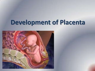
Placenta development
- 2. Objectives • Define placenta • Explain the development of placenta • State the gross anatomy of placenta at term • Describe the structures of placenta • Explain the placental circulation • State the placental ageing • List out the functions of placenta • Explain the umbilical cord • Describe the amniotic cavity, amnion and amniotic fluid • References 10/1/2012 9:41 AM 2
- 3. The placenta • Only the eutherian mammals possess placenta. The human placenta is discoid, haemochorial and deciduate. • The placenta is attached to the uterine wall and establishes connection between the mother and fetus through the umbilical cord. 10/1/2012 9:41 AM 3
- 4. Development of placenta • Developed from two sources. • The principal component is fetal which develops from the chorion frondosum and the maternal component consists of decidua basalis. 10/1/2012 9:41 AM 4
- 5. Development of placenta contd… • When the interstitial implantation is completed on 11th day, the blastocyst is surrounded on all sides by lacunar spaces around cords of syncytial cells, called trabaculae. • From the trabaculae develops the stem villi on 13th day which connect the chorionic plate with the basal plate. • Primary, secondary and tertiary villi are successively developed from the stem villi. • Arterio-capillary-venous system in the mesenchymal core of each villus is completed on 21st day. • This ultimately makes connection with the intraembryonic vascular systems through the body walls. 10/1/2012 9:41 AM 5
- 6. Development of placenta contd… • Simultaneously, lacunar spaces become confluent with one another and by 3rd-4th week, form a multilocular receptacle lined by syncytium and filled with maternal blood. • This space becomes the future intervillous space. • As the growth of the embryo proceeds, decidua capsularis becomes thinner beginning at 6th week and both the villi and the lacunar spaces in the abembryonic area get obliterated. This is compensated by – Exuberant growth and proliferation of the decidua basalis and – Enormous and exuberant division and subdivision of the chorionic villi in the embryonic pole (chorion frondosum). 10/1/2012 9:41 AM 6
- 7. Development of placenta contd… • These two i.e., chorion frondosum and the decidua basalis form the discrete placenta. It begins at 6th week and is completed by 12th week. • Until the end of 16th week, the placenta grows both in thickness and circumference 10/1/2012 9:41 AM 7
- 8. The placenta at term Gross anatomy • Circular disc with a diameter of 15-20 cm and thickness of about 2.5 cm at its center. • It thins off towards the edge. • It feels spongy and weight about 500 gm • Proportion to the weight of the baby being roughly 1:6 at term and occupies about 30% of the uterine wall. • It presents two surfaces, fetal and maternal, and a peripheral margin. 10/1/2012 9:41 AM 8
- 9. 10/1/2012 9:41 AM 9
- 10. The placenta at term contd… Fetal surface • Covered by the smooth and glistening amnion with the umbilical cord attached at or near its center. • Branches of the umbilical vessels are visible beneath the amnion. • The amnion can be peeled off from the underlying chorion except at the insertion of the cord. • At term, about four-fifths of the placenta is of fetal origin. 10/1/2012 9:41 AM 10
- 11. The placenta at term contd… Maternal surface • Rough and spongy • Dull red colour • A thin greyish, somewhat shaggy layer which is the remnant of decidua basalis and has come away with the placenta, may be visible. • 15-20 convex polygonal areas known as lobes or cotyledons which are limited by fissures. 10/1/2012 9:41 AM 11
- 12. Maternal surface of placenta contd… • Each fissure is occupied by the decidual septum. • Numerous small greyish spots are visible. • Amounts to less than one fifth of the total placenta. • Only the decidua basalis and the blood in the intervillous space are of maternal origin. 10/1/2012 9:41 AM 12
- 13. The placenta at term contd… • Margin: peripheral margin of the placenta is limited by the fused basal and chorionic plates and is continuous with the chorion leave and amnion. • Attachment: upper part of the body of the uterus encroaching to the fundus adjacent to the anterior or posterior wall with equal frequency. Separation: Placenta separates after the birth through the decidua spongiosum. 10/1/2012 9:41 AM 13
- 14. Structures of placenta • The placenta is limited internally by the amniotic membrane and chorionic plate; externally by the basal plate and in between these two lies the intervillous space containing the stem villi with their branches, the space being filled with maternal blood. • Amniotic membrane: It consists of single layer of cubical epithelium loosely attached to the adjacent chorionic plate. It takes no part in formation of placenta. 10/1/2012 9:41 AM 14
- 15. Structures of placenta contd… Chorionic plate: From within outwards, it consists of – Primitive mesenchymal tissue containing branches of umbilical vessels – A layer of cytotrophoblast and – Syncytiotrophoblast Basal plate: It consists of the following structures from outside inwards. – Part of the compact and spongy layer of the decidua basalis – Nitabuch's layer of fibrinoid degeneration of the outer syncytiotrophoblast at the junction of the cytotrophoblastic shell and decidua. – Cytotrophoblastic shell – Syncytiotrophoblast 10/1/2012 9:41 AM 15
- 16. 10/1/2012 9:41 AM 16
- 17. 10/1/2012 9:41 AM 17
- 18. Structures of placenta contd… • The basal plate is perforated by the spiral branches of the uterine vessels through which the maternal blood flows into the intervillous space but fail to reach the chorionic plate. • The septum consists of decidual elements convered by trophoblastic cells. • The areas between the septa are known as cotyledons (lobes), which are observed from the maternal surface, numbering 15-20. 10/1/2012 9:41 AM 18
- 19. Structures of placenta contd… • Intervillous space: It is bounded on the inner side by the chorionic plate and the outer side by the basal plate, limited on the periphery by the fusion of the two plates. • It is lined internally on all sides by the syncytiotrophoblast and is filled with slow flowing maternal blood. • Numerous branching villi which arise from the stem villi project into the space and constitute chief content of the intervillous space. 10/1/2012 9:41 AM 19
- 20. Structures of placenta contd… Stem villi • Arise from the chorionic plate and extend to the basal plate. • With the progressive development- primary, secondary and tertiary villi are formed. • Functional unit of the placenta is called the fetal cotyledon or placentome. • Major villi pass down through the intervillous space to anchor onto the basal plate. 10/1/2012 9:41 AM 20
- 21. Stem villi contd… • Functional subunit is called lobule which is derived from the tertiary stem villi. • About 60 stem villi persist in human placenta. • Thus, each cotyledon (totaling 15-29) contains 3-4 major stem villi. • The total surface, for exchange, varies between 4-14 square meters. • The fetal capillary system within the villi is almost 50 km long. 10/1/2012 9:41 AM 21
- 22. Structures of placenta contd… Structure of terminal villus: In the early placenta, each terminal villus has got the following structures from outside inward: – Outer syncytiotrophoblast – Cytotrophoblast – Basement membrane – Central stroma containing fetal capillaries, primitive mesenchymal cells, connective tissue and a few phagocytic (Hofbauer) cells. 10/1/2012 9:41 AM 22
- 23. Structure of terminal villus contd… • In the term placenta the syncytiotrophoblast becomes relatively thin at places overlying the fetal capillaries and thicker at other areas containing extensive endoplasmic reticulum. • The cytotrophoblast is relatively sparse. • Basement membrane becomes thicker. • Stroma contains dilated vessels along with all the constituents and few Hofbauer cells. • These cells have IgG surface receptors and can express class II MHC molecules. 10/1/2012 9:41 AM 23
- 24. Placental Circulation Placental circulation Utero- Feto-Placental Placental Circulation circulation 10/1/2012 9:41 AM 24
- 25. Uteroplacental circulation (Maternal circulation) • A mature placenta has a volume of about about 500 ml of blood; 350ml being occupied in the villi system and 150 ml lying in the intervillous space. • Intervillous blood flow at the term is estimated to be 500- 600 ml/minute, blood in the intervillous space is completely replaced about 3-4 times/minute. • Intervillous space pressure is about 10 to 15 mm Hg during uterine relaxation and 30-50 mm Hg during uterine contraction. • In contrast, the fetal capillary pressure in the villi is 20-40 mm Hg. 10/1/2012 9:41 AM 25
- 26. Utero-placental circulation contd… Arterial circulation: • About 120-200 spiral arteries open into the intervillous space. • Normally, there is cytotrophoblastic invasion into the spiral arteries initially upto the intra decidual portion within 12 weeks of pregnancy. • There is a secondary invasion of trophoblast between 12-16 weeks extending upto radial arteries within the myometrium, thus spiral arteries are converted to large bore uteroplacental arteries. • The net effect is funneling of arteries which reduce the pressure of the blood to 70-80 mm Hg before it reaches the intervillous space. 10/1/2012 9:41 AM 26
- 27. Utero-placental circulation contd… • Venous drainage: Through the uterine veins which pierce the basal plate randomly like the arteries. • Circulation of the intervillous space: The arterial blood enters the space under pressure. Lateral dispersion occurs, after it reaches the chorionic plate. Villi help in mixing and slowing of the blood flow. Mild stirring by the villi pulsation aided by uterine contraction help migration of the blood towards the basal plate and thence to uterine veins. 10/1/2012 9:41 AM 27
- 28. Fetoplacental circulation • The two umbilical arteries enter the chorionic plate underneath the amnion, each supplying one half of the placenta. • The arteries break up into small branches which enter the stems of the chorionic villi. • Each in turn divides into primary, secondary and tertiary vessels of the corresponding villi. • The blood flows into the corresponding venous channels either through the terminal capillary networks or through the shunts. 10/1/2012 9:41 AM 28
- 29. Fetal circulation contd… • Maternal and fetal blood streams flow side by side, but in opposite direction. • This counter current flow facilitates material exchange between the mother and fetus. • The villus capillary pressure varies from 20-40 mm Hg. • The fetal blood flow through the placenta is about 400 ml per minute. 10/1/2012 9:41 AM 29
- 30. Fetal circulation contd… • Placental membrane: Inspite of close proximity, there is no mixing of the maternal and fetal blood. The two are separated by tissues called placental membrane or barrier, consisting of the following. In the early pregnancy, it consists of – Syncytiotrophoblast – Cytotrophoblast – Basement membrane – Stromal tissue and – Endothelium of the fetal capillary wall with its basement membrane. It is about 0.025mm thick. • Near term, there is attenuation of the syncytial layer. Sparse cytotrophoblast and distended fetal capillaries almost fill the villus. The specialized zones of the villi where the syncytiotrophoblast is thin and anuclear known as Vasculo- Syncytial membrane. 10/1/2012 9:41 AM 30
- 31. 1. Intervillous space (with maternal blood) 1. Intervillous space 2. Placental barrier of a terminal villus 2. Syncytiotrophoblast 3. Fetal capillaries 3. Cytotrophoblast 4. Merged basal membranes of the fetal 4. Villus mesenchyma capillary and of the syncythiothrophoblast 5. Fetal capillaries 5. Endothelial cells 6. Hofbauer macrophage 6. Rare cytotrophoblast cells 7. Basal membrane of the capillaries 8. Basal membrane of the trophoblast portion 9. Syncytiotrophoblast with proliferation knots 10/1/2012 9:41 AM (nuclei rich region) 31
- 32. Placental ageing • As the placenta has got a life span, it is likely to undergo degenerative changes as a mark of senescence. The ageing process involves both the fetal and maternal components. 10/1/2012 9:41 AM 32
- 33. Placental ageing contd… Villi changes: The following changes are observed: • Decreasing thickness of the syncytium and appearance of syncytial knots. • Partial disappearance of Langhan's cells. • Decrease in the stromal tissue including Hofbauer cells. • Obliteration of some vessels and marked dilatation of the capillaries. • Thickening of the basement layer of the fetal endothelium and the cytotrophoblast. • Deposition of fibrin on the surface of the villi. 10/1/2012 9:41 AM 33
- 34. Placental ageing contd… • Decidual changes: There is an area of fibrinoid degeneration, where trophoblast cells meet the decidua. This zone is known as Nitabuch layer. The membrane is absent in placenta accreta. • Intervillous space: The syncytium, covering the villi and extending into the decidua or intervillous space, undergoes fibrinoid degeneration and form a mass entangling variable number of villi. These are called white infarcts. Calcification or even cyst formation may occur on it. There may be inconsistent deposition of fibrin called Rohr's stria at the bottom of the intervillous space and surrounding the fastening villi 10/1/2012 9:41 AM 34
- 35. The fetal membranes • It consists of two layers • Outer chorion and the inner amnion. Chorion: • Represents the remnant of chorion laeve. • Thicker than amnion • Friable and shaggy on both sides. • Internally, it is attached to the amnion by loose areolar tissue and remnant of primitive mesenchyme. • Externally, it is covered by vestiges of trophoblastic layer and the decidual cells of fused decidua capsularis and parietalis . • Contains no vessels or nerves. 10/1/2012 9:41 AM 35
- 36. The fetal membranes contd… Amnion: • Inner layer of the fetal membranes. • Internal surface is smooth and shiny and is in contact with liquor amnii. • The outer surface consists of a layer of connective tissue and is apposed to the similar tissue on the inner aspect of the chorion from which it can be peeled off. • The amnion can be peeled off except at the insertion of the umbilical cord. • Offers support to amniotic fluid and also produces the fluid. • In addition, it produces a phospholipid that initiates the formation of prostaglandins. 10/1/2012 9:41 AM 36
- 37. Functions of placenta • Transfer of nutrients and waste products • Enzymatic function • Barrier function • Immunological function • Storage • Endocrinal function 10/1/2012 9:41 AM 37
- 38. Functions of placenta 1. Transfer of nutrients and waste products between the mother and fetus. • The mechanism involved in the transfer of substances across the placenta are: – Simple diffusion – Facilitated diffusion (Carrier mediated) – Active transfer (against concentration gradient) – Endocytosis – Exocytosis – Leakage 10/1/2012 9:41 AM 38
- 39. Transfer function contd… Respiratory function: • Although, the fetal respiratory movements are observed as early as 11 weeks, there is no gaseous exchange. • Intake of oxygen and output of carbon dioxide take place by simple diffusion. • Partial pressure gradient is the driving force for exchange of gases. • The oxygen supply to the fetus is at the rate of 8 ml/kg/min and this is achieved with cord blood flow of 165-330ml/min. 10/1/2012 9:41 AM 39
- 40. Transfer function contd… Excretory function • Waste products like urea, uric acid and creatinine are excreted to the maternal blood by simple diffusion. • The main substance excreted from the fetus is carbon dioxide. • Bilirubin will also be excreted. 10/1/2012 9:41 AM 40
- 41. Transfer function contd… Nutritive function a. Glucose: • Principle source of energy • Transferred to the fetus by facilitated diffusion • Transfer rates decreases as maternal glucose concentration increases. b. Lipids • For fetal growth and development • Triglycerides and fatty acids are directly transported from the mother to the fetus in early pregnancy but are synthesized in the fetus later in the pregnancy. • EFA are transferred more than non EFA • Cholesterol is capable of direct transfer. 10/1/2012 9:41 AM 41
- 42. Transfer function contd… c. Amino acids • Transferred by active transport through enzymatic mechanism (ATPase). • Some proteins (IgG), cross by the process of endocytosis. • Fetal proteins are synthesized from the transferred amino acids and the level is lower than in mother. 10/1/2012 9:41 AM 42
- 43. Functions of placenta contd… d. Water and electrolytes • Sodium, potassium and chloride cross by simple diffusion, whereas calcium, phosphorus and iron cross by active transport. • Water soluble vitamins are transferred by active transport but the fat soluble vitamins are transferred slowly. e. Hormones- • Insulin, steroids from the adrenals, thyroid, chorionic gonadotrophin or placental lactogen cross the placenta at very slow rate. • Neither parathormone nor calcitonin crosses the placenta. 10/1/2012 9:41 AM 43
- 44. Functions of placenta contd… 2. Enzymatic function: • Diamine oxidase which inactivates the circulatory pressure amines • Oxytocinase which neutralizes the oxytocin • Phospholipase A2 which synthesizes arachidonic acid etc 3. Barrier function: 10/1/2012 9:41 AM 44
- 45. Functions of placenta contd… 4. Immunological function • Placental hormones, proteins (SP1), early pregnancy factor (EPF), PAPP-A, steroids and chorionic gonadotrophin, have got some immunosuppressive effect. • Villous trophoblasts do not express HLA Class I and class II molecules. Extravillous trophoblasts only express HLA class I molecules and no HLA class II molecules. • Though anti HLA antibodies and sentitised T cells against paternal antigens have been detected in the maternal serum, they have no significant effects on pregnancy. • There is shift of maternal response from cell mediated (T helper I) to humoral (T helper 2) immunity, which may be beneficial to pregnancy. 10/1/2012 9:41 AM 45
- 46. Functions of placenta contd… • Decidual natural killer (NK) cells and trophoblast (extravillous) HLA Class I molecules interact. • The immunological response of implantation and that of organ transplantation are different and not comparable. • Syncytiotrophoblast has got trophoblast lymphocytes cross reactive antigen (TLX). Consequently there is production of antibodies by the mother in response to this TLX. These blocking antibodies protect the fetus from rejection. 10/1/2012 9:41 AM 46
- 47. Functions of placenta contd… 5. Storage • The placenta metabolizes glucose, stores it in the form of glycogen and reconverts it to glucose as required. • It can also store iron and the fat soluble vitamins. 6. Endocrine Function • Human Chorionic Gonadotropin • Estrogen • Progesterone • Human Placental Lactogen 10/1/2012 9:41 AM 47
- 48. The Umbilical Cord • The umbilical cord is formed from the fetal membranes • Provides a circulatory pathway that connects the embryo to the chorionic villi of the placenta. • Its function is to transport oxygen and nutrients to the fetus from the placenta and to return waste products from the fetus to the placenta. • Measurement: It is about 53 cm (21 in) or 30-100 cm of variation in length at term and about 2 cm (3⁄4 in) thick. 10/1/2012 9:41 AM 48
- 49. Development of umbilical cord 10/1/2012 9:41 AM 49
- 50. The umbilical cord contd… • The bulk of the cord is a gelatinous mucopolysaccharide called Wharton’s jelly, which gives the cord body and prevents pressure on the vein and arteries that pass through it. • The outer surface is covered with amniotic membrane. • An umbilical cord contains only one vein but two arteries. 10/1/2012 9:41 AM 50
- 51. The umbilical cord contd… • The number of veins and arteries in the cord is always assessed and recorded at birth because about 1% to 5% of infants are born with a cord that contains only a single vein and artery. • From 15% to 20% of these infants are found to have accompanying chromosomal disorders or congenital anomalies, particularly of the kidney and heart. 10/1/2012 9:41 AM 51
- 52. The umbilical cord contd… • The rate of blood flow is 350 mL/min at term. • Blood can be withdrawn from the umbilical vein or transfused into the vein during intrauterine life for fetal assessment or treatment (termed percutaneous umbilical blood sampling [PUBS]). • The cord is unlikely to twist or knot to interfere with the fetal oxygen supply. • In about 20% of all births, a loose loop of cord is found around the fetal neck at birth. • The walls of the arteries are lined with smooth muscle. • It contains no nerve supply. 10/1/2012 9:41 AM 52
- 53. The umbilical cord contd… • Remnant of the umbilical vesicle (yolk sac) and its vitelline duct: May found as a small yellow body near the attachment of the cord to the placenta or on rare occasion, the proximal part of the duct persists as Meckel's diverticulum. • Allantois: A blind tubular structure may occasionally present near the fetal end which is continuous inside the fetus with its urachus and bladder. 10/1/2012 9:41 AM 53
- 54. The umbilical cord contd… Obliterated extra embryonic coelom: In the early period, intraembryonic coelom is continuous with extraembryonic coelom along with herniation of coils of intestine. The condition may persists as a congenital umbilical hernia or exomphalos. Attachment: • Early period- Cord is attached to the ventral surface of the embryo close to the caudal extremity • The point of attachment is moved permanently to the center of the abdomen at fourth month. • Unlike the fetal attachment, the placental attachment is inconsistent, either centre or eccentric. 10/1/2012 9:41 AM 54
- 55. Amniotic cavity, amnion and amniotic fluid Development: • Fluid accumulates slowly, at first, but ultimately the fluid filled cavity obliterates the chorionic cavity. • Initially the cavity is located on the dorsal surface of the embryonic disc. • With the formation of the head, tail and lateral folds, it comes to surround the fetus. • Its two growing margins merge into the body stalk. • Thus, the liquor amnii surrounds the fetus everywhere except at its attachment with the body stalk. 10/1/2012 9:41 AM 55
- 56. 10/1/2012 9:41 AM 56
- 57. 10/1/2012 9:41 AM 57
- 58. Structure of amnion • Fully formed amnion is 0.02-0.5 mm in thickness. • From within outwards the layers are: – Single layer of cuboidal epithelium – Basement membranes – Compact layer of reticular structure – Fibroblastic layer – Spongy layer • No blood supply, nerve supply nor any lymphatic supply. 10/1/2012 9:41 AM 58
- 59. Amniotic fluid • Origin of amniotic fluid: Mixed maternal and fetal origin. The following are the speculative theories: a. As a transudate across the umbilical cord or from fetal circulation in the placenta or secretion from the amniotic epithelium. b. Transudate of fetal plasma through the highly permeable fetal skin before it is keratinized at 20th week. c. Contribution from the fetus- Fetal daily urine output at term is about 400-1200 ml. The fetal swallows about 200-500 ml of liquor every day at term. 10/1/2012 9:41 AM 59
- 60. The origin and circulation of amniotic fluid Maternal circulation amniotic epithelium (Transudation) Secretes Placenta Amniotic fluid Fetal Skin Fetal Urine Exchange with Ingestion by the fetus respiratory tract Intestinal absorption of water Fetal Circulation 10/1/2012 9:41 AM 60
- 61. Amniotic fluid contd… Circulation: The water in the amniotic fluid is completely changed and replaced in every 3 hours. The presence of lanugo and epithelial scales in the meconium shows that the fluid is swallowed by the fetus and some of it passes from the gut into the fetal plasma. Volume: • 50ml at 12 weeks • 400 ml at 20 weeks and • reaches its peak of 1 litre at 36-38 weeks. • Thereafter the amount diminishes, till at term it measures about 600-800 ml. • As the pregnancy continues post term, further reduction occurs to the extent of about 200 ml at 43 weeks. 10/1/2012 9:41 AM 61
- 62. Amniotic fluid contd… Physical features: • Faintly alkaline with a pH of about 7.2 with low specific gravity of 0.010. • It becomes highly hypotonic to maternal serum at term pregnancy. • Osmolarity of 250 m Osmol/liter (fetal maturity). • Checking the pH of the fluid at the time of rupture helps to differentiate it from urine, which is acidic (pH 5.0–5.5). Colour: In early pregnancy, it is colourless but near term, it becomes pale straw colored due to presence of exfoliated lanugo and epidermal cells from the fetal skin. It may look turbid due to presence of vernix caseosa. 10/1/2012 9:41 AM 62
- 63. Abnormal colour of amniotic fluid • Meconium stained (green) is suggestive of fetal distress in presentation other than breech or transverse. Depending upon the degree and duration of the distress, it may be thin or thick or pea souped. • Golden colour in Rh incompatibility is due to excessive haemolysis of the fetal RBC and production of excess bilirubin. • Greenish yellow (saffron) in post maturity. • Dark colored in accidental hemorrhage is due to contamination of blood. • Dark brown (tobacco juice) amniotic fluid is found in IUD. The dark colour is due to frequent presence of old HbA. 10/1/2012 9:41 AM 63
- 64. Amniotic fluid contd… Composition: • first half of pregnancy, the composition of fluid is almost identical to a transudate of plasma. • late pregnancy, the composition is altered mainly due to contamination of fetal urinary metabolites. • The composition includes- water (98-99%) and solid (1-2%). • The following are the solid constituents: Organic- Protein- 0.3 mg% Uric acid- 4 mg% Glucose- 20 mg% Creatinine- 2 mg% Urea- 30 mg% Total lipids- 50 mg% NPN- 30 mg% Hormones (Prolactin, insulin and renin) 10/1/2012 9:41 AM 64
- 65. Amniotic fluid contd… Inorganic • The concentration of the sodium, chloride and potassium is almost the same as that found in maternal blood. Suspended particles: lanugo, exfoliated squamous epithelial cells from the fetal skin, vernix caseosa, cast off amniotic cells and cells from the respiratory tract, urinary bladder and vagina of the fetus. 10/1/2012 9:41 AM 65
- 66. Function of amniotic fluid • Its main function is to protect the fetus. During pregnancy: • It acts as a shock absorber. • Maintains an even temperature • The fluid distends the amniotic sac and thereby allows for growth and free movement of fetus and prevents adhesion between fetal parts and amniotic sac. • Its nutritive value is negligible however, water supply to the fetus in quite adequate. 10/1/2012 9:41 AM 66
- 67. Function of amniotic fluid contd… During labour: • The amnion and chorion are combined to form a hydrostatic wedge which helps in dilatation of cervix. • It prevents marked interference with the placental circulation during uterine contraction. • It flushes the birth canal at the end of first stage of labour and by its aseptic and bactericidal action protects the fetus and prevents ascending infection to the uterine cavity. 10/1/2012 9:41 AM 67
- 68. Clinical importance of amniotic fluid • Provides useful information about the well being and also maturity of the fetus. • Intra amniotic instillation of chemicals is used as method of induction of abortion. • Excess or less volume of liquor amnii is assessed by amniotic fluid index (AFI). It is used to diagnose the clinical condition of polyhydramnios or oligohydramnios respectively. • Rupture of the membranes with drainage of liquor is a helpful method in induction of labour. 10/1/2012 9:41 AM 68
- 69. References • Jacob A.A comprehensive textbook of midwifery and gynecological nursing.3rd edition.New Delhi:Jaypee;2012. • Fraser DM, Cooper MA.Myles textbook for midwives.15th edition. Philadelphia:churchill livingstone elsevier;2009 • Dutta DC.Textbook of obstetrics. 6th edition.Calcutta:New central book agency;2004 • Pillitteri A. Maternal and Child health nursing: Care of the childbearing and childrearing family. 6th edition. Lippicott Williams and Wilkins : Philadelphia; 2010 10/1/2012 9:41 AM 69
- 70. 10/1/2012 9:41 AM 70
- 71. 10/1/2012 9:41 AM 71
- 72. • The process of spiral artery remodeling during pregnancy. In early pregnancy, two types of extravillous trophoblasts are found outside the villous, endovascular and interstitial trophoblasts. Endovascular trophoblasts invade and transform spiral arteries during pregnancy to create low-resistance blood flow that is characteristic of the placenta. Interstitial trophoblasts invade the decidua and surround spiral arteries. 10/1/2012 9:41 AM 72
- 73. 10/1/2012 9:41 AM 73
- 74. 10/1/2012 9:41 AM 74