8 dermatology (skin diseases)
•
5 recomendaciones•235 vistas

8 Dermatology (Skin Diseases)

Recomendados
Más contenido relacionado
La actualidad más candente
La actualidad más candente (20)
Similar a 8 dermatology (skin diseases)
Similar a 8 dermatology (skin diseases) (20)
Más de karima Akool AlSalihi
Más de karima Akool AlSalihi (20)
Último
Último (20)
8 dermatology (skin diseases)
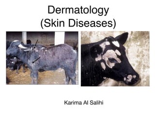
- 1. Dermatology (Skin Diseases) Karima Al Salihi
- 2. Dermatology (Skin Diseases) • The skin is a covering for the whole body so it is a mirror that reflects many systemic and local diseases. • The incidence of skin diseases in animals is high. • Many of the skin diseases are contagious therefore quick and accurate diagnosis is important in their control and eradication.
- 3. Function o f the skin (1) Protection from light, organisms and mechanical. (2) Immunologic function. (3) Vitamin D synthesis. (4) Heat regulation. (5) Secretary organ by sweating : e.g. wastes, urea. (6 ) Water reserve. (7) Sensory organs. (8 ) Reflection of internal feelings
- 4. Causes o f skin diseases: [1] Infectious: (1) Viral: 1) Contagious erythema. 2) Ulcerative dermatitis in sheep. 3) Lumpy skin diseases (in cattle). 4) Pox. 5) Vesicular papular dermatitis (in horse).
- 5. (2) Bacterial: 1) Bacterial dermatitis. 2) Tuberculosis. 3) Foot rot. (3) Mycotic: 1) Ring worm. 2) Epizootic lymphangitis. 3) Spirocheta. (4) Parasitic: 1) Protozoa e.g. Leishmaniasis. 2) Arthropodes as mange. 3) Helminthes as filariasis.
- 6. [2] Non infectious: (1) Nutritional deficiency of zinc, cobalt, iodine and copper. (2 ) Chemical injury as acids, alkalies and gases. (3) Histamine production: urticaria. (4) Lesions due to severe abrasion or friction. (5) Disturbance in circulation such as erythema. (6 ) Distribution in nervous tissue “itching”.
- 7. (7) Internal diseases (constipation, nephritis). (8 ) Neoplasms (wastes, papilloma, carcinoma). (9) Congenital defects (alopecia). (10) Over feeding of carbohydrate without enough exercise. (11) Traumatic (wounds). (12) Thermal, excessive heat, sun rays. (13) Long administration of some drugs as arsenic, mercury, serum causing skin rashes. (14) Endocrine disorders.
- 8. Scientific Term used in Skin Diseases [1] Primary lesions (less than 1 cm in size): (1) Macule: It is a flat circumscribed discoloration o f the skin or mucous membrane. (2) Papule: A solid elevation of the skin or mucous membrane extending deeper in the epidermis. (3) Nodule: A solid elevation of the skin or mucous membrane extending into the dermis (neoplastic or inflammatory). (4) Vesicle: A fluid filled superficial circumscribed elevation of skin or mucous membrane.
- 10. [2] Lesions more than 1 ml: Such as cyst, tumor, granuloma, plaque. [3] Primary lesions of varying sizes: (1)Pustule: Vesicle of the skin or mucous membrane which contain pus. (2)Wheal: An irregularly shaped, elevated transient lesion of the skin or mucous membrane due to edema.
- 12. [4] Secondary cutaneous lesions: (1) Erosion: Shallow ulcer of the epidermis doesn’t penetrate the entire epidermis. (2) Erythema: A redness of skin due to capillary congestion. (3) Hyperkeratosis: An increased thickening of the keratin layer of the epidermis (normal in the foot pads, nose of dog and cat). (4) Scab or crust: A collected dried exudate on the surface of the epidermis adherent to hairs.
- 14. (5) Scale: A collection of excessive keratin flacks upon the surface o f the skin. (6) Scar: A permenant depigmented area o f fibrous tissue without hair growth. (7) Parakeratosis: Retention of nuclei in the keratin layer (stratum comium) with absence of stratum granulosum characterized by scale formation. (8) Seborrhea: Over active disease of sebaceous glands result in oilness, crust or scales.
- 18. Principle s of treatment of skin diseases:( 1) Accurate diagnosis of the cause to choose the correct topical or systemic treatment by obtaining complete medical history and through medical examination. (2) Removal of hair coat and debris to enable topical application to come into contact with the causative agent. (3) In allergic diseases remove the causative agent. (4) In bacterial disease, sensitivity tests on culture of the organism are advisable
- 19. (5) Treat the primary cause and prevent secondary infection by: 1) Base of bacteriostatic ointment or dressing. 2) Administration of local anaesthetic ointments to prevent further damage from scratching. 3) Fluid therapy in case of fluid and electrolytes losses. 4) Sulpher containing amino acids or zinc or vitamin A facilitate the repair of skin tissues.
- 20. Drugs used for Treatment of Skin Diseases: (1) Stimulants: !These are compounds which have an action on the skin. !They are powders, aqueous sol., suspensions or mineral oils. !They applied externally on the skin lesion. (2) Sedatives: They used to depress irritation and it may be: 1) Emollients: 1- Oils: such as olive, caster, cotton seed oil. 2- Fats: such as wool fat. 3- Wax: such as bees wax. 4- Hydrocarbon: as liquid paraffin, cod-liver oil and glycerin. 2) Sedative powders: As chalk, talk, zinc carbonate and kaolin. (3) Astringent: It coagulates protein forming a protective layer. Such as copper sulphate, zinc sulphate and ferric chloride. (4) Corticosteroid: They inhibits fibroblasts and diminish the fibrosis of chronic inflammation, inhibit vasodilatation and increase permeability and exudate formation of acute inflammation.
- 21. (5) Counter irritant: They are substances which applied to the skin to cause local irritation and inflammation, e.g. iodine. (6) Caustics: They are agents which destroy tissue and excessive granulation and superficial tumors (warts), ex.: Nitric acid, caustic soda, phenol, mercury chloride.
- 22. (I) Diseases of the Epidermis and Dermis They include Dermatitis, Eczema, Urticaria, Photosensitization, Pityriasis, Parakeratosis and Hyperkeratosis. •Dermatitis •Definition: •It is inflammation of the dermis and epidermis. •Causes: •(1) In all species: •1) Mycotic dermatitis due to Dermatophilus congolensis in horses, cattle and sheep. •2) Staphylococcus aureus. 3) Ring worm.
- 23. 4) Photosensitive dermatitis. 5) Chemical irritation (contact dermatitis) topically. 6) Arsenic and systemic poisoning. 7) Mange mite infestation. 8 ) Biting flies. 9) Stephanofilaria sp. dermatitis. 10) Strongyloids sp. dermatitis.
- 24. (2) In cattle: 1) Udder impetigo (staphylococcus aureus). 2) Cow pox. 3) Lumpy skin disease, Foot and Mouth disease, Rinder pest, Bovine virus diarrhea.
- 25. (3) In sheep and goat: 1) Sheep pox. 2) Ulcerative dermatitis. 3) Fleece rot. 4) Ovine dermatitis. 5) Caprine idiopathic dermatitis. (4 ) In horses: 1) Horse pox. 2) Canadian horse pox. 3) Viscular stomatitis. 4) Dermatophytes including ringworm. Pathogenesis: (1) Inflammation of deeper layers of the skin involving blood vessels and lymphatics. (2) It may be acute, chronic, suppurative, weeping ulcerative or gangrenous. (3) Increased thickness, temperature, pain and itching.
- 27. Clinical findings: (1) Symptoms start by erythema, vesicles. (2) Edema of the skin and subcutaneous tissues. (3) Healing stage or scab formation or may be necrosis or gangrene of the affected area in severe cases. (4) Systemic reaction may occur when the affected skin area is extensive.
- 28. (5 ) Spread o f infection to subcutaneous may result in diffuse cellulitis. * (6) Shock with peripheral circulatory failure may be present in the early stages. (7) Toxemia due to absorption of tissue breakdown. Clinical pathology and diagnosis: (1) Skin scraping or swabs for parasites or bacterial examination. (2) Cellular and sensitivity tests for bacteria. (3) In allergic or parasitic states, accumulation of eosinophils in the inflammed area.
- 29. •Treatment: (1) Hygienic treatment: 1) Remove the injuries and harmful stimuli. 2) Remove the physical or chemical agent from environment. 3) Supply balanced diet to repair nutritional deficiency (vit. A, B, E, and D). (2) Medical treatment: •In infectious causes, identify the causative agent, make sensitivity test (in bacterial infection) to select the specific antibacterial drug. E.g. garamycin, terramycin ointement applied on the skin lesion.
- 30. (2) Systemic: 1)Antihistaminic: It is recommended when tissue destruction is extensive or the dermatitis is allergic in origin. 2)2) Calcium preparations 3) Fluid therapy: If shock is present. 4) Parental antibiotic and anti-fungal agents: 5) Anaesthetic drugs: •NB: If the lesions are extensive or secondary bacterial invasion is likely to occur, parental antibiotic or antifungal agents may be preferred to topical application. NB: The use of vaccination as prophylaxis in viral and bacterial dermatitis must not be neglected. Autogenous vaccines may be most satisfactory in bacterial infections.