5 diseases of the lung pneumonia
•
2 recomendaciones•201 vistas

5 Diseases of the lung - Pneumonia

Recomendados
Recomendados
Más contenido relacionado
La actualidad más candente
La actualidad más candente (19)
Similar a 5 diseases of the lung pneumonia
Similar a 5 diseases of the lung pneumonia (20)
Más de karima Akool AlSalihi
Más de karima Akool AlSalihi (20)
Último
Último (20)
5 diseases of the lung pneumonia
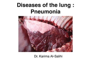
- 1. Diseases of the lung : Pneumonia Dr. Karima Al-Salihi
- 2. Definition: It is an inflammation of the lung tissue and usually accompanied by inflammation of the bronchioles and pleura. It is manifested clinically by an increase in respiratory rate, cough, abnormal breath sounds on auscultation and in most bacterial pneumonia, by evidence of toxaemia.
- 3. Aetiology: (1)Most o f the pneumonias in animals are bronchogenic in origin (airogenic) but some originated by the haematogenous and lymphatic route or by extension from neighbouring inflamed tissue. (2) Non infectious cases such as: 1) Exposure to cold and air currents with severe fatigue. 2) Bad ventilation stables. 3) Inhalation o f irritant gases and vapour. 4) Vitamin C and or vitamin A deficiency and a defect of immune protective mechanism.
- 4. 5) Over excretion as traveling for long distance. 6) Drenching pneumonia. 7) Too old and too young animals are more susceptible specially the debilitated ones. (3) Pneumonia may be caused by viruses, bacteria (or a combination of both), fungi and parasites. (4) Pneumonia are grouped here according to species:
- 5. (1)Cattle: 1) Pneumonic pasteurellosis (shipping fever): Pasteurella haemolytica, Pasteurella multocida. 2) Enzootic pneumonia of calves: parainfluenza-3, adenovirus 1,2 and 3, rhinovirus, bovine respiratory syncytial virus, bovine herps virus (IBR), Actinomyces, Corynbacterium pyogens and streptococcus sp. 3) Contagious bovine pleuropneumonia (Mycoplasma mycoides). 4) Lung worm pneumonia (Dictyocaulus viviparus). 5) Sporadically in tuberculosis caused by Mycobacterium bovis.
- 7. (2) Horses: 1) pleuropneumonia in mature horses, due to aerobic and anaerobic bacteria. 2) Newborn foals: Streptococcus sp., E.coli and Actinobacillus equi. 3) Older foals: Corynebacterium equi, Equine herpes virus and Equine influenza virus. 4) Dictycaulus arnfeldi and Parascaris equorum. 5) Galnders and epizootic lymphangitis.
- 8. (3) Sheep: 1) Pneumonic pasteurellosis (Pasteurella sp.) 2) Newborn lambs (uncommonly Streptococcus zooepidemicus, Salmonella abortus-ovis). 3) Severe pneumonia due to Mycoplasma sp. in lambs. 4) Symptomless pneumonias without secondary infection adenovirus, respiratory syncytial virus, reovirus and mycoplasma sp. 5) Corynebacterium pseudotuberculosis. 6) Lung worm (Dictyocaulus filaria).
- 9. Pathogenesis: ØThe process by which pneumonia develops varies with: 1) Causative agent and its virulence 2) Portal by which it is introduced into the lung
- 10. a primary bronchiolitis which spreads to involve surrounding pulmonary parenchyma Bacteria are introduced largely by way of the respiratory passages The reaction of the lung tissue may be in the form of: 1) An acute fibrinous process as in pasteurellosis. 2) Necrotizing lesions as in infection with Fusobacterium necrophorum. 3) As a more chronic caseous or granulomatous lesion in mycobacterial or mycotic infections.
- 11. 4) Spread o f the lesion through the lung occurs by: 1- Extension and passage of infective material along bronchioles and lymphatics. 2- The spread along air passages is facilitated by the normal movements of the bronchiolar epithelium and by coughing. NB: Hematogenous infection by bacteria results in a varying number o f septic foci which may enlarge to form lung abscesses. Pneumonia occurs when these abscesses rupture into air passages and spread as a secondary bronchopneumonia.
- 12. (3) Viral infection are also introduced chiefly by inhalation and cause a primary bronchiolitis but it is characterized by absence of acute inflammatory' reaction and toxaemia. 1) In viral infection, spread to alveoli causes enlargement and proliferation of the alveolar epithelium and the development o f alveolar edema. 2) Viral pneumonia is characterized by development of interstitial pneumonia results in consolidation of pulmonary parenchyma without involvement o f the bronchi and on auscultation loud bronchial sound is heard.
- 14. NB: The pathophysiology of all types of pneumonia is based upon the interference with gaseous exchanges between the alveolar air and the blood, because o f the obliteration of the alveolar air space and obstruction of air passage. The reduction o f oxygenation o f the blood is due to the failure o f a part of circulating blood to come in contact with oxygen so cyanosis is most likely to develop and this lead to anoxic anoxia and hypercapnia.
- 15. •Clinical signs: Signs depend on the stage o f the disease and secondary bacterial pneumonia. (1) In early stages of viral pneumonia. 1) Mild depression and anorexia. 2) Marked elevation in body temperature. 3) Serous to mucopurulent lacrimal and nasal discharges. 4) Cough and elevated respiratory rates in the early stage cough is dry, painful and harsh, but in late stage cough is moist and painless. 5) On auscultation of the lungs there may be an increase in breath sounds.
- 16. (2) In severe inflammation and toxemia (Bacterial pneumonia): 1) Fever of 40°C. 2) Deep laboured inspiration with extension of the head. 3) Dyspnoea in both respiratory and expiratory. 4) Ocular and nasal discharges progress from serous to mucopurulent. 5) Normal lung sounds are difficult to hear expect in calves, goats and sheep. The heavy chest wall of larger cattle makes it difficult to hear normal airway sounds. 6) By auscultation o f the anterior ventral lung fields reveals crackles and wheezes and an increase in bronchial sounds especially on inspiration.
- 17. 7) When ventral consolidation occurs, rough tracheal breathing is still audible ventrally. Pneumonia are present in three forms: (1) Acute lobar pneumonia. (2) Acute lobular pneumonia. (3) chronic interstitial pneumonia.
- 18. Acute Lobar Pneumonia (Croupous pneumonia) Definition It is the affection of the whole or the greater part of one lobe of the lung. If the two lobes are affected, it is called double lobar pneumonia. • •Clinical symptoms: (1) Rapid and shallow respiration. (2) Short, painful cough. (3) Inspiratory dyspnea, later on mixed dyspnea in severe cases. (4) Engorged eye capillaries and the mucous membranes are congested then cyanosed. (5) Sudden rise and continuous fever for about a week then gradually decreases.
- 20. (6) Nasal discharge. (7) The odor o f the breath may be informative (decay in pus formation or putrid gangrenous). (8) The heart is weak and the pulse is increased. (9) Percussion and auscultation on the lung area give abnormal sounds according to the stages o f pneumonia.
- 22. Acute Lobular Pneumonia (Catarrhal or bronchopneumonia) • It is a catarrhal inflammation of bronchi, bronchioles as well as alveoli (Broncho- pneumonia). It affects small group of lobules in one or both lobes o f the lung. It differs from acute lobar pneumonia in the following points: (1) Recurrent fever (Specific symptom): It shoots high and remain for 3 days then drop suddenly to normal and stay for few days, then shoots high again and so on, this is due to appearance o f a new foci o f infection in the lungs.
- 24. (2) Percussion and auscultation on the lung area reveal all sounds o f different stages o f pneumonia in different areas at the same time, in addition to the resonant and vesicular sound as well as rales. (3) Compensatory emphysema can be detected by percussion and auscultation around the affected areas specially the red consolidation stage.