Vitamin A and D Deficiency in Livestock
•
1 like•147 views

This document discusses vitamin A, D, and K deficiencies. It defines each vitamin, describes causes of deficiencies, pathogenesis, clinical signs, diagnosis, treatment and control. Vitamin A is important for vision, bone growth, and epithelial tissues. Deficiency can cause night blindness, skin issues, and susceptibility to infection. Vitamin D facilitates calcium and phosphorus absorption and bone mineralization. Deficiency impacts growth, reproduction and causes rickets. Vitamin K is required for blood clotting and deficiencies are rare but can occur with certain conditions that limit absorption or production of prothrombin.

Recommended
More Related Content
What's hot
What's hot (20)
Similar to Vitamin A and D Deficiency in Livestock
Similar to Vitamin A and D Deficiency in Livestock (20)
More from karima Akool AlSalihi
More from karima Akool AlSalihi (20)
Recently uploaded
Recently uploaded (20)
Vitamin A and D Deficiency in Livestock
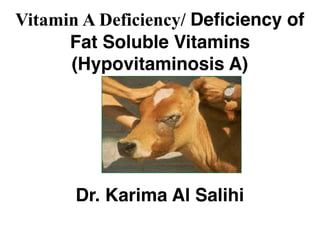
- 1. Vitamin A Deficiency/ Deficiency of Fat Soluble Vitamins (Hypovitaminosis A) Dr. Karima Al Salihi
- 2. Definition •It is an insufficient supply of vitamin A in the ration or its defective absorption from the alimentary canal.
- 3. Causes •Primary vitamin A deficiency •Lack of green feed or failure to add vitamin A supplement to diets. •Alcoholic form of vitamin A in green fodder doesn’t pass the placental barrier and a high intake of green pasture before parturition doesn’t increase the hepatic store of vitamin A in the newborn animals but only increase it in colostrum. •Ester form (in fish oil) passes through the barrier and feeding of these oils before parturition will an increase of the stores of the vitamin in fetal liver.
- 4. Secondary vitamin A deficiency It may occur in cases of: (1) Chronic disease in the liver or intestine because much of the conversion of carotene to vitamin A occurs in the intestinal epithelium while the liver is the main site storage of the vitamin. (2) Continued ingestion of mineral oil (liquid paraffin dissolves vitamin A and carotene resulting in it: malabsorption and excessive fecal excretion causing in severe depression of plasma carotene and vitamin / esters. (3) High cholorinated naphthalenes interfere with the conversion of carotene to vitamin A.
- 5. (4) Additional factors which increase the requirement of vitamin A such as: ! high environmental temperature, !high nitrate content in the feed which reduce the conversion of carotene to vitamin A. (5) Low phosphate diet facilitating storage of the vitamin A.
- 6. Pathogenesis Vitamin A is essential for the regeneration !of the visual purple necessary for dim light vision, !for normal bone growth ! for maintenance of normal epithelial tissues. There is a difference in tissue and organ response in difference species and particular clinical signs may occur: (1) Night vision: Ability to see in dim light and even light is reduced because of interference with regeneration of visual purple. (2) Bone growth: Vitamin A is necessary to maintain the normal position and activity of oesteoblasts and oesteoclasts.
- 7. •When deficiency occurs there is no retardation of endochondrial bone growth but there is incoordination of bone growth in that shaping. •Over growing of the cranial cavity occurs with resulting distortion and herniation of the brain and an increase in cerebrospinal fluid pressure up to 4 or 6 times normal.
- 8. (3) Epithelial tissue: Vitamin A deficiency leads to including the secretory and or a covering epithelium. !These secretory cells are gradually replaced by the stratified, keratinizing epithelial cells. !It occurs chiefly in the salivary glands, the urogenital tract (including placenta but not ovaries or renal tubules) and the para-ocular glands and teeth. !The secretion of thyroxin is markedly reduced.
- 9. Vitamin A deficiency affect the covering epithelial cells of: 1) Eye by thickening & striation of cornea causing corneal opacity in dog & calf. 2) Skin by thickening & keratinization causing dryness, bran like deposition & hyper keratinization (xerodermia). 3) GIT by degenerative changes causing enteritis. 4) Respiratory tract by degeneration & stratification causing respiratory infection. 5) Urinary tract by degeneration & cornification favoring the formation of renal stones. 6) Reproductive tract by degeneration impairing spermatogenesis in male & oogenesis in female as well as retention of fetal membranes & abortion. (7) Embryological development: Vitamin A is essential for organ formation during the growth of the fetus.
- 11. 6) Reproductive tract by degeneration impairing spermatogenesis in male & oogenesis in female as well as retention of fetal membranes & abortion. (7) Embryological development: Vitamin A is essential for organ formation during the growth of the fetus.
- 12. Clinical findings (1)Night blindness: Inability to see in dim light is the earliest signs in all species, Later on in day light. (2) Xerophthalmia (Xerox = dry, opthalmos = eye): It is a dryness of conjunctiva, bulbar conjunctiva due to nonfunctioning goblet cells. Thickening & striation of cornea giving opaque appearance (clouding), It occurs only in the dog and calf. In other species, a thin serous mucoid discharge from the eye occurs, followed by corneal keratinization, clouding and sometimes ulceration and photophobia (fright from light).
- 13. (3) Changes in the skin: In cattle, a heavy deposit of bran like scales on the skin are seen. Dry, scaly hooves with multiple, vertical cracks are particularly noticed in horses. (4) Emaciation, inappetence, weakness and stunted growth. (5) Loss of reproductive function: In the male, libido is retained but degeneration of the germinative epithelium of the seminiferous tubules causes reduction in the number of motile, normal spermatozoa produced. In young rams, the testicles may be visibly smaller than normal size and the placental degeneration occurs leading to abortion, birth of dead or weak young. Placental retention is also common.
- 14. (6) Nervous symptoms: Signs related to damage of the central nervous system include: 1) Paralysis of skeletal muscle due to damage of peripheral nerve roots. - 2) Encephalopathy due to increase intracranial' pressure. 3) Blindness due to constriction of optic nerve canal. 4) These defects occur at any age but most commonly in young growing animals. (7) Edema (anasarca) of legs and fore quarter is often recorded in feedlot. (8) A high incidence of otitis media, pneumonia and enteritis. (9) Increased susceptibility to infection due to low immunity.
- 15. Diagnosis (1) History (green feed or‘ vitamin A supplements aren’t being provided) and symptoms. (2) Testing for night-blindness is the early method. (3) Increase CSF pressure is the earliest measurable changes. (4) Estimations of vitamin A level in plasma and liver. (5) Histological examination of parotid salivary gland.
- 16. Differential diagnosis (1) Hypomagnesima in calves. (2) Rabies. (3) Lead poisoning •Treatment 1. Treat the real cause. 2. Source of vitamin A (green food, cod liver oil) at a dose rate equivalent to 10-20 times the daily maintenance. A. Orally: 440 IU/Kg in water emulsion B. Parenteral injection of an aqueous rather that the daily solution is preferred.
- 17. •Prognosis •A. The response to treatment in severe cases is often rapid and complete but the disease may be irreversible in chronic cases. •B. Calves with convulsive form due to increased CSF pressure will usually return to normal in 48 hours following treatment. • •C. Blind animal with ocular form will not respond to treatment and should be slaughtered. •Control •1. The minimum daily requirement in all species is 40 IU of Vitamin A/ Kg •2. During pregnancy, lactation or rapid growth the amount are usually increased by 50-75 % of the requirements.
- 18. (4) Vitamin D is produced from its precursor 7- dehydrocholesterol in mammalian skin and by natural irradiation with ultra- violet light. (5) Vitamin D4 and D5 occur naturally in the oils of some fish. •Causes (1) A lack of ultraviolet irradiation of the skin coupled with a deficiency of performed vitamin D complex in the diet lead to a deficiency of vitamin D in the tissues. (2) Cloudy, overcast sky, smoke, laden atmosphere and winter months exacerbates of the lack of irradiation. (3) Poor irradiation in animals with dark skin or heavy coats (sheep) and housed in doors for long periods. (4) Carotene when present in large quantity in certain feeds has antivitamin D potency.
- 19. Pathogenesis (1)Vitamin D3 is produced from its precursor 7- dehydrocholesterol in mammalian skin by netural irradiation with ultra-violet light. (2) Vitamin D2 is present in sun-cured hay and is produced by ultraviolet irradiation on plant sterols (calciferol). (3) Vitamins D4 and D5 occur naturally in the oil of some fish. (4) Calciferol or viosterol is produced commercially by the irradiation of yeast. (5) Dihydrotachysterol produced commercially by irradiation of yeast and is used in prevention of parturient paresis.
- 20. •Mode of action of vitamin D: •It facilitates deposition of Ca and ph in bone and increase absorption of these minerals from the alimentary canal. (1) When the Ca:ph ratio in ration is wider than the normal 1:1 to 2:1, vitamin D requirements are increased for good calcium and phosphorus retention and bone mineralization so that vitamin D deficiency with supplying an imbalance of Ca and ph might lead to disease. (2) When normal Ca and ph intake with the same degree of vitamin D deficiency, the disease does not occur.
- 21. •Clinical signs (1) Reduce productivity. (2) Decrease appetite and efficiency of food utilization cause poor weight gain in growing animals and poor production in adult. (3) Reproductive efficiency is reduced. (4) In late stage, lameness accompanied by signs of rickets in young animals and osteomalacia in adults. Treatment (1) Source of vitamin D2. (2) Adequate calcium and phosphorus. (3) A single IM injection of calciferol or vitamin D (1000 IU/kg BW) will meet the need of the animals for 3-6 months. (4) Orally vitamin D: 40-50 IU/kg/daily or single oral doses of two million units is an effective for 2 months in lambs.
- 22. Control (1) Orally vitamin D: 10 IU/kg daily requirement. (2) Sun dried hay is a good source. (3) Fish liver oils are high and rich in vitamin D4 and D5. (4) Irradiated dry yeast is the simplest and cheapest method of supplying vitamin D in mixed green feeds. (5) Single IM injection of vitamin D2 (calciferol) in oil to protect ruminants for some months (0.5-1 million are only recommended for sheep).
- 23. Vitamin K •Vitamin K is essential for the formation of prothrombin (in liver) which is essential for clotting of the blood. •Occurrence •Vitamin K deficiency is rare under natural conditions in domesticated animals because of the high content of substance with vitamin K activity in most plants and the synthesis of these substances by mineral activity in alimentary canal. Absorption of vitamin K from the intestine is dependent on the presence of bile and fat in the intestines. Storage is mainly in the liver and excretion is via the urinary tract.
- 24. •Therapeutic use of vitamin K •(1) Treatment of epistaxis. •(2) Coccidiosis. •(3) Sweet clover poisoning (toxic quantities of coumarin cause depress the prothrombin levels of blood and interferes with its clotting mechanism. •(4) Abomasal ulcer. •(5) Hepatitis and gastroenteritis: when vitamin K absorption is restricted or prothrombin formation impaired.