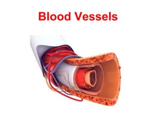
Histology of blood vessels
•
165 likes•65,603 views

Complete description of microscopic anatomy of blood vessels.

Recommended
More Related Content
What's hot
What's hot (20)
Similar to Histology of blood vessels
Similar to Histology of blood vessels (20)
More from Mehul Tandel
Recently uploaded
Recently uploaded (20)
Histology of blood vessels
- 2. Objectives • Introduction • Components of circulatory system • Blood vessels – Basic structure • Arteries: Elastic & Muscular • Arterioles, Meta-arteriole • Capillaries: Continuous, Fenestrated, Sinusoidal • Venules, Veins • Clinical Correlation
- 3. The transport system of the body that permit blood and lymph circulation to transport • Nutrients, oxygen, hormones etc, to the place where they are utilized (cells) and • The metabolites(waste products) and carbon dioxide are conveyed to appropriate places from where they are expelled. Circulatory System
- 4. Circulatory System Two sub systems: 1. Cardiovascular system: Heart, Arteries, Veins and capillaries. 2. The Lymphatic system: Lymphatic capillaries, lymphatic vessels, lymphoid organs and lymphatic ducts.
- 5. Cardio-vascular System • Central pumping organ - Heart • Conveying channels – Blood vessels • Conveying medium – Blood
- 6. Cardio-vascular System • Components of CVS Heart Arteries Veins Capillaries Sinusoids & Cavernous tissues
- 8. • Intricate network of hollow tubes that transport blood throughout the entire body • Two systems: • Arterial System: Transport blood away from heart • Arteries • Arterioles • Capillaries • Venous System: Transport blood towards the heart • Venules • Veins Blood Vessels
- 9. Basic Structure of Blood Vessels • All blood vessels except capillaries have 3 concentrically arranged basic Tunics or Coats
- 12. Tunica Intima Endothelium Basal Lamina Sub-endothelial CT Internal Elastic Lamina Fenestrated sheet of elastic fibers Fenestration provide passage for nutrients Simple Squamous epithelium Separates Tunica Intima from Media
- 13. Tunica Media Concentric layers of Smooth Muscle & Elastic Fibers Few collagen & reticular fibers & proteoglycans External Elastic Lamina Fenestrated sheet of elastic fibers Thinner than internal lamina Separates Tunica Media from Externa
- 14. Tunica Media Thicker in arteries than in veins of similar size Has relatively more smooth muscle and elastic fibres in arteries than in veins of same size
- 15. Tunica Adventitia Prevent excessive stretching Connective Tissue Numerous collagen fibers & few elastic fibers Continuous & fused with stroma of organs
- 16. Fibers Runs Longitudinally Mostly Elastic fibers Mostly collagen fibers • In Tunica intima & Tunica adventitia • In Tunica media Runs Circularly
- 17. Muscular Artery with Thickening of Tunica Intima Vessel tunics A. Tunica intima •Endothelium •Subendothelium Connective tissue Smooth muscle (Longitudinal fibers) •Internal elastic lamina B. Tunica media •Vascular smooth muscle •External elastic lamina (Circular fibers) C. Tunica adventitia •Connective tissue (Longitudinal)
- 18. Nourishment of Blood Vessels • Small & Medium sized Vessels: • All layers receive O2 & nutrition directly from blood in the lumen • Large Vessels: • Diffusion of O2 & nutrients possible up to tunica intima & inner part of tunica media only • Outer part of Tunica media & Adventitia: • Received nourishment from arterioles, capillaries & venules present in this layers “Vasa Vasorum” = Vessels of Vessel
- 20. Endothelium • Lines inner surface of heart & blood vessels • Simple squamous epithelium • Endothelial cells held together by cell junctions • Surrounded by basal lamina • Cells: polygonal & elongated • Cytoplasm: sparse & contains ER, mitochondria, MF & IF • Numerous pinocytotic vesicles - help in transport
- 21. Endothelium • Secrete substances involved in • Diapedesis, • Blood coagulation • Maintenance of tone of smooth muscles of vessels • Nitric oxide: vasodilation • Prostacyclin: vasodilation and inhibits platelet adhesion. • Thromboplastin and von Willebrand factor: involved in blood coagulation
- 22. Arterial System
- 23. Arteries • Thick walled blood vessels that carry blood away from the heart • Divide repeatedly like branch of a tree – Gradually become smaller in size – Decrease in the rate of blood flow – facilitate exchange through capillaries
- 24. Arteries The arterial system consists of: 1. Elastic arteries: – Lager arteries – More elastic fibres and less smooth muscles in TM 2. Muscular arteries: – Medium-sized arteries (0.5–10 mm in diameters) – More smooth muscles and less elastic fibres in TM
- 25. Arteries The arterial system consists of: 3. Arterioles: – 1 or 2 layers of smooth muscle cells in TM 4. Capillaries: – Only tunica Intima & Underlying basal lamina
- 26. Elastic Artery
- 27. Elastic Arteries • Lager arteries • Conducts blood from heart to smaller arteries – Conducting Arteries • Thickness of the wall is 1/10 the diameter of lumen
- 28. Tunica Intima - Elastic Arteries Endothelium with basal lamina Sub-endothelial CT Internal Elastic Lamina Not clearly distinguished • Longitudinally arranged collagen & elastic fibers • Elastic fibers are more as compared to muscular artery Same structure of tunica media
- 29. Tunica Media - Elastic Arteries Smooth Muscle & loose CT between elastic membranes Large amount of Elastic tissue in the form of 40-70 Fenestrated elastic membranes External Elastic Lamina not seen clearly Thinner than internal lamina
- 30. Tunica Adventitia - Elastic Arteries Very thin Contain elastic & collagen fibers running longitudinally Vasa Vasorum are present
- 31. Important Features of Elastic Artery • Large amount of elastic fibers in Tunica Media • Smooth Muscle & loose CT between elastic membranes • Elastic fibres in all layers the are often in the form of 40-70 Fenestrated Sheets (Holes in them) • Internal & External Elastic Lamina are not prominent because of the presence of large amount of elastin within tunica media • In Adventitia, small blood vessels may be found, termed Vasa Vasorum. They provide nourishment to the outer part of the vessel.
- 32. Elastic Artery • Presence of elastic fibres in the wall allows expansion during contraction (systole) and recoil during relaxation (diastole). • Maintains necessary Blood Pressure and permits the blood to flow more evenly through other arterial channels
- 33. Elastic Arteries - Examples • Aorta and its branches – Brachiocephalic – Carotid – Subclavian – Common iliac – Pulmonary – Vertebral
- 34. Large or Elastic Arteries A = tunica intima (interna) B = tunica media C = tunica adventitia (externa) Aorta–section – H&E – 4x objective
- 35. Elastic Artery (Aorta), Trichrome (10x Obj.) TI TM TA elastic fibers NOT vasa vasorum
- 36. Elastic Artery (Aorta), (40x obj.)TI TM TA Elastic fibers
- 37. Elastic Artery (Aorta), Trichrome (40x Obj.) TM TA Elastic fibers
- 38. Muscular Artery
- 39. Muscular Arteries • Medium-sized arteries • Arise from the large elastic arteries and distribute blood to various tissues and organs via arterioles & capillaries – Distributing Arteries • Maintain steady blood flow & blood pressure • Wall thickness: 1/4th of the luminal diameter
- 40. Tunica Intima - Muscular Arteries Endothelium with basal lamina Very thin Sub-endothelial CT Internal Elastic Lamina Well distinguished Appear as bright refractive membrane thrown into wavy folds due to contraction of the elastic membrane during the fixation process • Longitudinally arranged collagen(more) & elastic fibers
- 41. Tunica Media - Muscular Arteries Few collagen and elastic fibres intermixed with smooth muscle 40 layers of smooth muscle cells arranged circularly External Elastic Lamina is clearly seen
- 42. Tunica Adventitia - Muscular Arteries Thicker than elastic artery Contain collagen & elastic fibers running longitudinally Vasa Vasorum Sometimes may be equal to the thickness of the media
- 43. Muscular Arteries - Examples • Branches of external carotid artery • Radial & ulnar arteries • Femoral • Popliteal etc…
- 44. Muscular Artery
- 45. Vessel Tunics A. Tunica Intima •endothelium •subendothelium connective tissue smooth muscle (longitudinal fibers) •internal elastic lamina B. Tunica Media •vascular smooth muscle •external elastic lamina (circular fibers) C. Tunica Adventitia •connective tissue (longitudinal) Muscular artery (40x obj.)
- 46. muscular artery w/ intimal thickening (40x obj.) Vessel Tunics A. Tunica Intima •endothelium •subendothelium connective tissue smooth muscle (longitudinal fibers) •internal elastic lamina B. Tunica Media •vascular smooth muscle •external elastic lamina (circular fibers) C. Tunica Adventitia •connective tissue (longitudinal)
- 47. Muscular artery, trichrome (4x obj.)
- 48. Muscular artery, trichrome (40x obj.) tunica media internal elastic lamina elastic fibers tunica adventitia external elastic lamina
- 49. Difference between Elastic & Muscular artery Elastic Artery T Intima: is much thicker, sometimes 20% of the total wall thickness. • Subendothelial C T has more elastic fibres • IEL is not distinct T Media: Elastic fibres ++ T adventitia: Thin Muscular Artery T Intima: Thinner • Subendothelial C T has less elastic fibres • IEL is well demarcated. T Media: Smooth M + • EEL is also very prominent. T adventitia: Thickness is more and sometimes may be equal to the thickness of the media.
- 51. Elastic and Muscular artery
- 52. Arteriole
- 53. Arteriole • Small arteries with diameter - less than 100 μm – Larger Arterioles: 50-100 μm – Terminal Arterioles: < 50 μm • Arise from the muscular arteries & deliver blood to capillaries • Wall is relatively thicker than the lumen • Specialized for controlling blood flow – Regulate PVR • Major determinant of systemic blood pressure
- 54. Arteriole Tunica Intima - Thin No Sub-endothelial CT Internal Elastic Lamina – Absent or Very thin Tunica Media - Thin 1 – 3 layers of concentrically arranged smooth muscles Tunica Adventitia- Thin & poorly developed No external elastic lamina
- 56. Terminal Arteriole • < 50 μm in diameter – Smallest :– 12 μm • Have only a thin layer of muscle in their walls • Give off lateral branches k.a. : – Meta-arterioles • Initial segment of each lateral branch is surrounded by a few smooth muscle cells k.a.- – Pre-capillary sphincter
- 57. Metarteriole • Smallest arterioles. • Have discontinuous layers of smooth muscle • Terminate into capillaries. • Terminal portion o the metarterioles, just before the beginning of the capillaries, surrounded by a few smooth muscle cells k.a.- – Pre-capillary sphincter – Regulate the flow o blood to the capillaries
- 58. Capillaries
- 59. Capillaries • Smallest blood vessels • Thin-walled & form plexus which spread throughout the tissue & continuous with – Smallest arteriole at one end – Smallest venules at other end • Site of exchange of gases, nutrients and metabolic wastes • Abundant in tissue with high metabolic rate like – Kidney, liver and cardiac muscle • Average diameter – 8 μm
- 60. Structure of Capillaries The capillary wall is formed by Single layer of endothelial cells Glycoprotein layer of Basal lamina Out side the basal lamina- Contractile cells wrapped around the capillaries Pericytes Lacks T Media and therefore no smooth muscle cells Has only Tunica Intima
- 61. Types of Capillaries • According to the appearance under the electron microscope, there are 3 types of capillaries: 1. Continuous 2. Fenestrated 3. Sinusoidal – 6-10 μm D – 30-40 μm D
- 62. Pericyte Endothelial cell Continuous Basement membrane Tight junction Pinocytotic vesicles Least permeable, and most common E.G., skin, muscle, nervous tissues, etc… Continuous Capillaries
- 63. Pinocytotic vesicles Fenestrations (pores) Intercellular cleft Endothelial cell Continuous Basement membrane Large fenestrations (pores) increase permeability Occurs in areas of active absorption or filtration E.G., kidney, small intestine, endocrine glands etc… Fenestrated Capillaries
- 64. Endothelial cell Large intercellular cleft Discontinuous basement membrane Most permeable. Occurs in special locations E.G., liver, bone marrow, spleen Sinusoidal Capillaries Fenestrations (pores)
- 65. Vascular shunt Precapillary sphincters Metarteriole Thoroughfare channel Terminal arteriole True capillaries Postcapillary venule Sphincters open—blood flows through true capillaries Terminal arteriole Postcapillary venule Sphincters closed—blood flows through metarteriole – thoroughfare channel and bypasses true capillaries. Active Rest
- 66. Capillaries Arrowheads = capillaries Striated muscle – cross section – H&E – 40x objective
- 67. Capillaries Arrowhead = capillaries Capillaries have very small diameters and walls that consist of only a single layer of endothelium. Adipose tissue – cross section – H&E – 40x objective
- 68. Arteriole & Capillary within subcutaneous connective tissue Arteriole Capillary
- 69. Venous System
- 70. Venous System • Veins are thin walled • Carry blood from capillaries to heart • Large veins are formed by the union of smaller vein like tributary of a river • Often provided with valves – Prevent reflux of blood – Maintain unidirectional flow of blood
- 71. Types of Veins • 3 types: – Large sized Veins – Medium sized veins – Venules • Same 3 layered organisation as arterial system – Tunica intima – Tunica media – Tunica adventitia
- 72. Difference in Structure of Veins from the Arteries • Wall thickness to lumen diameter ratio is less – Wall of a vein is distinctly thinner than that of same sized artery • Tunica Media is thin and poorly developed – Contain more collagen & – Less elastic tissue – Veins are usually collapsed after death
- 73. Difference in Structure of Veins from the Arteries • Tunica Intima & adventitia are more prominent – Adventitia of veins is thicker than media • Internal and external elastic lamina are difficult to distinguish (Mainly in small veins) – No clear distinction between the tunica intima, media and adventitia
- 74. Artery Vein Wall Thickness to Lumen Diameter Ratio
- 75. Muscular artery Vein Intima Media Adventitia Vasa vasorum +++ Structure
- 76. Large Veins Tunica Intima - Well developed Endothelium & Sub-endothelial CT Tunica Media - Thin Less smooth muscle & elastic fibers & more connective tissue Tunica Adventitia Both internal & external elastic lamina are poorly defined ≥1 cm in diameter Well developed & thicker than the media Examples: SVC and IVC
- 77. Large Veins Tunica Adventitia ≥1 cm in diameter Longitudinal bundles of smooth muscle Facilitate shortening & elongation of vena cava with respiration
- 78. Large Vein (Inferior Vena Cava) Tunica intima Tunica media Tunica adventitia
- 79. Vein
- 80. Medium - Sized Veins Tunica Intima Very thin Endothelium & Thin Sub-endothelial CT Tunica Media Less muscular Few smooth muscle fibers Tunica Adventitia 1–10 mm in diameter Well developed & thicker than the media All layers are thinner than large vein Femoral vein & Superior mesenteric vein
- 81. Medium vein, H&E (4x obj.)
- 82. Medium vein, H&E (10x obj.) Tunica media • 3-15 layers of VSMCs • no external elastic lamina • Tunica adventitia • thicker than tunica media • larger veins have longitudinal smooth muscle in adventitia • Tunica intima • endothelium • subendothelium • IEL generally not evident
- 83. Medium vein, trichrome (4x obj.)
- 84. Medium vein, trichrome (10x obj.) Tunica adventitia Tunica media Elastic fibers
- 85. Differences between Medium sized Vein and a Medium sized Artery Medium sized vein • A collapsed larger lumen • Thin wall • No IEL • Tunica media has large quantity of collagen (few smooth muscles and less elastic fibers) • Tunica adventitia is thicker than Tunica media • Presence of valves to prevent back flow Medium sized artery • Lumen is patent & small • Thick wall • Internal Elastic Lamina is present • Tunica media is thicker than adventitia & has large quantity of smooth muscles & elastic fibers
- 86. Artery and Vein
- 88. small vein & artery, H&E (40x obj.) vein artery
- 89. small vein & artery, trichrome (40x obj.) vein artery
- 90. Venules • Smallest Veins • Capillaries drain into post-capillary venules (10–40 μm in diameter) • Post-capillary venules drain into large muscular venules (40–100 μm in diameter) • Important sites or exchange of metabolites
- 91. • Wall is thin • Large collapsed lumen • T intima: endothelium • T media: 1-2 layers of smooth muscle fibers • T adventitia: thick and composed of connective tissue rich in collagen fibres Venules
- 92. Arterioles, venules and capillaries within adipose tissue(200X)
- 93. Valves of Veins • Valve of a vein is composed of 2 leaflets • Each leaflet has a thin fold of the Tunica Intima • Components: Endothelium + Core of C.T. • Maintain unidirectional flow of blood towards the heart
- 94. Valves of Veins Valves are absent in • Very small veins; • Veins within the cranial cavity & vertebral canal • Venae cavae; SVC & IVC • In some other veins
- 95. Arterioles and venules, trichrome (40x obj.) arteriole venule
- 96. Arteriole and venule (?), trichrome (40x obj.) arteriole? venule? Remember, the change in vessel size is gradual, so you’ll find intermediate sizes. To make classification easy, focus on vessels that are clearly one type or another.
- 97. Arteriole and venule (?), trichrome (40x obj.) arteriole? (sectioned obliquely) venule?
- 98. Changes due to age in Artery • Thickening of Tunica Intima due to migration and proliferation of smooth muscle cells from T Media. • Formation of fibrofatty plaques by the deposition of fat and collagen in Tunica Intima (Atheroma) • Atheroma leads to narrowing of the arterial lumen and consequently reduced blood flow. • Calcification of Tunica Media (Arteriosclerosis) • Atherosclerosis (atheroma+arteriosclerosis)
- 99. Changes due to age in Artery • Damage to the endothelium can cause coagulation of blood forming a thrombus which can completely obstruct the artery leading to death of the tissue it supplies • If this happens with Myocardium: Coronary thrombosis leading to Myocardial Infarction (may manifest as heart attack) • In brain: Cerebral thrombosis leading to stroke and paralysis
- 100. Arteriosclerotic changes in a medium-sized artery Internal Elastic membrane: fragmented T. media T. adventitia External Elastic membrane Loss of elastic fibers apparent Lumen T. intima
- 101. Coronary artery arteriosclerosis & thrombosis T. Adventitia T. media T. intima thickened Thrombus Narrowed lumen of artery
- 102. Any Questions ?
- 103. Thank you