SlideShare una empresa de Scribd logo
1 de 5
Descargar para leer sin conexión
7
The Cranial Cavity
             BONY FEATURES                                        middle and posterior cranial fossae, each extending
                                                                  from one side to the other.
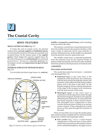
SKULL CAP OR CALVARIA (Fig. 7.1)                                      The anterior cranial fossa is separated posteriorly
     It forms the roof of cranial cavity. Its internal            from the middle fossa by the posterior borders of the
surface shows coronal, sagittal and lambdoid sutures              lesser wings of sphenoid which ends medially in
as it is formed by portions of frontal, two parietals and         anterior clinoid processes. In the middle it is separated
occipital bones. In the median plane is the sagittal              from the pituitary fossa by sphenoidal crest.
groove which lodges the superior sagittal sinus. By                   The middle cranial fossa is separated posteriorly
the side of the groove are arachnoid pits for arachnoid           from the posterior fossa by the superior border of
granulations. Numerous vascular grooves for                       petrous temporal laterally and dorsum sellae medially
meningeal vessels and impressions for gyri of brain               which has posterior clinoid processes at its lateral ends.
are visible.
                                                                  CONTENTS
INTERNAL SURFACE OF THE BASE OF SKULL
(Fig. 7.2)                                                        Dura matter and dural folds
    It is divisible into three large fossae viz, anterior,           The cranial dura mater has two layers — endosteal
                                                                  and meningeal (Fig. 7.3).
                                                                       K Endosteal layer or the outer layer is the
                                                                          endosteum of the skull bones. It is continuous
                                                                          with the pericranium through the sutures and
                                                                          foramina, and is adherent to the bones at these
                                                                          places and to the base of the skull. At the
                                                                          foramen magnum it fuses with the periosteum
                                                                          of the edge of the foramen to be continuous
                                                                          with the pericranium of the scalp.
                                                                       K Meningial layer or the inner layer is
                                                                          continuous with the spinal dura mater through
                                                                          the foramen magnum. It protects and supports
                                                                          different parts of the brain. In some places the
                                                                          two layers separate to form venous sinuses.
                                                                          The meningeal layer reduplicates to form
                                                                          curtains between parts of brain or to enclose
                                                                          venous sinuses. It also ensheaths the cranial
                                                                          nerves as they pass through the foramina of
                                                                          the skull.
                                                                      There are four folds, two occupy the median plane
                                                                  (falx cerebri and falx cerebelli) and two lie horizontally
                                                                  (tentorium cerebelli and diaphragm sellae).
                                                                       K Falx cerebri (Fig. 7.4) is a sickle-shaped
                                                                          reduplication of internal layer of dura mater
                                                                          which intervenes between the medial surfaces
  Fig. 7.1 Features on the interior aspect of skull cap.                  of two cerebral hemispheres. Its upper convex

                                                             41
42                                                               REGIONAL AND CLINICAL ANATOMY FOR DENTAL STUDENTS


                             Fig. 7.2 Upper surface of base of skull [basis cranii internal].

        edge is attached to the lips of the sagittal           sinus lying between the falx cerebri and the tentorium
        groove on the inner aspect of the vault of skull.      cerebelli.
        It is narrow in front where it is attached to the            K Falx cerebelli (Fig. 7.5) is a small, sickle shaped
        crista galli and becomes deeper posteriorly                     fold which separates the posterior parts of the
        where it has a straight inferior edge attached                  two cerebellar hemispheres. Superiorly, it is
        to the upper surface of the tentorium cerebelli.                attached to the under surface of the tentorium
    The superior sagittal sinus is enclosed within the                  cerebelli, and behind to the internal occipital
upper attached edge of falx cerebri. It begins at the                   crest. Its anterior border is free and concave.
crista galli anteriorly, and may be connected via an
emissary vein through foramen caecum with nasal
veins. Its chief tributaries are the superior cerebral
veins. It has two-to-three lateral prolongations known
as lacunae laterales (Fig. 7.3).
   The inferior sagittal sinus runs in the inferior
margin of the falx cerebri. It continues into the straight


                                                               Fig. 7.4 Falx cerebri, tentorium cerebelli and diaphragma
             Fig. 7.3 Duramater encephali.                              sellae.
THE CRANIAL CAVITY                                                                                                  43

         Its posterior border contains the occipital              The transverse sinus lies within its postero-lateral
         sinus, and its upper attached border is related      attachment to the lips of the sulcus on the side wall of
         to the posterior part of the straight sinus.         the posterior cranial fossa.
     K Tentorium cerebelli (Fig. 7.6) is a tent-like              The superior petrosal sinus lies within its antero-
         semilunar fold which roofs the posterior             lateral attachment to the superior border of the petrous
         cranial fossa and intervenes between the             temporal.
         cerebellum and occipital lobes of the brain. It
                                                                   K Diaphragma sellae (Fig. 7.4) is the fold roofing
         is attached from the apex of one petrous
                                                                      over the pituitary fossa with an aperture for
         temporal bone to the other along its upper
                                                                      infundibulum. On each side the layers of dura
         borders and horizontally along the inner
         surface of each side of the skull upto the                   mater separate to enclose the cavernous sinus.
         internal occipital protuberance.                             Anteriorly and posteriorly, the two layers.
                                                                      separate to enclose the small anterior and
    Its free margin is U-shaped and is known as the                   posterior intercavernous sinuses.
tentorial notch. Anteriorly, it is attached to the anterior
clinoid process, after crossing above the attached            Venous Sinuses
margin, which ends medially at the posterior clinoid              Intracranial venous sinuses differ from veins in
process.                                                      lacking muscular and adventitial walls. Their
    The straight sinus lies within the attachment of          endothelial lining is continuous with the veins
falx cerebri to the upper surface of the tentorium            connected with them. They do not have valves. Since
cerebelli.                                                    the meningeal layer of dura is tough and fibrous, the


                Fig. 7.5 Dural venous sinuses. Tentorium removed on one side to expose falx cerebelli.


                Fig. 7.6 Dural venous sinuses. Superior view of tentorium after removal of falx cerebri.
44                                                             REGIONAL AND CLINICAL ANATOMY FOR DENTAL STUDENTS

sinuses are always kept patent. All sinuses finally           Paired Sinuses (Fig. 7.7)
terminate directly or indirectly into the internal jugular         K Transverse sinuses lie along the posterolateral
vein.                                                                 edge of the tentorium cerebelli. On reaching
     Their tributaries are of three types:                            the petrous portion of the temporal bone, it is
     (i)   Veins from the brain                                       joined by the superior petrosal sinus to form
                                                                      the sigmoid sinus.
     (ii) Diploic veins from the dipole of the cranial
          bones and
     (iii) Emissary veins which connect them with
           extracranial veins.
     Some of the sinuses are unpaired, others paired.
Unpaired Sinuses (Fig. 7.5)
      K Superior sagittal sinus is situated in the
           midline along the superior border of falx
           cerebri. It extends from crista galli anteriorly
           to internal occipital protuberance posteriorly.
           Narrow at the beginning, it dilates as it
           proceeds backwards. At the internal occipital
           protuberance it usually turns to the right side
           to form the transverse sinus. Its tributaries
           are:
           — Superficial superior cerebral veins.
           — Venous spaces called lacunae lateralis.
           — Veins of frontal sinus through an emissary
             vein passing through foramen caecum.
           — Parietal emissary vein connects it to the
             occipital veins in the scalp.                    Fig. 7.7 Dural venous sinuses of posterior cranial fossa.
      K Inferior sagittal sinus runs in the posterior              K Sigmoid sinuses (Fig. 7.7) occupy the sigmoid
           two-thirds of the free edge of the falx cerebri.           sulcus in the posterior cranial fossa. It runs a
           At the anterior edge of the attachment of falx             sigmoid course to the lateral end of the jugular
           cerebri to the tentorium it joins the great                foramen where it ends in the superior bulb of
           cerebrial vein to form the straight sinus.                 the internal jugular vein. Mastoid emissary
      K Straight sinus runs along the intersection of                 vein, which emerges through the mastoid
           the falx cerebri and the tentorium cerebelli. It           process, connects the sigmoid sinus to the
           ends posteriorly at the internal occipital                 occipital or posterior auricular veins. Condylar
           protuberance, by turning laterally usually to
           the left to form the transverse sinus.
      K Occipital sinus runs in the posterior attached
           edge of falx cerebelli, between the internal
           occipital protuberance and the back of foramen
           magnum. It may split anteriorly to surround
           foramen magnum to get connected with the
           sigmoid sinus.
      K Basilar plexus lies on the clivus of skull. It
           connects the inferior petrosal sinuses
           anterolaterally with the cavernous sinuses
           anteriorly and the internal vertebral plexus
           posteriorly.
      K Anterior and posterior intercavernous sinuses
           lie in the corresponding margins of the
           diaphragma selle. Others pass across the floor     Fig. 7.8 Confluence of sinuses at internal occipital
           of the pituitary fossa.                                     protuberance.
THE CRANIAL CAVITY                                                                                                   45

        emissary vein, when present, passes through
                                                             CLINICAL APPLICATION
        the condylar canal to connect the lower end
        of the sigmoid sinus with the suboccipital               K The structure of the venous sinuses prevents
        venous plexus just below the base of the                    them from collapsing, so that if a sinus is torn
        skull.                                                      by a fracture of a cranial bone air can be sucked
                                                                    into the venous system sufficient to fill the
     K Superior petrosal sinuses lie within the                     right ventricle of the heart with froth to cause
        anterolateral attachment of the tentorium                   death.
        cerebelli. Medially they join the posterior end          K The sigmoid sinus is closely related to the
        of the corresponding cavernous sinus.                       middle ear, so it is liable to thrombosis due to
        Laterally they join the transverse sinus to                 an infection in the middle ear.
        form the sigmoid sinus.                                  K Sagittal sinus thrombosis can follow due to
     K Inferior petrosal sinuses begin at the                       infection of the skull, nose, face or scalp
        posterior end of the corresponding cavernous                because of its connections to these regions
        sinus. Each one of them runs backwards                      through diploic and emissary veins.
        laterally and downwards, in the groove                   K Sagittal sinus passes under the anterior
        between the temporal and occipital bones, to                fontanelle in the child. An intravenous
        the anterior compartment of the jugular                     infusion can be given into the sinus through
        foramen, through which it passes, to end in                 this fontanelle.
        the internal jugular vein. Its main tributaries
        are the labyrinthine veins from the internal            ANTERIOR CRANIAL FOSSA
        ear. Its other tributaries are the cerebellar and
                                                            BONY FEATURES
        pontine veins.
                                                                The anterior cranial fossa is limited laterally by the
     K Sphenoparietal sinuses (Fig. 7.13) run along
                                                            frontal bone, posteriorly by the posterior border of the
        the lesser wings of the sphenoid bone close to
                                                            lesser wings, anterior clinoid processes and anterior
        their posterior borders. Each ends in the
                                                            border of the sulcus chiasmaticus of the sphenoid bone.
        anterior part of the cavernous sinus.
                                                            The floor is formed by the orbital plates of frontal,
     K Cavernous sinsuses (Fig 7.13) lie lateral to         cribriform plate of the ethmoid and anterior part of the
        the body of sphenoid. It is unrelated to dural      body of sphenoid and its lesser wing.
        folds and is formed by the separation of the
                                                                The features of the fossa can be described as those
        two layers of dura mater. It extends from the
                                                            placed centrally and laterally (Fig. 7.9).
        superior orbital fissure to the petrous portion
        of the temporal bone. It is dealt with in detail        The median region is depressed area with the
        along with the contents of middle cranial           following features:
        fossa.                                                   K Crista galli (cocks comb) is a midline keel of
     K Middle meningeal veins (Fig. 7.13) may be                    bone projecting upwards. It is the upper end of
        regarded as venous sinuses. They join the                   the perpendicular plate of ethmoid, and gives
        spheno-parietal sinuses.                                    attachment to falx cerebri.
    At the internal occipital protuberance superior              K Frontal crest is a raised ridge running
sagittal, straight, two transverse and occipital sinuses            upwards in the part of frontal bone lying in
meet to form a common pool known as the confluence                  front of the crista galli. It also gives attachment
of sinuses.                                                         to falx cerebri.


                                    Fig. 7.9 Anterior cranial fossa—bony features.

Más contenido relacionado

La actualidad más candente (20)

Infra temporal fossa
Infra temporal fossaInfra temporal fossa
Infra temporal fossa
 
Facial artery dr gosai
Facial artery dr gosaiFacial artery dr gosai
Facial artery dr gosai
 
Norma basalis
Norma basalis  Norma basalis
Norma basalis
 
Anatomy of the face face
Anatomy of the face faceAnatomy of the face face
Anatomy of the face face
 
brachial arches and derivatives .
brachial arches  and derivatives .brachial arches  and derivatives .
brachial arches and derivatives .
 
Cranial cavity ( Department of Anatomy)
Cranial cavity ( Department of Anatomy)Cranial cavity ( Department of Anatomy)
Cranial cavity ( Department of Anatomy)
 
Submandibular Region (Anatomy of the Neck)
Submandibular Region (Anatomy of the Neck)Submandibular Region (Anatomy of the Neck)
Submandibular Region (Anatomy of the Neck)
 
Carotid Arteries (Anatomy of the Neck)
Carotid Arteries (Anatomy of the Neck)Carotid Arteries (Anatomy of the Neck)
Carotid Arteries (Anatomy of the Neck)
 
Pharyngeal arches
Pharyngeal archesPharyngeal arches
Pharyngeal arches
 
Mandibular nerve
Mandibular nerve Mandibular nerve
Mandibular nerve
 
Cavernous sinus
Cavernous sinusCavernous sinus
Cavernous sinus
 
Anatomy of para nasal sinuses
Anatomy of para nasal sinusesAnatomy of para nasal sinuses
Anatomy of para nasal sinuses
 
Pharyngeal apparatus
Pharyngeal apparatusPharyngeal apparatus
Pharyngeal apparatus
 
soft palate
soft palatesoft palate
soft palate
 
Muscles of the Tongue
Muscles of the TongueMuscles of the Tongue
Muscles of the Tongue
 
Segments of internal carotid artery
Segments of internal carotid arterySegments of internal carotid artery
Segments of internal carotid artery
 
Cavernous sinus
Cavernous sinusCavernous sinus
Cavernous sinus
 
Cranial nerve nuclei
 Cranial nerve nuclei  Cranial nerve nuclei
Cranial nerve nuclei
 
Maxillary artery
Maxillary arteryMaxillary artery
Maxillary artery
 
Lateral wall of nose
Lateral wall of noseLateral wall of nose
Lateral wall of nose
 

Similar a Cranial cavity

Anatomy of meninges, ventricles, cerebrospinal fluid
Anatomy of meninges, ventricles, cerebrospinal fluidAnatomy of meninges, ventricles, cerebrospinal fluid
Anatomy of meninges, ventricles, cerebrospinal fluidMBBS IMS MSU
 
Meninges of brain and spinal cord
Meninges of brain and spinal cordMeninges of brain and spinal cord
Meninges of brain and spinal cordmunnam37
 
Envelopes of the brain
Envelopes of the brainEnvelopes of the brain
Envelopes of the brainJSlinkyNY
 
Development Of Vertebral Column Group 1
Development Of Vertebral Column Group 1Development Of Vertebral Column Group 1
Development Of Vertebral Column Group 1Anan
 
3rd class dr.ali boresliy
3rd class dr.ali boresliy3rd class dr.ali boresliy
3rd class dr.ali boresliyAHS_Physio
 
1. Match the decription with the corresponding cranial dural septum.pdf
1. Match the decription with the corresponding cranial dural septum.pdf1. Match the decription with the corresponding cranial dural septum.pdf
1. Match the decription with the corresponding cranial dural septum.pdfarrowit1
 
envelopes-of-the-brain
envelopes-of-the-brainenvelopes-of-the-brain
envelopes-of-the-brainYoAmoNYC
 
7938186 envelopes-of-the-brain
7938186 envelopes-of-the-brain7938186 envelopes-of-the-brain
7938186 envelopes-of-the-brainabctutor
 
Anatomy of cerebellum
Anatomy of cerebellumAnatomy of cerebellum
Anatomy of cerebellumMBBS IMS MSU
 
Cerebellar anatomy as applied to cerebellar microsurgical resections
Cerebellar  anatomy as applied to cerebellar microsurgical resectionsCerebellar  anatomy as applied to cerebellar microsurgical resections
Cerebellar anatomy as applied to cerebellar microsurgical resectionsINUB
 
Base of skull by dr kifayat
Base of skull by dr kifayatBase of skull by dr kifayat
Base of skull by dr kifayatKifayat Khan
 
Nervous System- Comparative account of Brain.pdf
Nervous System- Comparative account of Brain.pdfNervous System- Comparative account of Brain.pdf
Nervous System- Comparative account of Brain.pdfPRIMEPRIME4
 
Head and neck_dev_07
Head and neck_dev_07Head and neck_dev_07
Head and neck_dev_07Mpdodz
 
Dural folds and cavernous sinus
Dural  folds and cavernous sinusDural  folds and cavernous sinus
Dural folds and cavernous sinusAravind Murugesan
 
Gwowth cranial base /fixed orthodontic courses
Gwowth cranial base /fixed orthodontic coursesGwowth cranial base /fixed orthodontic courses
Gwowth cranial base /fixed orthodontic coursesIndian dental academy
 

Similar a Cranial cavity (20)

Anatomy of meninges, ventricles, cerebrospinal fluid
Anatomy of meninges, ventricles, cerebrospinal fluidAnatomy of meninges, ventricles, cerebrospinal fluid
Anatomy of meninges, ventricles, cerebrospinal fluid
 
Meninges of brain and spinal cord
Meninges of brain and spinal cordMeninges of brain and spinal cord
Meninges of brain and spinal cord
 
Cranial cavity part 1
Cranial cavity part 1Cranial cavity part 1
Cranial cavity part 1
 
Envelopes of the brain
Envelopes of the brainEnvelopes of the brain
Envelopes of the brain
 
Development Of Vertebral Column Group 1
Development Of Vertebral Column Group 1Development Of Vertebral Column Group 1
Development Of Vertebral Column Group 1
 
3rd class dr.ali boresliy
3rd class dr.ali boresliy3rd class dr.ali boresliy
3rd class dr.ali boresliy
 
1. Match the decription with the corresponding cranial dural septum.pdf
1. Match the decription with the corresponding cranial dural septum.pdf1. Match the decription with the corresponding cranial dural septum.pdf
1. Match the decription with the corresponding cranial dural septum.pdf
 
2. brain
2. brain2. brain
2. brain
 
envelopes-of-the-brain
envelopes-of-the-brainenvelopes-of-the-brain
envelopes-of-the-brain
 
7938186 envelopes-of-the-brain
7938186 envelopes-of-the-brain7938186 envelopes-of-the-brain
7938186 envelopes-of-the-brain
 
Meninges
MeningesMeninges
Meninges
 
Anatomy of cerebellum
Anatomy of cerebellumAnatomy of cerebellum
Anatomy of cerebellum
 
Nervous systems in vertebrates: T.Y.B.Sc. Sem VI Notes
Nervous systems in vertebrates: T.Y.B.Sc. Sem VI NotesNervous systems in vertebrates: T.Y.B.Sc. Sem VI Notes
Nervous systems in vertebrates: T.Y.B.Sc. Sem VI Notes
 
Cerebellar anatomy as applied to cerebellar microsurgical resections
Cerebellar  anatomy as applied to cerebellar microsurgical resectionsCerebellar  anatomy as applied to cerebellar microsurgical resections
Cerebellar anatomy as applied to cerebellar microsurgical resections
 
Base of skull by dr kifayat
Base of skull by dr kifayatBase of skull by dr kifayat
Base of skull by dr kifayat
 
Meninges - Anatomy of the Meninges & CSF
Meninges - Anatomy of the Meninges & CSFMeninges - Anatomy of the Meninges & CSF
Meninges - Anatomy of the Meninges & CSF
 
Nervous System- Comparative account of Brain.pdf
Nervous System- Comparative account of Brain.pdfNervous System- Comparative account of Brain.pdf
Nervous System- Comparative account of Brain.pdf
 
Head and neck_dev_07
Head and neck_dev_07Head and neck_dev_07
Head and neck_dev_07
 
Dural folds and cavernous sinus
Dural  folds and cavernous sinusDural  folds and cavernous sinus
Dural folds and cavernous sinus
 
Gwowth cranial base /fixed orthodontic courses
Gwowth cranial base /fixed orthodontic coursesGwowth cranial base /fixed orthodontic courses
Gwowth cranial base /fixed orthodontic courses
 

Más de Knowlittle Matharu (20)

Hospital management
Hospital managementHospital management
Hospital management
 
Tqm
TqmTqm
Tqm
 
Dialysis unit
Dialysis unitDialysis unit
Dialysis unit
 
Advertisingagencyanditsfunctions 110214051140-phpapp02
Advertisingagencyanditsfunctions 110214051140-phpapp02Advertisingagencyanditsfunctions 110214051140-phpapp02
Advertisingagencyanditsfunctions 110214051140-phpapp02
 
Green hospitals
Green hospitalsGreen hospitals
Green hospitals
 
Green hospitals
Green hospitalsGreen hospitals
Green hospitals
 
Role of it in scm
Role of it in scmRole of it in scm
Role of it in scm
 
Attachments (1)
Attachments (1)Attachments (1)
Attachments (1)
 
Dermatology
DermatologyDermatology
Dermatology
 
Dermatology 2008 trial
Dermatology 2008 trialDermatology 2008 trial
Dermatology 2008 trial
 
Diabetes update
Diabetes updateDiabetes update
Diabetes update
 
Chemical safety bio-medical_waste_management_self_learning_document_for_docto...
Chemical safety bio-medical_waste_management_self_learning_document_for_docto...Chemical safety bio-medical_waste_management_self_learning_document_for_docto...
Chemical safety bio-medical_waste_management_self_learning_document_for_docto...
 
Chapter10
Chapter10Chapter10
Chapter10
 
Chapter9
Chapter9Chapter9
Chapter9
 
Chapter8
Chapter8Chapter8
Chapter8
 
Chapter7
Chapter7Chapter7
Chapter7
 
Chapter4
Chapter4Chapter4
Chapter4
 
Chapter3
Chapter3Chapter3
Chapter3
 
Chapter2
Chapter2Chapter2
Chapter2
 
Chapter1
Chapter1Chapter1
Chapter1
 

Último

METHODS OF ACQUIRING KNOWLEDGE IN NURSING.pptx by navdeep kaur
METHODS OF ACQUIRING KNOWLEDGE IN NURSING.pptx by navdeep kaurMETHODS OF ACQUIRING KNOWLEDGE IN NURSING.pptx by navdeep kaur
METHODS OF ACQUIRING KNOWLEDGE IN NURSING.pptx by navdeep kaurNavdeep Kaur
 
ANTI-DIABETICS DRUGS - PTEROCARPUS AND GYMNEMA
ANTI-DIABETICS DRUGS - PTEROCARPUS AND GYMNEMAANTI-DIABETICS DRUGS - PTEROCARPUS AND GYMNEMA
ANTI-DIABETICS DRUGS - PTEROCARPUS AND GYMNEMADivya Kanojiya
 
Radiation Dosimetry Parameters and Isodose Curves.pptx
Radiation Dosimetry Parameters and Isodose Curves.pptxRadiation Dosimetry Parameters and Isodose Curves.pptx
Radiation Dosimetry Parameters and Isodose Curves.pptxDr. Dheeraj Kumar
 
maternal mortality and its causes and how to reduce maternal mortality
maternal mortality and its causes and how to reduce maternal mortalitymaternal mortality and its causes and how to reduce maternal mortality
maternal mortality and its causes and how to reduce maternal mortalityhardikdabas3
 
MedDRA-A-Comprehensive-Guide-to-Standardized-Medical-Terminology.pdf
MedDRA-A-Comprehensive-Guide-to-Standardized-Medical-Terminology.pdfMedDRA-A-Comprehensive-Guide-to-Standardized-Medical-Terminology.pdf
MedDRA-A-Comprehensive-Guide-to-Standardized-Medical-Terminology.pdfSasikiranMarri
 
PERFECT BUT PAINFUL TKR -ROLE OF SYNOVECTOMY.pptx
PERFECT BUT PAINFUL TKR -ROLE OF SYNOVECTOMY.pptxPERFECT BUT PAINFUL TKR -ROLE OF SYNOVECTOMY.pptx
PERFECT BUT PAINFUL TKR -ROLE OF SYNOVECTOMY.pptxdrashraf369
 
Introduction to Sports Injuries by- Dr. Anjali Rai
Introduction to Sports Injuries by- Dr. Anjali RaiIntroduction to Sports Injuries by- Dr. Anjali Rai
Introduction to Sports Injuries by- Dr. Anjali RaiGoogle
 
LUNG TUMORS AND ITS CLASSIFICATIONS.pdf
LUNG TUMORS AND ITS  CLASSIFICATIONS.pdfLUNG TUMORS AND ITS  CLASSIFICATIONS.pdf
LUNG TUMORS AND ITS CLASSIFICATIONS.pdfDolisha Warbi
 
Culture and Health Disorders Social change.pptx
Culture and Health Disorders Social change.pptxCulture and Health Disorders Social change.pptx
Culture and Health Disorders Social change.pptxDr. Dheeraj Kumar
 
Clinical Pharmacotherapy of Scabies Disease
Clinical Pharmacotherapy of Scabies DiseaseClinical Pharmacotherapy of Scabies Disease
Clinical Pharmacotherapy of Scabies DiseaseSreenivasa Reddy Thalla
 
COVID-19 (NOVEL CORONA VIRUS DISEASE PANDEMIC ).pptx
COVID-19  (NOVEL CORONA  VIRUS DISEASE PANDEMIC ).pptxCOVID-19  (NOVEL CORONA  VIRUS DISEASE PANDEMIC ).pptx
COVID-19 (NOVEL CORONA VIRUS DISEASE PANDEMIC ).pptxBibekananda shah
 
Presentation on General Anesthetics pdf.
Presentation on General Anesthetics pdf.Presentation on General Anesthetics pdf.
Presentation on General Anesthetics pdf.Prerana Jadhav
 
Basic principles involved in the traditional systems of medicine PDF.pdf
Basic principles involved in the traditional systems of medicine PDF.pdfBasic principles involved in the traditional systems of medicine PDF.pdf
Basic principles involved in the traditional systems of medicine PDF.pdfDivya Kanojiya
 
Biomechanics- Shoulder Joint!!!!!!!!!!!!
Biomechanics- Shoulder Joint!!!!!!!!!!!!Biomechanics- Shoulder Joint!!!!!!!!!!!!
Biomechanics- Shoulder Joint!!!!!!!!!!!!ibtesaam huma
 
The next social challenge to public health: the information environment.pptx
The next social challenge to public health:  the information environment.pptxThe next social challenge to public health:  the information environment.pptx
The next social challenge to public health: the information environment.pptxTina Purnat
 
Presentació "Real-Life VR Integration for Mild Cognitive Impairment Rehabilit...
Presentació "Real-Life VR Integration for Mild Cognitive Impairment Rehabilit...Presentació "Real-Life VR Integration for Mild Cognitive Impairment Rehabilit...
Presentació "Real-Life VR Integration for Mild Cognitive Impairment Rehabilit...Badalona Serveis Assistencials
 
Presentation for Bella Mahl 2024-03-28-24-MW-Overview-Bella.pptx
Presentation for Bella Mahl 2024-03-28-24-MW-Overview-Bella.pptxPresentation for Bella Mahl 2024-03-28-24-MW-Overview-Bella.pptx
Presentation for Bella Mahl 2024-03-28-24-MW-Overview-Bella.pptxpdamico1
 
systemic bacteriology (7)............pptx
systemic bacteriology (7)............pptxsystemic bacteriology (7)............pptx
systemic bacteriology (7)............pptxEyobAlemu11
 
Case Report Peripartum Cardiomyopathy.pptx
Case Report Peripartum Cardiomyopathy.pptxCase Report Peripartum Cardiomyopathy.pptx
Case Report Peripartum Cardiomyopathy.pptxNiranjan Chavan
 

Último (20)

METHODS OF ACQUIRING KNOWLEDGE IN NURSING.pptx by navdeep kaur
METHODS OF ACQUIRING KNOWLEDGE IN NURSING.pptx by navdeep kaurMETHODS OF ACQUIRING KNOWLEDGE IN NURSING.pptx by navdeep kaur
METHODS OF ACQUIRING KNOWLEDGE IN NURSING.pptx by navdeep kaur
 
ANTI-DIABETICS DRUGS - PTEROCARPUS AND GYMNEMA
ANTI-DIABETICS DRUGS - PTEROCARPUS AND GYMNEMAANTI-DIABETICS DRUGS - PTEROCARPUS AND GYMNEMA
ANTI-DIABETICS DRUGS - PTEROCARPUS AND GYMNEMA
 
Radiation Dosimetry Parameters and Isodose Curves.pptx
Radiation Dosimetry Parameters and Isodose Curves.pptxRadiation Dosimetry Parameters and Isodose Curves.pptx
Radiation Dosimetry Parameters and Isodose Curves.pptx
 
maternal mortality and its causes and how to reduce maternal mortality
maternal mortality and its causes and how to reduce maternal mortalitymaternal mortality and its causes and how to reduce maternal mortality
maternal mortality and its causes and how to reduce maternal mortality
 
MedDRA-A-Comprehensive-Guide-to-Standardized-Medical-Terminology.pdf
MedDRA-A-Comprehensive-Guide-to-Standardized-Medical-Terminology.pdfMedDRA-A-Comprehensive-Guide-to-Standardized-Medical-Terminology.pdf
MedDRA-A-Comprehensive-Guide-to-Standardized-Medical-Terminology.pdf
 
PERFECT BUT PAINFUL TKR -ROLE OF SYNOVECTOMY.pptx
PERFECT BUT PAINFUL TKR -ROLE OF SYNOVECTOMY.pptxPERFECT BUT PAINFUL TKR -ROLE OF SYNOVECTOMY.pptx
PERFECT BUT PAINFUL TKR -ROLE OF SYNOVECTOMY.pptx
 
Introduction to Sports Injuries by- Dr. Anjali Rai
Introduction to Sports Injuries by- Dr. Anjali RaiIntroduction to Sports Injuries by- Dr. Anjali Rai
Introduction to Sports Injuries by- Dr. Anjali Rai
 
LUNG TUMORS AND ITS CLASSIFICATIONS.pdf
LUNG TUMORS AND ITS  CLASSIFICATIONS.pdfLUNG TUMORS AND ITS  CLASSIFICATIONS.pdf
LUNG TUMORS AND ITS CLASSIFICATIONS.pdf
 
Culture and Health Disorders Social change.pptx
Culture and Health Disorders Social change.pptxCulture and Health Disorders Social change.pptx
Culture and Health Disorders Social change.pptx
 
Clinical Pharmacotherapy of Scabies Disease
Clinical Pharmacotherapy of Scabies DiseaseClinical Pharmacotherapy of Scabies Disease
Clinical Pharmacotherapy of Scabies Disease
 
Epilepsy
EpilepsyEpilepsy
Epilepsy
 
COVID-19 (NOVEL CORONA VIRUS DISEASE PANDEMIC ).pptx
COVID-19  (NOVEL CORONA  VIRUS DISEASE PANDEMIC ).pptxCOVID-19  (NOVEL CORONA  VIRUS DISEASE PANDEMIC ).pptx
COVID-19 (NOVEL CORONA VIRUS DISEASE PANDEMIC ).pptx
 
Presentation on General Anesthetics pdf.
Presentation on General Anesthetics pdf.Presentation on General Anesthetics pdf.
Presentation on General Anesthetics pdf.
 
Basic principles involved in the traditional systems of medicine PDF.pdf
Basic principles involved in the traditional systems of medicine PDF.pdfBasic principles involved in the traditional systems of medicine PDF.pdf
Basic principles involved in the traditional systems of medicine PDF.pdf
 
Biomechanics- Shoulder Joint!!!!!!!!!!!!
Biomechanics- Shoulder Joint!!!!!!!!!!!!Biomechanics- Shoulder Joint!!!!!!!!!!!!
Biomechanics- Shoulder Joint!!!!!!!!!!!!
 
The next social challenge to public health: the information environment.pptx
The next social challenge to public health:  the information environment.pptxThe next social challenge to public health:  the information environment.pptx
The next social challenge to public health: the information environment.pptx
 
Presentació "Real-Life VR Integration for Mild Cognitive Impairment Rehabilit...
Presentació "Real-Life VR Integration for Mild Cognitive Impairment Rehabilit...Presentació "Real-Life VR Integration for Mild Cognitive Impairment Rehabilit...
Presentació "Real-Life VR Integration for Mild Cognitive Impairment Rehabilit...
 
Presentation for Bella Mahl 2024-03-28-24-MW-Overview-Bella.pptx
Presentation for Bella Mahl 2024-03-28-24-MW-Overview-Bella.pptxPresentation for Bella Mahl 2024-03-28-24-MW-Overview-Bella.pptx
Presentation for Bella Mahl 2024-03-28-24-MW-Overview-Bella.pptx
 
systemic bacteriology (7)............pptx
systemic bacteriology (7)............pptxsystemic bacteriology (7)............pptx
systemic bacteriology (7)............pptx
 
Case Report Peripartum Cardiomyopathy.pptx
Case Report Peripartum Cardiomyopathy.pptxCase Report Peripartum Cardiomyopathy.pptx
Case Report Peripartum Cardiomyopathy.pptx
 

Cranial cavity

  • 1. 7 The Cranial Cavity BONY FEATURES middle and posterior cranial fossae, each extending from one side to the other. SKULL CAP OR CALVARIA (Fig. 7.1) The anterior cranial fossa is separated posteriorly It forms the roof of cranial cavity. Its internal from the middle fossa by the posterior borders of the surface shows coronal, sagittal and lambdoid sutures lesser wings of sphenoid which ends medially in as it is formed by portions of frontal, two parietals and anterior clinoid processes. In the middle it is separated occipital bones. In the median plane is the sagittal from the pituitary fossa by sphenoidal crest. groove which lodges the superior sagittal sinus. By The middle cranial fossa is separated posteriorly the side of the groove are arachnoid pits for arachnoid from the posterior fossa by the superior border of granulations. Numerous vascular grooves for petrous temporal laterally and dorsum sellae medially meningeal vessels and impressions for gyri of brain which has posterior clinoid processes at its lateral ends. are visible. CONTENTS INTERNAL SURFACE OF THE BASE OF SKULL (Fig. 7.2) Dura matter and dural folds It is divisible into three large fossae viz, anterior, The cranial dura mater has two layers — endosteal and meningeal (Fig. 7.3). K Endosteal layer or the outer layer is the endosteum of the skull bones. It is continuous with the pericranium through the sutures and foramina, and is adherent to the bones at these places and to the base of the skull. At the foramen magnum it fuses with the periosteum of the edge of the foramen to be continuous with the pericranium of the scalp. K Meningial layer or the inner layer is continuous with the spinal dura mater through the foramen magnum. It protects and supports different parts of the brain. In some places the two layers separate to form venous sinuses. The meningeal layer reduplicates to form curtains between parts of brain or to enclose venous sinuses. It also ensheaths the cranial nerves as they pass through the foramina of the skull. There are four folds, two occupy the median plane (falx cerebri and falx cerebelli) and two lie horizontally (tentorium cerebelli and diaphragm sellae). K Falx cerebri (Fig. 7.4) is a sickle-shaped reduplication of internal layer of dura mater which intervenes between the medial surfaces Fig. 7.1 Features on the interior aspect of skull cap. of two cerebral hemispheres. Its upper convex 41
  • 2. 42 REGIONAL AND CLINICAL ANATOMY FOR DENTAL STUDENTS Fig. 7.2 Upper surface of base of skull [basis cranii internal]. edge is attached to the lips of the sagittal sinus lying between the falx cerebri and the tentorium groove on the inner aspect of the vault of skull. cerebelli. It is narrow in front where it is attached to the K Falx cerebelli (Fig. 7.5) is a small, sickle shaped crista galli and becomes deeper posteriorly fold which separates the posterior parts of the where it has a straight inferior edge attached two cerebellar hemispheres. Superiorly, it is to the upper surface of the tentorium cerebelli. attached to the under surface of the tentorium The superior sagittal sinus is enclosed within the cerebelli, and behind to the internal occipital upper attached edge of falx cerebri. It begins at the crest. Its anterior border is free and concave. crista galli anteriorly, and may be connected via an emissary vein through foramen caecum with nasal veins. Its chief tributaries are the superior cerebral veins. It has two-to-three lateral prolongations known as lacunae laterales (Fig. 7.3). The inferior sagittal sinus runs in the inferior margin of the falx cerebri. It continues into the straight Fig. 7.4 Falx cerebri, tentorium cerebelli and diaphragma Fig. 7.3 Duramater encephali. sellae.
  • 3. THE CRANIAL CAVITY 43 Its posterior border contains the occipital The transverse sinus lies within its postero-lateral sinus, and its upper attached border is related attachment to the lips of the sulcus on the side wall of to the posterior part of the straight sinus. the posterior cranial fossa. K Tentorium cerebelli (Fig. 7.6) is a tent-like The superior petrosal sinus lies within its antero- semilunar fold which roofs the posterior lateral attachment to the superior border of the petrous cranial fossa and intervenes between the temporal. cerebellum and occipital lobes of the brain. It K Diaphragma sellae (Fig. 7.4) is the fold roofing is attached from the apex of one petrous over the pituitary fossa with an aperture for temporal bone to the other along its upper infundibulum. On each side the layers of dura borders and horizontally along the inner surface of each side of the skull upto the mater separate to enclose the cavernous sinus. internal occipital protuberance. Anteriorly and posteriorly, the two layers. separate to enclose the small anterior and Its free margin is U-shaped and is known as the posterior intercavernous sinuses. tentorial notch. Anteriorly, it is attached to the anterior clinoid process, after crossing above the attached Venous Sinuses margin, which ends medially at the posterior clinoid Intracranial venous sinuses differ from veins in process. lacking muscular and adventitial walls. Their The straight sinus lies within the attachment of endothelial lining is continuous with the veins falx cerebri to the upper surface of the tentorium connected with them. They do not have valves. Since cerebelli. the meningeal layer of dura is tough and fibrous, the Fig. 7.5 Dural venous sinuses. Tentorium removed on one side to expose falx cerebelli. Fig. 7.6 Dural venous sinuses. Superior view of tentorium after removal of falx cerebri.
  • 4. 44 REGIONAL AND CLINICAL ANATOMY FOR DENTAL STUDENTS sinuses are always kept patent. All sinuses finally Paired Sinuses (Fig. 7.7) terminate directly or indirectly into the internal jugular K Transverse sinuses lie along the posterolateral vein. edge of the tentorium cerebelli. On reaching Their tributaries are of three types: the petrous portion of the temporal bone, it is (i) Veins from the brain joined by the superior petrosal sinus to form the sigmoid sinus. (ii) Diploic veins from the dipole of the cranial bones and (iii) Emissary veins which connect them with extracranial veins. Some of the sinuses are unpaired, others paired. Unpaired Sinuses (Fig. 7.5) K Superior sagittal sinus is situated in the midline along the superior border of falx cerebri. It extends from crista galli anteriorly to internal occipital protuberance posteriorly. Narrow at the beginning, it dilates as it proceeds backwards. At the internal occipital protuberance it usually turns to the right side to form the transverse sinus. Its tributaries are: — Superficial superior cerebral veins. — Venous spaces called lacunae lateralis. — Veins of frontal sinus through an emissary vein passing through foramen caecum. — Parietal emissary vein connects it to the occipital veins in the scalp. Fig. 7.7 Dural venous sinuses of posterior cranial fossa. K Inferior sagittal sinus runs in the posterior K Sigmoid sinuses (Fig. 7.7) occupy the sigmoid two-thirds of the free edge of the falx cerebri. sulcus in the posterior cranial fossa. It runs a At the anterior edge of the attachment of falx sigmoid course to the lateral end of the jugular cerebri to the tentorium it joins the great foramen where it ends in the superior bulb of cerebrial vein to form the straight sinus. the internal jugular vein. Mastoid emissary K Straight sinus runs along the intersection of vein, which emerges through the mastoid the falx cerebri and the tentorium cerebelli. It process, connects the sigmoid sinus to the ends posteriorly at the internal occipital occipital or posterior auricular veins. Condylar protuberance, by turning laterally usually to the left to form the transverse sinus. K Occipital sinus runs in the posterior attached edge of falx cerebelli, between the internal occipital protuberance and the back of foramen magnum. It may split anteriorly to surround foramen magnum to get connected with the sigmoid sinus. K Basilar plexus lies on the clivus of skull. It connects the inferior petrosal sinuses anterolaterally with the cavernous sinuses anteriorly and the internal vertebral plexus posteriorly. K Anterior and posterior intercavernous sinuses lie in the corresponding margins of the diaphragma selle. Others pass across the floor Fig. 7.8 Confluence of sinuses at internal occipital of the pituitary fossa. protuberance.
  • 5. THE CRANIAL CAVITY 45 emissary vein, when present, passes through CLINICAL APPLICATION the condylar canal to connect the lower end of the sigmoid sinus with the suboccipital K The structure of the venous sinuses prevents venous plexus just below the base of the them from collapsing, so that if a sinus is torn skull. by a fracture of a cranial bone air can be sucked into the venous system sufficient to fill the K Superior petrosal sinuses lie within the right ventricle of the heart with froth to cause anterolateral attachment of the tentorium death. cerebelli. Medially they join the posterior end K The sigmoid sinus is closely related to the of the corresponding cavernous sinus. middle ear, so it is liable to thrombosis due to Laterally they join the transverse sinus to an infection in the middle ear. form the sigmoid sinus. K Sagittal sinus thrombosis can follow due to K Inferior petrosal sinuses begin at the infection of the skull, nose, face or scalp posterior end of the corresponding cavernous because of its connections to these regions sinus. Each one of them runs backwards through diploic and emissary veins. laterally and downwards, in the groove K Sagittal sinus passes under the anterior between the temporal and occipital bones, to fontanelle in the child. An intravenous the anterior compartment of the jugular infusion can be given into the sinus through foramen, through which it passes, to end in this fontanelle. the internal jugular vein. Its main tributaries are the labyrinthine veins from the internal ANTERIOR CRANIAL FOSSA ear. Its other tributaries are the cerebellar and BONY FEATURES pontine veins. The anterior cranial fossa is limited laterally by the K Sphenoparietal sinuses (Fig. 7.13) run along frontal bone, posteriorly by the posterior border of the the lesser wings of the sphenoid bone close to lesser wings, anterior clinoid processes and anterior their posterior borders. Each ends in the border of the sulcus chiasmaticus of the sphenoid bone. anterior part of the cavernous sinus. The floor is formed by the orbital plates of frontal, K Cavernous sinsuses (Fig 7.13) lie lateral to cribriform plate of the ethmoid and anterior part of the the body of sphenoid. It is unrelated to dural body of sphenoid and its lesser wing. folds and is formed by the separation of the The features of the fossa can be described as those two layers of dura mater. It extends from the placed centrally and laterally (Fig. 7.9). superior orbital fissure to the petrous portion of the temporal bone. It is dealt with in detail The median region is depressed area with the along with the contents of middle cranial following features: fossa. K Crista galli (cocks comb) is a midline keel of K Middle meningeal veins (Fig. 7.13) may be bone projecting upwards. It is the upper end of regarded as venous sinuses. They join the the perpendicular plate of ethmoid, and gives spheno-parietal sinuses. attachment to falx cerebri. At the internal occipital protuberance superior K Frontal crest is a raised ridge running sagittal, straight, two transverse and occipital sinuses upwards in the part of frontal bone lying in meet to form a common pool known as the confluence front of the crista galli. It also gives attachment of sinuses. to falx cerebri. Fig. 7.9 Anterior cranial fossa—bony features.