SlideShare una empresa de Scribd logo
1 de 18
Descargar para leer sin conexión
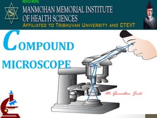
COMPOUND
MICROSCOPE
Mr. Govardhan Joshi
Microscope
• Microscope is the combination of two
words; "micro" meaning small and
"scope" meaning view.
• Compound deals with the microscope
having more than one lens.
History
• Optical
microscopes are
the oldest and
simplest of the
microscopes.
A 1879 Carl Zeiss Jena Optical microscope
The History
• Hans and Zacharias Janssen of Holland in the
59 ’s reated the first o pou d i ros ope
• Anthony van Leeuwenhoek and Robert Hooke
made improvements by working on the lenses
Anthony van Leeuwenhoek Hooke Microscope Robert Hooke
The History
Zacharias Jansen The First Mi ros ope
DiagramShowingLight
TravelingThrough The
Microscope
Parts of Microscope
Parts and function of
Microscope
Mechanical supporting system
Optical system
Illumination system
Adjustment system
Mechanical supporting system
• Base: Support microscope
• Arm: Supports the tube and connects it to the base
• Revolving nose piece/ turret: holds two or more objective
lenses and can be rotated to easily change power
• Stage: horizontal platform with central opening
• Mechanical stage: gives controlled movement to the object on
the slide
Optical system
• Objective:
• one or more lens held in the position by hollow metal cylinder
• Magnify the object
• They almost always consist of
• 4X- scanning objective
• 10X- low power objective
• 40X- high power objective- retractable
• 100X- oil immersion objective- retractable
• Eyepiece:
• Consist of upper eye lens and lower field lens
• They are usually 10X or 15X power
Illumination system
• Source of light: day light and electric light
• Mirror:
• Reflect light rays from light source to object
• One side is concave and other side is plane
• Concave mirror- used for near light source
• Plane mirror- used for distant source of lighted day light
• Condenser: focus the light onto the specimen
• Diaphragm: used to regulate amount of light passing into the
condenser
• Filter: contain blue and white filter below condenser
Adjustment system
• Coarse adjustment: raises / lowers stage to bring image into
focus
• Fine adjustment: brings image into sharp focus
Other parts
• Power Switch
• Stage Clips
• Stage Stop
• Stage adjustment
• Condenser adjustment
• Rheostat control knob
Use of microscope
Principle:
A focused beam of light passes through the
material under study into the microscope.
Parts of the specimen that are optically
dense and having high refractive index or
are colored when stain cast a potential
shadow which is magnified in to main stage
as it passes into the o server’s eye.
Focusing under low power
• Put the slide on the stage and bring specimen over central
aperture
• Lower the condenser and partly close the diaphragm
• Focus the object using coarse adjustment
• Use fine adjustment as required
Focusing under high power
• Rotate the nose piece to high power
• Adjust the condenser and open the diaphragm to admit
enough light
• Use fine adjustment as required
Focusing under oil immersion
• Focus the object first under low power
• Rotate nose piece to oil immersion objective
• Raise the condenser completely
• Open the iris diaphragm
• Keep a drop of immersion oil
• Use fine adjustment as required

Más contenido relacionado

La actualidad más candente (20)

Phase contrast microscope
Phase contrast microscopePhase contrast microscope
Phase contrast microscope
 
Compound microscope
Compound microscopeCompound microscope
Compound microscope
 
Simple microscope
Simple microscopeSimple microscope
Simple microscope
 
Introduction to microscopy
Introduction to microscopyIntroduction to microscopy
Introduction to microscopy
 
Types of microscope
Types of microscopeTypes of microscope
Types of microscope
 
Types of Microscope
Types of MicroscopeTypes of Microscope
Types of Microscope
 
Care of microscope
Care of microscopeCare of microscope
Care of microscope
 
Principles and application of light, phase constrast and fluorescence microscope
Principles and application of light, phase constrast and fluorescence microscopePrinciples and application of light, phase constrast and fluorescence microscope
Principles and application of light, phase constrast and fluorescence microscope
 
Microscopy
MicroscopyMicroscopy
Microscopy
 
Light microscope
Light microscopeLight microscope
Light microscope
 
DARK FIELD MICROSCOPY
DARK FIELD MICROSCOPYDARK FIELD MICROSCOPY
DARK FIELD MICROSCOPY
 
Fluorescence Microscopy
Fluorescence MicroscopyFluorescence Microscopy
Fluorescence Microscopy
 
Phase contrast microscopy
Phase contrast microscopyPhase contrast microscopy
Phase contrast microscopy
 
Compound microscope
Compound microscopeCompound microscope
Compound microscope
 
phase contrast microscope
phase contrast microscopephase contrast microscope
phase contrast microscope
 
Compound microscope
Compound microscopeCompound microscope
Compound microscope
 
Microscopy
MicroscopyMicroscopy
Microscopy
 
Microscope and its types
Microscope and its typesMicroscope and its types
Microscope and its types
 
centrifuge
centrifugecentrifuge
centrifuge
 
Light microscope
Light microscopeLight microscope
Light microscope
 

Similar a Compound microscope

compoundmicroscope-140728185207-phpapp01.pptx
compoundmicroscope-140728185207-phpapp01.pptxcompoundmicroscope-140728185207-phpapp01.pptx
compoundmicroscope-140728185207-phpapp01.pptxMohanSinghDhakad1
 
microscopepartsandfunctions ppt.pdf
microscopepartsandfunctions ppt.pdfmicroscopepartsandfunctions ppt.pdf
microscopepartsandfunctions ppt.pdfomarmendez83
 
Slit lamp biomicroscopy and illumination techniques
Slit lamp biomicroscopy and illumination techniquesSlit lamp biomicroscopy and illumination techniques
Slit lamp biomicroscopy and illumination techniquesLoknath Goswami
 
partsandfunctionsofacompoundmicroscope-120920041156-phpapp02-1.pptx
partsandfunctionsofacompoundmicroscope-120920041156-phpapp02-1.pptxpartsandfunctionsofacompoundmicroscope-120920041156-phpapp02-1.pptx
partsandfunctionsofacompoundmicroscope-120920041156-phpapp02-1.pptxJanetBarcimo1
 
Microscopepartsandfunctions
MicroscopepartsandfunctionsMicroscopepartsandfunctions
MicroscopepartsandfunctionsBrandi Shoemaker
 
partsandfunctionsofacompoundmicroscope-120920041156-phpapp02.pdf
partsandfunctionsofacompoundmicroscope-120920041156-phpapp02.pdfpartsandfunctionsofacompoundmicroscope-120920041156-phpapp02.pdf
partsandfunctionsofacompoundmicroscope-120920041156-phpapp02.pdfJoycePerez27
 
partsandfunctionsofacompoundmicroscope-120920041156-phpapp02 (1).pptx
partsandfunctionsofacompoundmicroscope-120920041156-phpapp02 (1).pptxpartsandfunctionsofacompoundmicroscope-120920041156-phpapp02 (1).pptx
partsandfunctionsofacompoundmicroscope-120920041156-phpapp02 (1).pptxJoycePerez27
 
Review of the microscope
Review of the microscopeReview of the microscope
Review of the microscopeSuki Savier
 
Q2 module-1-2 -microscope
Q2 module-1-2 -microscopeQ2 module-1-2 -microscope
Q2 module-1-2 -microscopeDee Bayn
 
Q2 module-1-2 -microscope
Q2 module-1-2 -microscopeQ2 module-1-2 -microscope
Q2 module-1-2 -microscopeDee Bayn
 

Similar a Compound microscope (20)

compoundmicroscope-140728185207-phpapp01.pptx
compoundmicroscope-140728185207-phpapp01.pptxcompoundmicroscope-140728185207-phpapp01.pptx
compoundmicroscope-140728185207-phpapp01.pptx
 
Microscope 1
Microscope 1Microscope 1
Microscope 1
 
microscopepartsandfunctions ppt.pdf
microscopepartsandfunctions ppt.pdfmicroscopepartsandfunctions ppt.pdf
microscopepartsandfunctions ppt.pdf
 
Microscope .pptx
Microscope  .pptxMicroscope  .pptx
Microscope .pptx
 
The_Microscope.ppt
The_Microscope.pptThe_Microscope.ppt
The_Microscope.ppt
 
Slit lamp biomicroscopy and illumination techniques
Slit lamp biomicroscopy and illumination techniquesSlit lamp biomicroscopy and illumination techniques
Slit lamp biomicroscopy and illumination techniques
 
partsandfunctionsofacompoundmicroscope-120920041156-phpapp02-1.pptx
partsandfunctionsofacompoundmicroscope-120920041156-phpapp02-1.pptxpartsandfunctionsofacompoundmicroscope-120920041156-phpapp02-1.pptx
partsandfunctionsofacompoundmicroscope-120920041156-phpapp02-1.pptx
 
Parts and functions of a compound microscope
Parts and functions of a compound microscopeParts and functions of a compound microscope
Parts and functions of a compound microscope
 
Microscopepartsandfunctions
MicroscopepartsandfunctionsMicroscopepartsandfunctions
Microscopepartsandfunctions
 
Compound microscope
Compound microscopeCompound microscope
Compound microscope
 
Microscope
MicroscopeMicroscope
Microscope
 
partsandfunctionsofacompoundmicroscope-120920041156-phpapp02.pdf
partsandfunctionsofacompoundmicroscope-120920041156-phpapp02.pdfpartsandfunctionsofacompoundmicroscope-120920041156-phpapp02.pdf
partsandfunctionsofacompoundmicroscope-120920041156-phpapp02.pdf
 
Microscopy-1.pptx
Microscopy-1.pptxMicroscopy-1.pptx
Microscopy-1.pptx
 
Slit lamp biomicroscope
Slit lamp biomicroscopeSlit lamp biomicroscope
Slit lamp biomicroscope
 
partsandfunctionsofacompoundmicroscope-120920041156-phpapp02 (1).pptx
partsandfunctionsofacompoundmicroscope-120920041156-phpapp02 (1).pptxpartsandfunctionsofacompoundmicroscope-120920041156-phpapp02 (1).pptx
partsandfunctionsofacompoundmicroscope-120920041156-phpapp02 (1).pptx
 
Microscopy
MicroscopyMicroscopy
Microscopy
 
Review of the microscope
Review of the microscopeReview of the microscope
Review of the microscope
 
Q2 module-1-2 -microscope
Q2 module-1-2 -microscopeQ2 module-1-2 -microscope
Q2 module-1-2 -microscope
 
Q2 module-1-2 -microscope
Q2 module-1-2 -microscopeQ2 module-1-2 -microscope
Q2 module-1-2 -microscope
 
Lab 0-B Microscope Training
Lab 0-B Microscope TrainingLab 0-B Microscope Training
Lab 0-B Microscope Training
 

Más de Govardhan Joshi

Más de Govardhan Joshi (11)

Immunoassay
ImmunoassayImmunoassay
Immunoassay
 
Erythrocyte Sedimentation Rate
Erythrocyte Sedimentation RateErythrocyte Sedimentation Rate
Erythrocyte Sedimentation Rate
 
Respiratory tract cytology
Respiratory tract cytologyRespiratory tract cytology
Respiratory tract cytology
 
Lupus erythematosus cell
Lupus erythematosus cellLupus erythematosus cell
Lupus erythematosus cell
 
Smear preparation techniques
Smear preparation techniquesSmear preparation techniques
Smear preparation techniques
 
Cytology of urine
Cytology of urineCytology of urine
Cytology of urine
 
Skin puncture method
Skin puncture methodSkin puncture method
Skin puncture method
 
Blood indices
Blood indicesBlood indices
Blood indices
 
Hemocytometer
HemocytometerHemocytometer
Hemocytometer
 
Atomic absorption spectrophotometry
Atomic absorption spectrophotometryAtomic absorption spectrophotometry
Atomic absorption spectrophotometry
 
Total leucocyte count
Total leucocyte countTotal leucocyte count
Total leucocyte count
 

Compound microscope

  • 2. Microscope • Microscope is the combination of two words; "micro" meaning small and "scope" meaning view. • Compound deals with the microscope having more than one lens.
  • 3.
  • 4. History • Optical microscopes are the oldest and simplest of the microscopes. A 1879 Carl Zeiss Jena Optical microscope
  • 5. The History • Hans and Zacharias Janssen of Holland in the 59 ’s reated the first o pou d i ros ope • Anthony van Leeuwenhoek and Robert Hooke made improvements by working on the lenses Anthony van Leeuwenhoek Hooke Microscope Robert Hooke
  • 6. The History Zacharias Jansen The First Mi ros ope
  • 9. Parts and function of Microscope Mechanical supporting system Optical system Illumination system Adjustment system
  • 10. Mechanical supporting system • Base: Support microscope • Arm: Supports the tube and connects it to the base • Revolving nose piece/ turret: holds two or more objective lenses and can be rotated to easily change power • Stage: horizontal platform with central opening • Mechanical stage: gives controlled movement to the object on the slide
  • 11. Optical system • Objective: • one or more lens held in the position by hollow metal cylinder • Magnify the object • They almost always consist of • 4X- scanning objective • 10X- low power objective • 40X- high power objective- retractable • 100X- oil immersion objective- retractable • Eyepiece: • Consist of upper eye lens and lower field lens • They are usually 10X or 15X power
  • 12. Illumination system • Source of light: day light and electric light • Mirror: • Reflect light rays from light source to object • One side is concave and other side is plane • Concave mirror- used for near light source • Plane mirror- used for distant source of lighted day light • Condenser: focus the light onto the specimen • Diaphragm: used to regulate amount of light passing into the condenser • Filter: contain blue and white filter below condenser
  • 13. Adjustment system • Coarse adjustment: raises / lowers stage to bring image into focus • Fine adjustment: brings image into sharp focus
  • 14. Other parts • Power Switch • Stage Clips • Stage Stop • Stage adjustment • Condenser adjustment • Rheostat control knob
  • 15. Use of microscope Principle: A focused beam of light passes through the material under study into the microscope. Parts of the specimen that are optically dense and having high refractive index or are colored when stain cast a potential shadow which is magnified in to main stage as it passes into the o server’s eye.
  • 16. Focusing under low power • Put the slide on the stage and bring specimen over central aperture • Lower the condenser and partly close the diaphragm • Focus the object using coarse adjustment • Use fine adjustment as required
  • 17. Focusing under high power • Rotate the nose piece to high power • Adjust the condenser and open the diaphragm to admit enough light • Use fine adjustment as required
  • 18. Focusing under oil immersion • Focus the object first under low power • Rotate nose piece to oil immersion objective • Raise the condenser completely • Open the iris diaphragm • Keep a drop of immersion oil • Use fine adjustment as required