2. GOALS
1. To have standardized terminology and approach in
interpretation of CTG
2. To identify abnormal CTG and appropriate
intervention
3. Risk management and documentation
5. • 17th century - 1818 Francis-Isaac
• Professor John Ferguson described fetal heart sound in 1827
Hohl fetal stethoscope Depaul fetal stethoscope
6. Procedure Rationale
1 Perform Leopold’s manoeuver by palpating
the maternal abdomen
To identify fetal presentation and
position
2 Place the listening device over the area of
maximum intensity, which is usually over the
back of the fetus, and clarity of the fetal heart
sound
To obtain the clearest and loudest
sound, this is easier to count.
3 Count the maternal radial pulse To differentiate it from the fetal
rate
4 Palpate the abdomen for the absence of
uterine activity
To be able to count FHR between
contraction
5 Count the FHR for 30 to 60 seconds between
contractions
To identify the basal heart rate
(BHR) which can only be assessed
during the absence of uterine
activity
6 Auscultate the FHR during contraction, if
possible, and for 30 seconds after the end of
the contraction
To identify the FHR during the
contraction and as a response to
the contraction
7 When there are distinct discrepancies in FHR
during or between listening periods,
auscultate for a longer period during, after
and between contraction
To identify changes from the
baseline that indicate the need for
another mode of FHR monitoring
7. FREQUENCY OF AUSCULTATION
Stageoflabour Lowrisk Highrisk
Latentphase Every30-60minutes Every30minutes
Activephase Every30minutes Every15minutes
Secondstage Every15minutes Every5minutes
12. DEFINITION
(CTG) is a technical means of recording (-graphy) the
fetal heartbeat (cardio-) and the uterine contractions (-
toco-) during pregnancy
The machine used to perform the monitoring is called a
cardiotocograph (CTG), more commonly known as an
electronic fetal monitor (EFM)
15. ADVANTAGES
FHR & contraction can be
monitored & recorded at
the same time
Reduce rates of seizure in
newborn
DISADVANTAGES
Prevent mother from moving
Unable to change position
Increase interventions
(instrumental deliveris or C-sec)
16. TYPES
EXTERNAL CTG
An ultrasound transducer
over the abdomen that will
pick up the baby's
heartbeat. The heartbeat
will be recorded
continuously on a paper
strip.
Tocogram- a pressure
gauge that measures the
frequency of your
contractions.
INTERNAL CTG
This method can only be
used if membranes (fore-
waters) and your cervix
have ruptured either
spontaneously or
artificially.
An electrode is placed on
the baby’s scalp to directly
monitor the fetal heart
rate. An electrode is called
a fetal scalp electrode (FSE)
17.
18. The length of the CTG strip depends on the paper speed. In
the UK it is usually 1cm/min.
Each vertical division on the paper is 1cm and therefore 1
min.
SALSO 2015
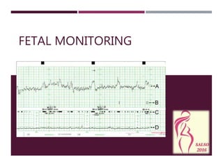
ELECTRICAL FETAL MONITORING: TERMINOLOGY
21. DR DEFINE RISK
C CONTRACTION
BR BASELINE HEART
RATE
A ACCELARATION
VA VARIABILITY
D DECELARATION
O OVERALL
INTERPRETATION OF CTG: DR C BR A VA DO
22. The process of birth is the most dangerous journey any
individual undertakes
Assess from the history and examination either low
risk or high risk pregnancy
Admission CTG performed, then intermittent ascultation vs
continuous monitoring
1. DR = DEFINE RISK
23. *ALL ANTENATAL PATIENT CAME TO LABOUR ROOM WILL
HAVE AN ADMISSION CTG AT LEAST 20 MINS TRACING
RISK CTG MONITORING
LOW - Intermittent tracing 2-4hly
- Continous CTG is unnecessary
MODERATE - Intermittent CTG trace to be decide by registra or
specialist
- Frequency & length of CTG tracing depends on
individual case & previous trace
HIGH - Intermittent CTG trace to be decide by registrar or
specialist
- Continous CTG may be needed
24. Assess contraction pain from the tocogram- quantifying the number
of contractions present in a 10-minute window
There are several factors used in assessing uterine activity.
Frequency
Duration
Normal- less than or equal to 5 contractions in 10 minutes, averaged
over a 30-minute window
Tachysystole- more than 5 contractions in 10 minutes, averaged over
a 30-minute window
2. C = CONTRACTION
27. Level of the fetal heart rate when this stable with
accelerations and decelerations excluded. Determined
over a period of 5 or 10 min and expressed in bpm.
Normal range is 100-160bpm.
3. BR = BASELINE FETAL HEART RATE
31. Bradycardia- baseline fetal heart rate < 100bpm
* A stable basline fetal heart rate between 90-99
bpm with normal baseline variability may be a
normal variation
32. Transient increase in heart rate of 15bpm or more and
lasting 15s or more.
The recording of at least 2 accelerations in a 20 min
period is considered a reactive trace
Accelerations are the hallmark of fetal health.
4. A = ACCELERATION
33. NICE GUIDELINES 2014
Acceleration is not a feature to categorise CTG
Fetal heart rate accelerations is generally a sign that
unborn baby is healthy
Abscence of accelerations in an otherwise normal CTG
does not indicate acidosis
If FBS is indicated and sample cannot be obtained,but
scalp stimulation results in fetal heart rate
acceleration,decide whether to continue labour or
expedite the birth in light of the clinical circumstances and
in discussion with the woman
34. Baseline varies within a particular band width excluding
accelerations and decelerations. It indicates the integrity of the
autonomic nervous system
Silent , 0-5bpm
Reduced, 5-
10bpm
Normal, 10-25bpm
Saltatory, >25bpm
5. VA = VARIABILITY
43. Normal/
reassuring
Non-
reassuring
Abnormal
None or early Variable deceleration dropping from
baseline less than 60 beats
AND
taking less than 60 sec to recover
Present over 90min
Occuring over 50% of contraction
Non-reassuring variable
deceleration
Still observed 30min after starting
conservative measures
Variable deceleration dropping from
baseline >60 bpm
OR
taking > 60sec to recover
Present for up to 30min
Occuring over 50% of contractions
Bradycardia or single prolonged
deceleration lasting 3min or more
Late deceleration present for
up to 30min
Occuring over 50% of contractions
Late deceleration present for
>30 min
Occuring over 50% of contraction
Do not improve with conservative
measures
44. Decelerations (variable or late) accompanied by
fetal tachycardia or reduced baseline variability
Take action sooner than 30min
46. 4 FEATURES OF CTG
1) Acceleration – no longer include
2) Baseline fetal heart rate
3) Baseline variability
4) Deceleration
Used to
categorise CTG
47. CTG catogories
Normal /reassuring All 3 features are normal
Non-reassuring 1 non-reasuring feature
AND 2 normal/reassuring
features
• Conservative measures
Abnormal
(need for conservative
measures AND further
testing)
1 abnormal feature
OR
2 non-reasuring features
• Conservative measures
• Offer FBS
or
Expedite birth if FBS
cannot be obtained and no
accelerations are seen as a
result of scalp stimulation
Abnormal
(need for urgent intervention)
Bradycardia or single
prolonged deceleration with
baseline below 100bpm
persisting 3min or more
• Start Conservative measure
• Prepare for urgent birth
• Expedite birth if persist for
9min
• If heart rate recovers before
9 min, reassess decision to
expedite birth in discussion
with the woman
48. OVERALL CARE
Do not make any decision about woman’s care in labour on the basis of
CTG findings alone
Make a documented systematic assessment of the condition of woman
and the unborn baby hourly or more frequently if there is concern
49. HOW TO MANAGE??
DEPENDS ON VARIOUS FACTORS
1.Severity of abnormal trace
2.Antenatal risk
3.Patient’s age & parity
4.How advance she is in labour
5.Progress of labour
6.Colour of liquour
7.Patient’s opinion
50. CTG GOALS DO DON’T
Recurrent Type 2
decelerations
Improve placenta
perfusion
-Left lateral
position
-Reducing
frequency of
uterine
contractions
-Administer O2
-IV Fluid bolus
Prolonged
deceleration or
bradycardia
Improve placenta
perfusion
-Tocolytic agents
Minimal or absent
variability
Improve placenta
perfusion
-Lateral position
-Fetal stimulation
Hyperstimulation Reduce uterine
contractility
-Stop oxytocin
-Tocolytic agents
Recurrent variable
deceleration
Reduce cord
compression
-Maternal position Amnioinfussion
51. ** In an emergency setting-
acceptable time for decision to the
delivery of the baby is 30 minutes
52. SECOND STAGE CTG
Most of the time CTG is abnormal
Early deceleration or variable deceleration is
acceptable
Which CTG changes require interventions?
Prolonged bradycardia
Reduced variability
54. Fetal scalp PH testing is essentially an invasive vaginal
procedure performed when a woman is in active labour
to determine if the baby is getting enough oxygen
If possible should be performed before a decission of C-
Sec is made where the CTG changes are not conclusive or
suspicious
55. RESULT…
pH RESULT ACTION
NORMAL 7.25-7.35 REPEAT IF
ABNORMALITY
PERSIST
BORDERLINE 7.20-7.25 REPEAT TEST EVERY 30
MINS TILL DELIVERY IF
VAGINAL DELIVERY IS
EXPECTED SOON
ABNORMAL < 7.20 EXPEDITE DELIVERY
56. RISKS
Bleeding from puncture site
Infection
Bruising on fetal’s scalp
CONTRAINDICATIONS
Infections- hep C or HIV
Thrombocytopenia
Non cephalic presentation
Prematurity <34 weeks
57. RISK MANAGEMENT
Proper documentation in case notes- date,
time, signature
Document paired cord blood gases & accurate
Apgar Score
Photocopy the CTG and store properly
Frequent training of staff