
Recommended
More Related Content
What's hot
What's hot (20)
Similar to Vascular Anomalies: Vascular Tumours and Vascular malformations
Similar to Vascular Anomalies: Vascular Tumours and Vascular malformations (20)
More from Dr Sumita Shankar
More from Dr Sumita Shankar (7)
Recently uploaded
Recently uploaded (20)
Vascular Anomalies: Vascular Tumours and Vascular malformations
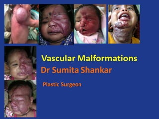
- 1. Vascular Malformations Dr Sumita Shankar Plastic Surgeon
- 2. Content • Introduction • Historical background • Classification • Hemangiomas and other common congenital vascular tumor • Vascular Malformation • Diagnosis • Management
- 3. Introduction • Vascular Anomalies are congenital lesions of abnormal vascular development • “Anomalies” signifies any deviation in normal cutaneous vasculature, either congenital or acquired • “Hemangioma” is commonly used in a generic sense to describe a variety of vascular lesions – congenital or acquired
- 4. Introduction : Vascular Anomalies • Most Commonly divided in to Hemangioma & Malformations • Vascular tumors and Malformations may occur anywhere on the body. • Importance: cause significant Functional Aesthetic impairment Anxiety
- 6. Anatomico-Pathologic Classification Virchow(1863) a) Angioma Angioma simplex: system composed of capillaries Angioma cavernosum: a replacement of normal vasculature with large channels Angioma racemosum: tissue consisted of markedly dilated interconnected vessels. b) Lymphangioma
- 8. Biologic Classification Mulliken and Glowacki (1982) On basis of cell kinetics, there are two major types of vascular anomalies: Hemangiomas: lesions demonstrating endothelial hyperplasia. Vascular Malformations : lesions with normal endothelial turnover. • Low flow Capilary / Lymphatic / Venous • High flow Arterial / Arterio-venous / Capillary-lymphatic Capillary-venous / Lymphatico-venous Capillary-lymphatico-venous
- 10. ISSVA Classification • 1996 Classification modified by International society for the study of vascular anomalies • differentiates vascular tumors from vascular malformations. • VASCULAR TUMORS VASCULAR MALFORMATIONS 1) Benign tumors 1) Slow/low flow Infantile Hemangioma Venular, venous, lymphatic Congenital Hemangioma 2) Intermediate tumors 2) Fast /high flow Kaposiform hemangioendothelioma AVM Spindle cell hemangioendothelioma AVF
- 12. ISSVA Classification of Vascular Tumours
- 14. Hemangiomas • The word "hemangioma" comes from the Greek haema-, "blood"; angeio , "vessel"; -oma , "tumor". • A hemangioma is a benign and usually self-involuting tumor of the endothelial cells that line blood vessels, and is characterised by increased number of normal or abnormal vessels filled with blood. • Exhibits rapid early growth until 6-8 months of age, followed by regression by 5-9 years of age. • Capillary Hemangioma: Cavernous Hemangioma the most common type. made up of small capillaries is made up of larger blood vessels that are normal in size and diameter, that are dilated. They are not as but high in number. are typically closely packed and the spaces Bright red in colour (or "caverns") between them are filled with blood.
- 15. Infantile Hemangioma • Present shortly after birth • Well demarcated, flat & erythematous red patches • Can be confused with several red birth marks • Rapid proliferation & vertical growth – pathognomic feature • Appear during 1st to 4th week of birth, rapid post- natal growth till 5 -6 month, after 1 year growth rate of lesion is equivalent to child growth rate • Five distinct developmental phase PRODORMAL PHASE INITIAL PHASE PROLIFERATION PHASE MATURATION PHASE REGRESSION PHASE
- 16. • PRODORMAL PHASE Precursor lesion – telengectasia, anemic reddish-blue, port-wine stain Some times can not be detected • INITIAL PHASE Appear with in few days Infiltrating to surrounding tissue Clearly protrude from skin – light reddish , shine brightly • PROLIFERATIVE PHASE Exophytic or endophytic subcutaneous growth CCDS (color coded duplex sonography) – hypercapilarization • MATURATION PHASE Reduction of bulk along with wrinkles Gray regression area in dark red hemangioma • REGRESSION PHASE Usually accomplished by 6th birthday of child Small lesions can regress without destroying surrounding tissue Large lesions leave – telengectasia, areas of atrophy, skin wrinkles, hypopigmentation
- 17. Associated malformative anomalies • IH can arise in association with other structural anomalies • Female predilection (9:1) • Not necessarily vascular • Acronym – PHACE Posterior fossa anomalies Hemangioma of face Arterial abnormalities Cardiac defect & coarcation Eye anomalies • VASCULAR ANOMALIES – persistent embryonic extra / intra cranial arteries Hypoplasia / absence of ipsilateral carotid vessels Aneurysmal dilation of CA Coarcation & Rt sided aortic notch • Cerebroocclusive changes – seizure and stroke in infancy • Chance of Sternal nonunion or Supraumbilical Raphe - PHACES
- 18. Congenital Hemangiomas • RICH • NICH • others
- 19. CONGENITAL HEMANGIOMA RICH (Rapid Involuting Congenital Hemangioma) • Red-violaceous color • Central telengectasia • Peripheral pale halo (occasional) • Superficial ulceration • Central nodularity • Transient Thrombocytopenia • Platelet level increase with regression • Mainly seen in trunk & extrimities • Rapidly involute during first week/ month
- 20. Congenital Hemangioma NICH (Non Involuting Congenital Hemangioma) • Ovoid/ macular/ slightly raised • Light gray in colour • Prominent coarse telengectasia • Warm on palpation • Predisposition in mandibular border • Well circumscribed – 5-6 cm diameter • Commonly diagnosed in late childhood • Sometime expand in adolescence & exhibit polypoid excrescence
- 21. RICH & NICH • radiologicaly & histologically overlap with IH • There are more similarities rather than dissimilarities between His, RICHs, and NICHs • They all can coexist with His
- 22. Evolution scheme of untreated IH, RICH, and NICH. IH appears after birth, grows generally until the end of the first year of life, then stabilizes and gradually involutes most significantly during years 1 to 2 and continues to improve over the ensuing years. Michel Wassef et al. Pediatrics 2015;136:e203-e214
- 23. Tufted Angioma • Develops in Infancy or early childhood on neck or upper trunk • Dull red macules with a mottled appearance • Histo: Clusters of angiosomes tufts and lobules scattered in the demis in a so called ‘Cannonball’ pattern • There is still a controversy that whether this lesion is a separate type or a variety of NICH • US reveal – separate lobular structure with vessels along the edges • It is a rare lesion of head and neck region
- 24. Complication of IH • Ulceration : <5% shows ulceration common in lip, anogenital region • Obstruction Vision : obstruction of visual axis, diplopia, can distort growing cornea – error in refractive index Respiratory : Hm in nasal tip can obstruct the vestibular passage, subglottic hemangioma cause sever airway obstruction Auditory : can obstruct auditory canal – conductive hearing loss Bleeding : spontaneous bleeding from small puncture of lesion can occur – occasionally associated with ulceration Congestive Heart Failure • Skeletal deformation Deviation of nasal pyramid Minor indentation of calvaria Orbital enlargement
- 25. Pyogenic Granuloma • Solitary, red papule, grows rapidly, forming a stalk. • Lesions less than 1 cm in diameter • Presentation is inversely correlated with age
- 26. Uncommon Vascular Tumors • Kaposiform Hemangio endothelioma • Associated with Kasabach-meritt syndrome Hemolytic Anaemia Thrombocytopenia Hypofibrogenemia
- 27. Angiosarcoma • Rare • Aggressive malignant neoplasm • Female preponderance • Average age onset 3.7 yrs • Poor prognosis
- 29. Vascular Malformations • Present at birth. • No rapid ‘proliferative phase’ and do not ‘involute’. They grow commensurately with the patient. • # 31% are in the head and neck region. • Interruption at a particular stage of development of a vessel may result in VM. • The type of VM depends on stage at which normal morphogenesis is interrupted.
- 30. Development of Vascular Malformations • ENDOTHELIAL stage multiple endothelium lined lakes are formed. • RETIFORM stage, capillary communication develops between these lakes. Some of these interconnecting channels have muscular sheaths and others do not. • MATURATION stage, channels that have muscular lining differentiate to become arteries and those without muscle become veins. Abnormal development of either arterial or the venous side of vascular network during this phase of development may result in vascular malformation.
- 31. Progression of Vascular Malformations • Trauma, infection, and hormonal fluctuation (pregnancy or puberty) increases growth of VM. • The mechanism of growth is due to alteration in the flow dynamics within and around the lesion”. • • This results in recruitment of “collateral vessels” and dilatation of involved vessels.
- 32. Capillary Malformations (Portwine Stain) • Naevus flammeus neonatorum “Angel’s kiss” or Stork bite • appear as reddish-pink macules over facial & nuchal region • dermatomes may be smooth initially but become more “ pebble – like” as the patient grows. • in 1999 Waner and Suen based on their identification of the anomalies re-categorized them as Venular Malformations
- 33. Portwine Stain
- 34. Associated Syndromes with CMc • Sturge-Weber Syndrome SWS encephalotrigeminal angiomatosis consists of congenital Hamartomatous Malformations that may affect the eye, the skin, and the central nervous system at different times. • Cutis Marmorata Telengiectatica Congenita • Macrocephaly Capillary Malformation
- 35. Venous Malformations - Low-flow lesions • “Cavernous Hemangioma” a misnomer • Bluish, soft and easily compressible, no bruits. • No “pulsations or a thrill” • They are sequestered from the main vessel • Undergoes a spontaneous continuous cycle of thrombosis and thrombolysis, • may undergo calcifications to form Phleboliths that are painful on palpation and could be a radiologic marker in some types of VeM
- 36. Classification of Venous malformation Type Description • Type I Isolated malformation without peripheral drainage • Type II Malformation that drains into normal veins • Type III Malformation that drains into dysplastic veins • Type IV Venous ectasia
- 37. Genuine Diffuse Phlebactasia of Bockenheimer • Associated with Limb Discrepancy Extensive venous malformtion of Limb • Klippel-Trenaunay –Weber syndrome Triad of capillary malformations, bone hypertrophy and venous malformations.
- 38. VeM Left Eyebrow
- 40. Associated Syndromes with VeM • Glomuvenous Malformation • Cutaneomucous Venous Malformation • Blue Rubber Bleb Nevus Syndrome
- 41. Lymphatic Malformations -Low flow lesions • Obstruction or sequestration of the primitive lymphatic vessels during embryogenesis • Within the oral cavity the LMs are more commonly present on anterior 2/3 of tongue, followed by palate, gingiva and oral mucosa. • Predilection for head and neck and the axilla, where embryonic lymph sacs are located
- 42. Lymphatic Malformation (LM) • Common (cystic) LMs Macrocystic LM Microcystic LM Mixed cystic LM • Generalized lymphatic anomaly (GLA) LM in Gorham-Stout disease Channel-type LM • Primary lymphedema • Others
- 44. Lymphatic Malformations Macrocystic LMs (outdated terms include Lymphangioma Cavernosum, Cystic Hygroma, Lymphangioma Cysticum) • Multiple cysts of >2 cm • Commonly found in posterior triangle of the neck and in the cervical area • Localized painless non-pulsatile swelling • No bruit or thrill • A rubbery compressible consistency • Normal appearing skin unless hemorrhage which can produce a blue discoloration.
- 45. Macrocystic LMs
- 46. de Serres Classification of Lymphatic Malformation of Head and Neck Stages Location • I Unilateral infrahyoid • II Unilateral suprahyoid • III Unilateral infra & supra hyoid • IV Bilateral suprahyoid • V Bilateral supra & infra hyoid
- 47. Microcystic LM (Outdated term Lymphangioma) In the oral cavity appear as multiple translucent non- compressible cysts or vesicles of <2 cm. containing viscous clear fluid, producing a pebbly or warty surface resembling “frog spawn” or “tapioca pudding”.
- 48. Congenital / Primary Lymphedma • ‘Milroy’s Disease • Anomalous Lymphatic Channels • Compression, Suction lipectomy • Lymphovenous Shunts
- 49. Arterial Malformation • Aneurysms • Fistulae • Ectasias • Stenoses
- 50. Arteriovenous Malformation • Congenital High-Flow • Growth triggered by Puberty /Trauma • Pathogenesis – TGF- beta & a genetic two hit hypothesis (knudson’s)
- 52. Arterial / Arteriovenous Malformations (AVM) “High-flow lesions” ( outdated terms – Cirsoid aneurysm, arteriovenous aneurysm) • Direct communication between the arterial and venous systems through a nidus formed by arteriovenous shunts, along with hypertrophy of the afferent arterial and efferent venous system. • AVM is present at birth • May become clinically apparent only during the 4-5th decade of life and is often misdiagnosed due to delay in clinical presentation. • The most common site for AVM is the brain, followed by the head, neck, limbs, trunk, and viscera. • The majority of the head and neck lesions occur on the cheek, followed by the ear, nose, forehead and upper lip.
- 53. AVM: Clinical Presentation • They are purple-blue raised painful macule, are pulsatile with thrill and bruit, warm to touch. • Do not empty fully on compression, and refill quickly on reliving digital pressure. • Can have embolism, pain, bleeding, ulceration, and congestive cardiac failure due to increased cardiac load. • Often a patient presents with severe bleeding as the first sign that a high flow-lesion is present. They may also complain of recurrent gingival bleeding and loose or depressible teeth
- 54. Staging of arterio-venous malformations Schobinger Staging of AVM • Stage 1 (Quiescence) : A blue-skin blush, warmth and AVM on Ultrasound • Stage2 (Expansion) : A mass with a pulsation,bruit and a thrill • Stage 3 (Destruction) :A mass with ulceration, bleeding and pain • • Stage 4 (Decompensation) :lesions producing heart failure
- 55. Combined Vascular Malformation • CM + VM Capillary-venous malformation • CM + LM Capillary-lymphatic malformation • CM + AVMCapillary-arteriovenous malformation • LM + VM Lymphatic-venous malformation • CM + LM + VM Capillary-lymphatic-venous malformation • CM + LM + AVM Capillary-lymphatic-arteriovenous malformation • CM + VM + AVM Capillary-venous-arteriovenous malformation • CM + LM + VM + AVM Capillary-lymphatic-venous- arteriovenous malformation
- 56. Vascular Malformations Associated With Other Anomalies • Klippel-Trenaunay syndrome: CM + VM +/− LM + limb overgrowth • Parkes-Weber syndrome: CM + AVF + limb overgrowth • Servelle-Martorell syndrome: limb VM + bone undergrowth • Sturge-Weber syndrome: facial + leptomeningeal CM + ocular anomalies +/− bone and/or soft tissue overgrowth • Limb CM + congenital nonprogressive limb hypertrophy • Maffucci syndrome: VM +/− spindle cell hemangioma + enchondroma • Macrocephaly-CM (M-CM)/megalencephaly-CM-polymicrogyria (MCAP) • Microcephaly-CM (MICCAP) • CLOVES syndrome: LM + VM + CM +/− AVM + lipomatous overgrowth • Proteus syndrome: CM, VM and/or LM + asymmetric somatic overgrowth • Bannayan-Riley-Ruvalcaba syndrome: AVM + VM + macrocephaly, lipomatous overgrowth • CLOVES, congenital, lipomatous, overgrowth, vascular malformations, epidermal nevi and spinal/skeletal anomalies and/or scoliosis.
- 57. Hereditay Haemorrhagic Telengiectasis (Rendu Osler Weber disease) • Congenital hereditary disease • Numerous telangiectatic or angiomatous areas. • Triad of telangiectasia, recurrent epistaxis, and a positive family history. • KASABACK – MERRITT SYNDROME Hemolytic anaemia , thrombocytopenia • coagulopathy.
- 58. Kippel- Trenaunay Syndrome • Also knownas Capillary- Lymphatico- Venous Malformation (CLVM)-slow flow type • Sporadic • Association with gene not yet known • Limb hypertrophy present at birth which • Progressively worsens with growth • Complicatio-recurrent infection, • Fast flow associated with- Parkes – Weber syndrome
- 59. Diagnosis • Clinical Presentation • Ultrasound Doppler • CT • MRI • Arteriography • Nuclear Scan
- 60. Investigation
- 61. Computed tomography • To define –bony architecture, identify phleboliths and other dystrophic calcification, to describe hepatic lesions • CECT / Helical CT – More informative in Arteriovenous malformations- arterial,nidus and venous structures.
- 64. MRI • Excellent for evaluating low flow malformation and soft tissue • Protocol-spin echoor fast spin echo T1 weighted imaging- for baseline • T2 weighted short tau inversion recovery images- hemosiderin, dystrophic calcification or phlebolith
- 66. ARTERIOGRAPHY • An invasive diagnostic test that uses x-rays to take pictures of blood vessels. • A long flexible catheter is inserted through the femoral artery to deliver dye (Iodine & Barium compounds) into the arteries making them visible on the x-ray. • This test can help diagnose an arteriovenous malformation, tumor, clots, and arterial stenosis
- 68. Upper lip AVM
- 69. Nuclear Medicine in diagnostic of Vascular Malformation • Whole Body Blood Pool Sinctigraphy (WBBPS) • Make red blood cell visible through physiological contrast medium • High &/or altered “hematic” signal indicate the presence of VM • Advantage Discrimination can be done between high and low flow lesions • Limitation • Poorly detailed image • Do not allow discrimination between venous and arterial structures • Lymphosinctigraphy
- 70. Management of Vascular Malformation Multi-disciplinary team
- 72. Conservative Management • Proper skin care • Local wound care- prevent hemorrhage. • Compression Therapy. • Life style modification and appropriate physical therapy- orthopaedic footwear • Psychological support
- 73. Drug Therapy • Propranolol • Corticosteroids • Vincristine and Interferon alfa-2a &2b • Cyclophosphamide
- 74. Propanolol • A serendipity – 2008 • Newborn with proliferative hemangioma with cardiovascular ds- on propranolol therapy • Shows rapid regression of lesion
- 75. PROPRANOLOL • for IH • Leaute – Labreze and colleagues, 2008, reported the regressing effect of propranolol on infantile hemangioma • It has effect on growing hemangiom by 3 different molecular mechanism • Vasoconstriction • Inhibition of angiogenesis • Induction of apoptosis • Doses: 1-3 mg/kg/day for 3 to 12 month depending on severity • Currently it was suggested to tapper the doses over 2-3 wk at the end of treatment to avoid REBOUND TACHYCARDIA
- 78. Vascular tumour left Labia Majora
- 79. STEROID THERAPY • When a large facial hemangioma began to shrink coincidently with steroid administration for thrombocytopenia. • The response is reported to be in range of 30-90% (Edgerton 1976). • Effect of corticosteroid Induction of apoptosis / Inhibition of angiogenesis • Recommended dosage Prednisolone : 1-5 mg/kg/day • Initial higher dosage for life threatening condition • According to recent studies 2mg/kg/day in two divided dose for 3 month, followed by 6-9 month tapering dose
- 80. Vincristine therapy • used for corticosteroid resistant hemangiomas • It is naturally occurring vinca alkaloid isolated from the leaves of periwinkle plant , Catharanthus roseus • Interfere with mitotic spindle microtubules by binding to tubulin – inhibit mitosis • Dose : 1mg/sq.m or 0.05mg/kg – used by IV route in tapering dose
- 81. INTERFERON THERAPY • Giant hemangiomas that have been unresponsive to corticosteroid therapy • Interferon therapy, which inhibits angiogenesis, • It is administered subcutaneously and daily. • Ezekowitz et al demonstrated 50% reduction in lesion size in cases which were refractory to corticosteroid therapy. • Dose : 1-3 x 10^6 unit/sq.m body surface/day – via s/c route
- 82. Cyclophosphamide • Alkylating agent – used for VM treatment – reviewed by Hurvitz et al • Side effect • Nausea • Vomiting • Reversible alopecia
- 83. Sclerotherapy • Sclerotherapy is of irritant solution into lumen of a vessel- causes thrombosis and subsequently fibrosis • Ideal Sclerosing solution -Painless to inject - Free of adverse effects - Specific to affected vessel
- 85. Ethanol Sclerotherapy • Gold Standard – widely used and accepted percutaneous sclerosant • Readily available, inexpensive, easy to use. Long shelf life • Potent Irritant-transmural destruction of vessel wall • Avoided near to- large nerves, skin, mucosa, genitalia, finger / toes. • Not exceed 1ml/kg at any one visit. • Ethylcellulose and Ethibloc
- 86. Bleomycin sclerotherapy • Glycopeptide antibiotic- Streptomyces verticillus • Most comonly used slerosing agent for the treatment of vascular anomalies in china • Sclerotherapy has no great pain or nerve injury- the cervical-facial region • USG guide is most effective method
- 87. Sodium tetradecyl sulfate • An anionic surfactant • Widely used infor sclerosis of esophageal varices and varicose veins • Now also in Vascular Malformation • Not effective as ethano • Cause urticaria, anaphylaxis, hemolysis and hematuria
- 90. Sclerotherapy for Lymphatic Malformation • Newer agent particular for LMs-OK432 • Lyophilized powder made Streptococcus pyogens (Grp A, type 3, Su strain) inoculated with benzylpenicillin. • Multi-institutional randomised trial for Ok-432 • 95% of complete for macrocystic and 0-80% for micro cystic or cavernous subtype
- 91. Side effects and Complications • Allergic reaction • Matting- is the appearance of fine telangiectasis which disappears over a period of time • Cutaneous necrosis • Superficial thromboplebitis • Hematoma formation • Hyperpigmentation • Damage to surrounding nerve • Pulmonary embolism ?
- 92. Embolotherapy • Embolic Agents • Liquid – dehydrated ethanol • Semiliquid-N-butyl cyanoacrylate glue and ethylene vinyl alcohol copolymer (Onyx) • Solid (e.g., coils, particles) embolic agents • Dominant draining veins-coils,glue and etha • AVFs use-coils and plugs.
- 94. Endovascular Management • In management of truncular Malformation • Thermal Laser Ablation • Thermal Ablation- mainly used for venous incompetence • Minimal role in other Vascular Malformation
- 95. LASER THERAPY • Apfelberg advocated the use of Argon Laser treatment for hemangiomas in proliferative phase. • Argon Laser penetrates the skin, and the blue- green light is absorbed by red cells with in the hemangioma and normal vessels in the papillary dermis. • Disadvantages – Thermal damage with in the skin may cause ulceration of the superficial portion of the hemangioma; the end result is scar. • persistence of deep hemangioma because currently available argon lasers do not penetrate deeper than 1.5mm into the skin. • Laser is useful in treating capillary dermal malformations
- 97. Endoscopic Laser
- 98. OPERATIVE THERAPY • If every hemangioma began as a localized nest of cells, the ideal t/t would logically be early excision before the tumor extended into the surrounding dermis ( Modlin,1955; Andrews et al,1957) • It is usually best to wait until the child is 8-12 years of age before trimming the residual that exists after regression. • There is usually sufficient extra skin remnant after involution for linear closure.
- 99. Indication of Surgical Excision • Rapidly growing • Affecting anatomical site where scar would be easily hidden • Non involutive congenital Hm • Hm of nose producing secondary cartilage deformity • Ulcered and bleeding Hm not responding to corticosteroids and/or laser treatment
- 100. Vascular Tumours
- 101. Vascular Tumours- Excision Forehead Flap
- 104. Forehead Hemangioma
- 105. SS Plasty
- 106. Debulking of capillary malformation
- 107. CM causing Macrotia
- 108. Intramuscuar VeM Intra oral VeM Upper Lip oral VeM
- 109. Venous Malformations
- 113. LVM
- 115. AVM Nose & eyelid
- 116. AVM Floor mouth
- 117. AVM Left Lat. Canthal region
- 118. AVM : Upper Lip
- 122. Work Flow For Vascular Anomalies
- 123. Conclusion • Inspite of a developed classification – misnomers are still used • Don’t intervene – till the nature of lesion is not known. • Controlled aggressive approach is required for management of vascular malformation. • Use of minimal invasive technique always have better outcomes until complication not occurred. • Most of the Extratruncular lesions almost always requires multimodality of treatment. Truncular mostly require surgery • Vascular Malformation with complication or at life threatening location required urgent intervention.
- 124. Thank You…
Editor's Notes
- I am honoured to be here . I thank the organisers for selecting me an d be a part of this. Bottom line her eis I am aplasti c surgeon and even though I have a very large series of to my credit by virtue of my experience oi worjing at Nilofer hospita, events like these alosgive us an opprtunity to learn and upgrade our knowledge
- This is a brief overview of my presentation
- In common practice any lesion which is vascular is called as hemangiomas. Even though we have come a long way historically speaking. I think if we see the latest classification, we will get a lot of clarity. VA which all the disease related to vessels which are abnormal. Which are congenital or acquired Importace:
- In short it is important to understand difference between vascular tumours and malformations Importace: they can cause functional /aesthetic problems and most importantly is anxiety
- Anatomically we have artery, capillary veins and lymph channels and pathologially it can be just simple involvement or complex involvement
- 1982 mulliken tried to simplify it further in to hemangiomas and malformations
- Which is called as biologic classification With or without endothelial hyperplasia As you see here it is further claasifiede as low flow and hih flow depending on acticvity with in lesion
- 1193 jaclson tried to separate lymphatic malformation separately which is not though accepted universally but it ease out in separating things
- 1996 ISSVA formed a classifictaimn by studying the nature of disease in to tumors and malformations
- Which is further modified in to adding of other complex syndromes and asso anomalies. As it was found that you can have benign tumours , locally aggressive lesions and malignant tumours. Similarly malformations can be simple ,combined or syndromic or other unclassified ones
- Tumour of blood Further classified originally as capillry or cavernoue type which are not so red
- iF is present at bith they are generally smally macules or patches which rapidkey priliferate ad grow Grow till 1 year
- Evolution scheme of untreated IH, RICH, and NICH. IH appears after birth, grows generally until the end of the first year of life, then stabilizes and gradually involutes most significantly during years 1 to 2 and continues to improve over the ensuing years. CHs are fully grown at birth and rapidly involute (RICH) or persist indefinitely (NICH). Reprinted with permission from Mulliken JB and Enjolras O.27 NICH, noninvoluting congenital hemangioma; RICH, rapidly involuting congenital hemangioma. The Y axis show relative size.