Approach to TOF physiology
•
93 recomendaciones•8,515 vistas

Clinical approach to TOF physiology - cyanotic congenital heart disease with decreased pulmonary blood flow

Recomendados
Más contenido relacionado
La actualidad más candente
La actualidad más candente (20)
Destacado
Destacado (20)
Similar a Approach to TOF physiology
Similar a Approach to TOF physiology (20)
Más de Satyam Rajvanshi
Más de Satyam Rajvanshi (7)
Último
Último (20)
Approach to TOF physiology
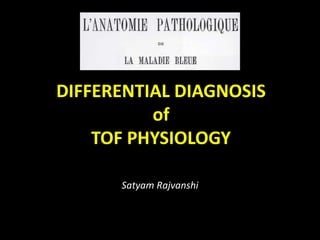
- 1. DIFFERENTIAL DIAGNOSIS of TOF PHYSIOLOGY Satyam Rajvanshi
- 3. Cyanotic CHD CLASSIFICATION Anatomico-pathological Physiological
- 4. Cyanotic CHD with Increased PBF • (5 Ts and 2 Ss) – TAPVC – Single Ventricle – TGA – Single (Common) – Taussig-Bing Atrium – Tricuspid Atresia – Truncus Arteriosus
- 5. Cyanotic CHD with Increased PBF • (5 Ts and 2 Ss) Admixture Physiology (Intercirculatory mixing) • PRE-TRICUSPID • Venous level – TAPVC • Atrial level – Single atrium, Tricuspid atresia, HLHS • POST-TRICUSPID • Ventricular level – Single Ventricle • Arterial level – Truncus Transposition Physiology • TGA • Taussig Bing
- 6. Cyanotic CHD with Decreased PBF RV Dominance PAH Acyanotic CHD with ES VSD PDA, AP window ASD Cyanotic CHD with PAH DORV TGA TAPVC Truncus No PAH RVOTO Quiet Precordium Single S2 ± PS murmur TOF DORV WITH PS TGA WITH PS L-TGA WITH PS ASD with PS
- 7. Cyanotic CHD with Decreased PBF LV dominance LV type Apex (Exam/CXR) No Parasternal Heave LAD ± LV dominance on ECG Tricuspid Atresia with PS Pulmonary Atresia with intact IVS Single Ventricle with PS Ebstein’s
- 8. Cyanotic CHD with Near Normal PBF • Pulmonary AV Fistula • Unroofed coronary sinus into LA • Anomalous drainage of vena cava to LA
- 9. CCHD Physiological classification • TOF Physiology • Transposition Physiology • Admixture Physiology • Eisenmenger Physiology • Duct-dependent Physiology – For PBF – Pulmonary atresia – For SBF – Aortic atresia/HLHS
- 10. CCHD Physiological classification • TOF Physiology • Transposition Physiology • Admixture Physiology • Eisenmenger Physiology • Duct-dependent Physiology – For PBF – Pulmonary atresia – For SBF – Aortic atresia/HLHS • Near-normal Physiology – Pulmonary AVFs • Miscellaneous – Ebstein’s – ASD with PS – Unroofed CS into LA
- 11. • Symptom complex – guide to PHYSIOLOGY • Examination – guide to ANATOMY (Physical findings) • Radiology, ECG – add to BOTH
- 13. TOF PHYSIOLOGY • Cyanotic CHD with decreased PBF having 2 key components anatomically – Severe RVOTO – Decreasing PBF – Large VSD – causing equalization of RV and LV pressure with right to left shunt due to outflow obstruction (Acyanotic TOF not included)
- 14. TOF PHYSIOLOGY • Cyanotic CHD with decreased PBF having 2 key symptoms Physiologically – History of Spells – History of Squatting – No CHF symptoms
- 15. Cyanotic CHD with Decreased PBF RV Dominance PAH Acyanotic CHD with ES VSD PDA, AP window ASD Cyanotic CHD with PAH DORV TGA TAPVC Truncus No PAH RVOTO Quiet Precordium Single S2 ± PS murmur TOF DORV WITH PS TGA WITH PS L-TGA WITH PS ASD with PS
- 16. Cyanotic CHD with Decreased PBF LV dominance LV type Apex (Exam/CXR) No Parasternal Heave LAD ± LV dominance on ECG Tricuspid Atresia with PS Pulmonary Atresia with intact IVS Single Ventricle with PS Ebstein’s
- 17. Cyanotic CHD with Decreased PBF LV dominance LV type Apex (Exam/CXR) No Parasternal Heave LAD ± LV dominance on ECG Tricuspid Atresia with PS Pulmonary Atresia with intact IVS Single Ventricle with PS Ebstein’s
- 18. TOF
- 19. “Tetralogy of Fallot” History • 1671: First reported by Niels Stenson a.k.a Nicholas Steno • 1777; 1784; 1839; 1866; 1872 Similar Case reports
- 20. • 1888: Etienne Louis Arthur Fallot – Anatomic diagnosis at bedside – Confirmed at postmortem – Coined term Tetralogie (Fr.)
- 21. • 1894: Pierre Marie (French), first used term “Tetralogie de Fallot” • 1924: Maude Abbott, first used term “Tetralogy of Fallot” & “Fallot’s Tetralogy”
- 22. Pathology • Van Praagh called TOF “Monology of Stenson” • Central pathology – Underdevelopment/hypoplasia of Subpulmonary infundibulum • Gives rise to 4 components of ‘tetralogy’ – Obstructive RVOT – Large Malaligned VSD – Aortic override – Dominant RV hypertrophy
- 23. Pathology • Van Praagh called TOF “Monology of Stenson” • Central pathology – Underdevelopment/hypoplasia of Subpulmonary infundibulum • Gives rise to 4 components of ‘tetralogy’ – Obstructive RVOT – Large Malaligned VSD – Aortic override – Dominant RV hypertrophy
- 24. TOF • 1 in 3600 live births • M=F
- 25. Natural History • Survival – 66% 1st yr – 50% 3rd yr – 25% 1st decade • Poor survival with PA – 50% 1st yr – 10% 1st decade
- 26. Symptoms/Presentation • Cyanosis – 1-2 weeks after birth – More severe the PS, earlier the presentation • Hypercyanotic Spells – 2 months to 2 years of age • Exertional dyspnoea – Older child • Squatting – To alleviate a spell or dyspnoea
- 27. Physical Exam • Physically underdeveloped • Cyanosis (Depending on PBF) • Pulse – NORMAL (irrespective of PS severity) – Wide PP – only in Large MAPCA/Severe AR
- 28. Physical Exam • JVP – NORMAL (Height and waveform) (RAP stays normal unless significant TR present)
- 29. Physical Exam Palpation • RV impulse – Gentle; like normal neonatal RV; but stays like that even as child grows – 4th LICS – 5th LICS & Subxyphoid (if Sub-Infundibular stenosis)
- 30. Physical Exam Palpation • LV impulse – ABSENT (Conspicuous feature) – Absent even if MAPCAs present
- 31. Physical Exam Palpation • 2nd / 3rd LICS – A2 maybe palpable – Left upper ICS (Not right) (due to hypoplastic PA)
- 32. Physical Exam Palpation • Right sternoclavicular joint pulsation – Right sided Aortic arch • No thrill due to RVOTO – BF goes uninterrupted to dilated Aorta
- 33. Physical Exam Auscultation • Aortic area (2nd RICS) – Loud aortic EC from aortic root – Maximum in expiration • Pulmonary area – Very delayed and soft P2 – EC and P2 almost inaudible (Bicuspid PV – decreased mobility)
- 34. Physical Exam Auscultation • 3rd LICS – Superficial murmur starting with S1 Duration and intensity decreases with severity of PS • No S4 (RA contraction not forceful, RAP normal) • No S3 (No RVF)
- 35. Physical Exam Auscultation • Continuous murmurs – In case of Pulmonary atresia/MAPCAs • AR murmur ± • PR murmur ±
- 36. ECG • P wave – Height normal, peaked – Duration short (LA underfilled) • PR – Normal
- 37. ECG • QRS – RAD (Like newborns) – Clockwise depn – Normal duration – No notching – RVH • Tall R in V1 • Sudden transition V1-2 • Q in V5,6 - PBF
- 38. ECG • T wave – Maybe upright/inverted – Deep inversion rare (RVSP never suprasystemic)
- 39. CXR
- 40. CXR • Reduced PBF markings • Lacy appearance in Pulmonary Atresia • Dilated Asc Ao • Rt Ao arch 20-30%
- 41. CXR • Coer-en-Sabot • Heart resembling a wooden boot – Concave PA bay – Small underfilled LV above a horizontal IVS – Concentric RVH
- 42. CXR • Coer-en-Sabot • Intrauterine life LV is normal – so boot shape develops after 1-2 mos of birth • TOF-PA – Boot even in neonates if low PBF
- 43. DORV-VSD-PS
- 44. DORV • Exact incidence unknown – Less than TOF • Subaortic VSD 40-50% DORV • PS 40-70% Subaortic VSD
- 45. DORV-PS • History • Physical Exam • CXR Same as TOF
- 46. DORV-PS • Exceptions - If restrictive VSD present (Subaortic stenosis) • Long decrescendo systolic murmur at LPS area (obligatory flow murmur) • ECG - LVH
- 47. DORV-PS • Differentiation from TOF - ECG - PR Prolonged - Counterclockwise depn - RVH but no sudden transition
- 48. D-TGA – VSD – PS
- 49. D-TGA • 1 in 2500-5000 live births • M>F 4:1 • VSD is most common communication • PS (LVOTO) present in 15%
- 50. Symptoms/Presentation • Cyanosis – Since birth – 1st day of life • Hypercyanotic Spells – May be present occasionally • Squatting – Rare
- 51. Natural History • Mortality rate in TGA without PS – 30% 1st week – 50% 1st month – 90% 1st year • Better survival with PS – may survive adolescence
- 52. Physical Exam • Birth weight > normal (Contrast to other CHDs) • Deep Cyanosis • Scalp & Arm varicose veins (Systemic volume overload with desaturated blood)
- 53. • Pulse – Full volume bounding pulse – Warm extremities • JVP – Elevated RAP with dominant A wave (Systemic volume overload with desaturated blood)
- 54. • Palpation – RV impulse gentle at birth – Soon after 1 week – Prominent RV impulse (Systemic volume overload with desaturated blood) – Rt sternoclavicular impulse – Rt arch (11-16% in TGA-VSD-PS) Just like TOF
- 55. • Auscultation – Just like TOF
- 56. ECG • P wave – Tall, Peaked (Hypervolemic RA) • RAD, RVH • T – Usually positive in ALL precordial leads – Taller in right precordial leads • Counterclockwise depn
- 57. CXR • Thymic shadow absent (after 12 hrs of birth) • Characteristic narrow pedicle (egg on side) ABSENT in severe PS – due to dilated right anterior aorta • CXR like TOF
- 58. L-TGA – VSD – PS
- 59. L-TGA • 1 in 13000 live births • M>F 1.5:1 • VSD 80% L-TGA • PS 50% L-TGA 80% of PS a/w VSD
- 60. Symptoms/Presentation • Cyanosis appearance according to PS severity • Spells and squatting uncommon • Older adults – Stokes-Adams & Syncope (High degree AV blocks)
- 61. Natural History • Better survival with VSD-PS – may survive adolescence
- 62. Physical Exam • Like TOF
- 63. • Pulse – Normal – Bradycardia/Blocks • JVP – Normal – 1st /2nd /3rd degree AV blocks
- 64. • Palpation – IVS almost vertical and parallel to left sternal border – Morph. RV – anterior & left position forming apex – Systolic RV Impulse – Morph. LV – posterior & right position behind sternum – LV Impulse non-palpable (even if enlarged) – Ao EC & A2 palpable 2nd LICS
- 65. • Auscultation – Just like TOF – In case of blocks • Soft S1 – in 1st degree AVB • Variable S1 – High degree AVB
- 66. ECG • AV Blocks maybe present – CHB – ventricular activation sequence normal; QRS narrow • LAD (d/t LAFB) • RVH
- 67. • Q /q – Present in right precordial leads / absent in left (Septal activation right to left directed) – Present in III, avf (III>avf) / absent in I, aVL (Septal activation superiorly directed) • T – Usually positive in ALL precordial leads • Clockwise depn
- 68. CXR • Thymic shadow absent • Ao & PA side by side • Ao left and Anterior • Straight left border • Hump shaped heart • RPA and LPA at same level
- 69. TRICUSPID ATRESIA – PS
- 70. Tricuspid Atresia (TA) • 1 in 17000 live births • 90% TA – No TGA • 90% have Restrictive VSD – Physiologically PS • 10% TA – TGA • 90% have no PS – Increased PBF
- 72. Natural History • TA-NRGA-PS – 80% mortality in 1st yr – Already restrictive VSD – decreases in size and closes! (Like a PM-VSD!) – Acquired Pulmonary atresia without embryological collaterals – fatal!
- 73. Physical Exam • Like TOF • No left precordial bulge – RV underdeveloped
- 74. • Pulse – Normal • JVP – Height increased – A prom – PFO/Restrictive ASD/Decr LV compliance – V prom – MR
- 75. • Palpation – IVS almost vertical and parallel to left sternal border – Systolic LV Impulse (present even if low PBF) – RV Impulse non-palpable
- 76. • Auscultation – Single S1 (M1) – Single S2 (A2); P2 soft & delayed – maybe heard – LVS4 if unrestrictive VSD/LVH – Obligatory VSD holosystolic murmur • 3rd-4th LICS • Radiates to 2nd LICS • Duration shortens in closing VSD
- 77. ECG • P wave – Tall, Peaked (RAE) • LAD • LVH with adult precordial progression • Counterclockwise depn
- 78. CXR • RAE – prominent rt. Upper border • Small RV – Flat receding rt. Inferior border • LV apex • Prominent Ao/small PA
- 79. SINGLE VENTRICLE – PS
- 80. Single ventricle (SV) • 1 in 20000 live births • M>F 2-4:1 • 80% SLV • 10% SRV • 10% Indeterminate • SLV OC inverted (left); Noninverted (right) TGA present (Ao from OC; PA from SLV)
- 81. Symptoms/Presentation • Cyanosis since birth • Spells and squatting maybe present
- 82. Natural History • SLV 50% mortality before 14 yrs • SRV 50% mortality before 4 yrs
- 83. Physical Exam • Like TOF
- 84. • Pulse – Normal • JVP – Normal – V prom – if Rt AV valve regurgitant
- 85. • Palpation – Systolic LV Impulse at apex – 3rd LICS – palpable impulse due to inverted OC maybe present
- 86. • Auscultation – Like TOF
- 87. ECG • P wave – Tall, Peaked (RAE) • Inverted OC – RAD, Clockwise depn • Noninverted OC – LAD, Counterclockwise depn • Stereotyped rS or RS complexes V2-V5 • Tall R in V1 despite LVH
- 88. CXR • Non-inverted OC • Doesn’t form left border – just like D-TGA • LV apex • Characteristic waterfall appearance of RPA – NOT present in PS – Low PBF
- 89. CXR • Inverted OC • Form left border – just like L-TGA • Maybe visible as convexity on left • Left straight border • LV apex
- 90. APPROACH?
- 91. • Incidence acccording to age and natural history – Age of presentation? – Survival?
- 92. • PRESENTATION – Timing of cyanosis appearance? – Characteristic spells/squatting?
- 93. • Physical Exam – LV or RV predominent?
- 94. • ECG – LAD/RAD – Clock/Counterclock – LVH/RVH – AV Blocks
- 95. • CXR – Less helpful – LV/RV apex – Left Straightening