Musculoskeletal changes in pregnancy
•Descargar como PPTX, PDF•
64 recomendaciones•36,087 vistas


Recomendados
Más contenido relacionado
La actualidad más candente
La actualidad más candente (20)
Destacado
Destacado (20)
Similar a Musculoskeletal changes in pregnancy
Similar a Musculoskeletal changes in pregnancy (20)
Más de amrit kaur
Último
Último (20)
Musculoskeletal changes in pregnancy
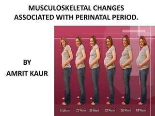
- 1. MUSCULOSKELETAL CHANGES ASSOCIATED WITH PERINATAL PERIOD. BY AMRIT KAUR
- 2. MUSCULOSKELETAL CHANGES DURING PREGNANCY One of the most obvious changes in pregnancy is the alteration of the woman's body. Mechanical changes related to the weight of growing breasts, uterus and fetus, as well as an increase in lumbar lordosis, result in a shift in the woman's center of gravity, which may cause problems with balance.
- 3. • Pregnancy wt. gain - 9 to 14kg. • Stretching of abdominal muscles • Decrease in ligamentous tensile strength. • Hyper mobility of joints due to ligamentous laxity. • Pelvic floor drops as much as 2.5 cm • COG shifts upwards & forwards. • posture – shoulder girdle becomes rounded, scapular protraction, increase in cervical lordosis. knee hyperextension. increase in lumber lordosis. balance – pt. walks with wider BOS.
- 4. NEUROMUSCULAR AND ARTICULAR RELATIONSHIP • The musculoskeletal system is composed of 3 sub systems- the muscular, articular and neural system. • Changes in degree of passive restraint offered by the collagenous tissue surrounding the articulation. • Oestrogen causes proliferation of connective tissue and that relaxin acts to provide vascularization and softening of this connective tissue. • Increase in joint laxity in multiple joints during pregnancy.
- 5. • Such a changes in passive restraint of any joint is bound to have significant influences on the afferent input to the to the spinal cord and higher cortical centers. • Muscle stiffness is also reduced, leading to decrease in muscle responsiveness to pertubations. • Increase in articular pressure, sometimes pinching of articular capsule. • Proprioceptive acuity of hypermobile joints is having lesser sensitivity than normal.
- 6. • Injury of the capsule and /or ligament influences muscle activity not only in the muscle which cross the injured joint, but also in those which are remote from it. • Interruption of the flow of impulses from the mechanoreceptor in a joint capsule into central nervous system result in clinically evident disturbances of the perception of joint position and movement and of the reflexes concerned with posture and gait. • The arthrokinetic reflex might be considered as a triggering factor which would initiate a whole chain of adaptation reactions, eventually resulting in a changed movement pattern .
- 7. POSTURAL CHANGES ASSOCIATED WITH PREGNANCY • The most obvious physical features in pregnancy influencing the women's posture are the alterations in body mass and the consequent changes in the center of gravity. • The physiotherapist must place emphasis on directing and training women to ensure that their newly assumed posture does not overstress any body segment since the outcome of this could be fatigue , micro trauma and pain.
- 8. • There is tendency to an increased anterior displacement of the line of gravity • This natural tendency to anterior displacement may be counterbalanced by a number of options such as increased activation of gastrocnemius and soleus muscle, an active posterior displacement of the body ,extension of hip joint or posterior displacement of upper trunk. • The mechanism that may be used to achieve an active posterior displacement of the body may include increase of the lumbosacral angle, and increase in lumbar curvature or a displacement of pelvis anteriorly and shoulder posteriorly, which may be reflected in greater degree of thoracic kyphosis.
- 9. SPINAL INSTABILITY DURING PREGNANCY 3 subsystems • Control subsystem –neural feedback from various forces and motion transducers, located in ligaments, tendon and muscles, and neural control centers. • Passive subsystem –vertebrae, facet articulations, inter vertebral discs, spinal ligaments and joint capsules. • Active subsystem- muscles and tendons surrounding vertebral column .
- 10. • In pregnancy there is rapid change in passive sub system, due to the change in circulating hormones. • Possible cause for spinal instability is either increase ROM not restrained by the passive subsystem (the neutral zone) or a decrease control in this area by the active subsystem. • In either situation , a portion of the normal range of motion is not controlled and this may lead to increased shear forces, micro trauma and pain.
- 11. SPINAL PAIN DURING PREGNANCY CAUSES • Weight gain • Rapid postural changes • Vascular effects • Previous h/o back pain • Repetitive lifting /banding • Pelvic insufficiency due to hormonal changes
- 12. 3 major region • Pain above lumbar region only • Pain in lumbar region with or without radiation to one or both legs. • Pain over the sacroiliac area sometimes with radiation to the legs.
- 13. BIOMECHANICS OF THE BODY • Biomechanics includes the bones and muscles, and how they work together to make the body move. The brain receives a continuous supply of information about the changing posture of the pregnant body and eventually accepts the altered arrangement and balance of body parts. Feedback from the skin, joints, and muscles are involved in this process. By the end of pregnancy, the brain has re- configured its image of the body in balance. • The major changes that occur with respect to biomechanics include: • The change in the center of gravity as the baby grows in size during the second half of pregnancy.The center of gravity changes from the center of the pelvis with surrounding body weight evenly distributed in all directions to a point forward and slightly up from the center of the pelvis. The increased weight positioned forward of the pelvic midpoint causes forward gravitational pull.
- 14. • Movement of joints due to changes in weight distribution. • Balance of muscle strength changes around the joint to accommodate the new weight distribution. To compensate for the forward gravitational pull, the posture changes to maintain a balanced erect position. The back of the waist curves in, the top of the pelvis tilts forward and the curves in the upper spine increase. These changes put stress on muscles and joints. • Spinal curves increase, placing a greater load on the vertebrae. • Joint laxity causes a greater risk for injury. Hormone changes (increased levels of relaxin and elastin) that affect the looseness of ligaments and tendons make the joints more mobile. Although this helps with the birth of the baby, it can stress joints and muscles during movement. • There can be more structural discomfort such as low back pain. • There is increased potential for nerve compression and blood vessel entrapment, such as sciatica or carpal tunnel syndrome.
- 15. Uterus and uterine ligaments • In pregnancy, the uterus grows to a weight of about 2.5 pounds and has a capacity of approximately 1.5 to 2.5 gallons. It enlarges through the stretching of muscle fiber to the size of a watermelon. The muscle fibers lengthen 7-11 times and widen 2-7 times. It also increases the number and size of its blood vessels and nerves. • After the first trimester (first 13 weeks of pregnancy), the uterus begins to prepare for labor and delivery by contracting involuntarily and irregularly. These are called Braxton-Hicks contractions. They are: prelabor contractions that work toward shortening and widening the cervix and stretching the bottom of the uterus ,soften the cervix and prepare it for labor • usually are not painful but can be uncomfortable • In labor, the uterus contracts with increasing frequency and intensity and pushes the baby through the birth canal.
- 16. • The uterine ligaments hold the uterus in place and undergo prolonged stretching during pregnancy. • The round ligaments attach the uterus to the pubic bone in the front and help maintain the uterus in the center of the pelvis. Either a sharp pain or a dull ache near the hip joint is common when they are stretched upward. • The broad ligaments connect the uterus to the sacrum and are often involved in backaches during pregnancy. Pain in the low back is the result of weak abdominal muscles, poor posture, and the weight of the abdomen pulling on the back ligaments.
- 17. MUSCLE CHANGES • As the uterus enlarges the abdominal muscles are stretched. The top layer of the abdominal muscles are the rectus abdominal muscles. They run from the rib cage to the pelvic bone. As the abdomen gets larger these muscles can separate. This abdominal muscle separation is called a diastasis, and can lead to a loss of abdominal strength. • Strectching of pelvic floor muscles. • Some muscle will go for tightness, some will be lengthen.
- 18. • Overstretching and weakening of gluteal muscles and hamstrings (buttocks and back of leg). • Overstretching and weakening of abdominal muscles and pelvic floor muscles. • Overstretching and weakening of upper back muscles (shoulders forward). • Shortening and tightening of hip flexor muscles. • Shortening of upper back flexors and pectoral muscles (chest caves in).