SlideShare una empresa de Scribd logo
1 de 43
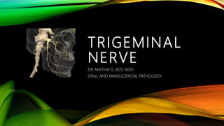
TRIGEMINAL
NERVE
DR AMITHA G, BDS, MDS
ORAL AND MAXILLOFACIAL PATHOLOGY
TRIGEMINAL NERVE
- Fifth cranial nerve
- Have a large sensory root and a small motor root.
- Motor root arises – arises from the lateral aspect of lower pons (cranially) 
the motor root cross the apex of the petrous temporal bone beneath the
superior petrosal sinus, to enter the middle cranial fossa.
- Sensory root – arises from the lateral aspect of lower pons (caudally).
TRIGEMINAL GANGLION
- Sensory ganglion of fifth cranial nerve.
- Homologous with the dorsal nerve root ganglia of spinal
nerves.
- All such ganglia are made of pseudounipolar nerve cells,
with a T shaped arrangement of their process; one process
arises from the cell body which then divides into a central
and peripheral process.
- Ganglion is cresentic or semilunar in shape with its
convexity directed anterolaterally.
- 3 divisions of trigeminal nerve emerge from this convexity.
- Posterior concavity of the ganglion receives the sensory root of the nerve.
- Situation and meningeal relations
- Ganglion lies on the trigeminal impression, on the anterior surface of the petrous temporal
bone near its apex.
- It occupies a special space of dura matter called the trigeminal or Meckel’s cave.
- There are 2 layers of the dura below the ganglion.
- The cave is lined by pia arachnoid, so that ganglion along with the motor root of the
trigeminal nerve is surrounded by CSF.
- The ganglion lies at a depth of about 5cm from the preauricular point.
RELATIONS
- Medially
(a) internal carotid artery
(b) posterior part of cavernous sinus
- Laterally - middle meningeal artery
- Superiorly - parahippocampal gyrus
- Inferiorly
(a) motor root of trigeminal nerve
(b) greater petrosal nerve
(c) apex of the petrous temporal bone
(d) foramen lacerum.
Associated root and branches
- Central processes of ganglion cells form the large sensory root of the trigeminal nerve
which is attached to the pons at its junction with the middle cerebellar peduncle.
- Peripheral processes of the ganglion cells form 3 divisions of the trigeminal nerve, namely
ophthalmic, maxillary and mandibular.
- Small motor root of the trigeminal nerve is attached to the pons superomedial to the
sensory root.
- It passes under the ganglion from its medial to the lateral side, and join the mandibular
nerve at the foramen ovale.
OPTHALIMIC DIVISION
Terminal branches of Ophthalmic division of trigeminal nerve, are
1. Frontal
 Supratrochlear
 Supraorbital
2. Nasociliary
 Branch of ciliray ganglion
 2-3 long ciliary nerves
 Posterior ethmoidal
 Infratrochlear
 Anterior ethmoidal
3. Lacrimal
LACRIMAL NERVE
- Smallest of the 3 terminal branches of
ophthalmic nerve
- It enters the orbit through the lateral part of the
superior orbital fissure and runs forwards along
the upper border of the lateral rectus muscle in
company with the lacrimal artery.
- Anteriorly it receives communication from the
zygomaticotemporal nerve, passes deep to the
lacrimal gland and ends in the lateral part of the
upper eyelid.
Supplies
- Lacrimal gland, conjunctiva, upper eyelid.
- Its own fibers to the gland are sensory.
- Secretomotor fibers to the gland come from the greater petrosal nerve through its
communication with zygomaticotemporal nerve.
FRONTAL NERVE
- Is the largest of the 3 terminal branches of the
ophthalmic nerve.
- Begins in the lateral wall of the anterior part of
cavernous sinus.
- It enters the orbit though the lateral part of the
superior orbital fissure, and runs forwards on the
superior surface of levator palpebrae superioris.
- At the middle of the orbit it divides into small
supratrochlear branch and large supraorbital
branch.
NASOCILIARY NERVE
- One of the terminal branches of the ophthalmic division of trigeminal nerve
- Begins in the lateral wall of the anterior part of the cavernous sinus.
- It enters the orbit through the middle part of the superior orbital fissure between the two
divisions of the occulomotor nerve.
- It crosses above the optic nerve from lateral to medial side along with ophthalmic artery and
runs along the medial wall of the orbit between the superior oblique and the medial rectus. It
ends at anterior ethmoidal foramen by dividing into the infratrochlear and anterior ethmoidal
nerves.
- Its branches are as follows
1. A communicating branch to ciliary ganglion – forms the sensory root of the ganglion. It
is often mixed with the sympathetic root.
2. Two to three long ciliary nerves run on the medial side of the optic nerve, pierce the
sclera, and supply sensory nerves to the cornea, the iriss and the ciliary body.
They also carry sympathetic nerves to dilator papillae.
3. Posterior ethmoidal nerve – passes through the posterior ethmoidal foramen and
supplies the ethmoidal and sphenoidal air sinuses. It is frequently absent.
4. Infratrochlear nerve – smaller terminal branch of the nasociliary nerve given off at the
anterior ethmoidal foramen. It emerges from the orbit below the trochlear for the tendon
of the superior oblique and appears on the face above the medial angle of the eye. It
supplies the conjunctiva, the lacrimal sac and caruncle, the medial ends of the eyelids and
the upper half of the external nose.
5. Anterior ethmoidal nerve – is the larger terminal branch of the nasociliary nerve. It
leaves the orbit by passing through the anterior ethmoidal foramen. It appears for a very
short distance, in the anterior cranial fossa, above the cribriform plate of the ethmoid
bone. It then descends into the nose through a slit at the side of the anterior part of the
crista galli. In the nasal cavity, it lies deep to the nasal bone. It gives off 2 internal nasal
branches medial and lateral to the mucosa of the nose. Finally, it emerges at the lower
border of the nasal bone as the external nasal nerve which supplies the skin of the lower
half of the nose.
MAXILLARY NERVE
MAXILLARY NERVE
- Arises from the trigeminal ganglion
- Runs forward in the lateral wall of the cavernous sinus below the ophthalmic nerve and leaves
the middle cranial fossa by passing through the foramen rotandum .
- Next, the nerve crosses the upper part of the pterygopalatine fossa, beyond which it is
continues as the infraorbital nerve.
- In the pterygopalatine fossa the nerve is intimately related to the pterygopalatine ganglion,
and gives off zygomatic and posterior superior alveolar arteries.
- The posterior superior alveolar nerve enters the posterior surface of the body of the maxilla,
and supplies the three upper molar teeth and the adjoining part of gum.
PTERYGOPALATINE GANGLION
- Is the largest parasympathetic peripheral ganglion.
- It serves as a relay station for secretomotor fibers to the lacrimal gland and to the mucous
glands of the nose, paranasal sinuses the palate and pharynx.
- Topographically relatedo the maxillary nerve, but functionally connected to the facial nerve
through its greater petrosal branch.
- Location – the flattened ganglion lies in the pterygopalatine fossa just below the maxillary
nerve, in front of the pterygoid canal and lateral to the sphenopalatine foramen.
Connections
1. Motor or parasympathetic root of the ganglion
- By the nerve of pterygoid canal.
- It carries preganglionic fibers that arise from neurons present near the superior
salivatory and lacrimatory nuclei, and pass through the nervus intermedius, the facial
nerve, the geniculate ganglion, greater petrosal nerve, nerve of pterygoid canal, to
reach the ganglion.
- The fibers relay in the ganglion.
- Post ganglionic fibers arise in the ganglion to supply secretomotor nerves to the lacrimal
gland and to mucous gland of the nose, paranasal sinuses, palate and nasopharynx.
2. Sympathetic root
- Is also derived from the nerve of pterygoid canal.
- It contains postganglionic fibers arising in the superior cervical sympathetic ganglion which
pass through the internal carotid plexus, deep petrosal nerve, nerve of pterygoid canal to
reach the ganglion .
- The fibers pass through the ganglion without relay and supply vasomotor nerves to the
mucous membrane of the nose, the paranasal sinuses, palate an dnasopharynx.
3. Sensory root
- Comes from the maxillary nerve
- Its fibers pass through the ganglion without relay.
- They emerge in the branches
Branches
- The branches of the ganglion are actually branches of he maxillary nerve.
- They also carry parasympathetic and sympathetic fibers which pass through the ganglion.
- The branches are
a. Orbital branches
Pass through the inferior orbital fissure
Supply the periosteum of orbit and orbitalis muscle
b. Palatine branches
The greater or anterior palatine nerve descends through the greater palatine canal, and
supplies the hard palate and the lateral wasll of the nose, i.e inferior conchaand adjoining
meatuses.
The lesser or middle and posterior palatine nerves supply the soft palate and the tonsil.
c. Nasal branches
Enter the nasal cavity through the sphenopalatine foramen.
The lateral posterior superior nasal nerves (about 6 in number), supply the posterior part
of the roof of the nose and of the nasal septum.
The largest oof these nerves are called nasaopalatine nerve, which descends upto the
anterior part of the hard palate through the incisive foramen.
d. Pharyngeal branch
Passes through the palatogingival canal and supplies the part of the nasopharynx behind
the auditory tube
e. Lacrimal branch
The postganglionuc fubers pass back into the maxillary nerve to leave it through its
zygomatic nerve and its zygomatic temporal branch.
It is a communicating branch to lacrimal nerve to supply secretomotor fibers to the
lacrimal gland.
Preganglionic fibers have their origin in the lacrimatory nucleus.
INFRAORBITAL NERVE
- It is the continuation of the maxillary nerve.
- It enters the orbit through the inferior orbital fissure.
- It then runs forwards on the floor of the orbit or the roof of the maxillary sinus, at the first in
the infraorbital groove and then in the infraorbital canal remaining outside the periosteum of
the orbit.
- It emerges on the face through the infraorbital foramen and terminates by dividing into
palpebral, nasal and labial branches.
- The nerve is accompanied by the infraorbital branch of the third part of the maxillary artery
and the accompanying vein.
Branches
a. Middle superior alveolar nerve
b. Anterior superior anterior superior alveolar nerve arise in the infraorbital canal and runs in a
sinuous canal having a complicated course in the anterior wall of the maxillary sinus. It
supplies the upper incisor and canine teeth, the maxillary sinus and the antero inferior part
of the nasal cavity.
c. Terminal branches – palpebral, nasal and labial – supply a large area of the skin on the face.
They also supply the mucous membrane of the upper lip and cheek.
ZYGOMATIC NERVE
- It ia branch of the maxillary nerv, given off in the pterygopalatine fossa.
- It enters the orbit through the lateral end of the inferior orbital fissue, and runs along the
lateral wall, outside the periosteum, to enter the zygomatic bone.
- Just before or after entering the bone it divides into its 2 terminal branches, the
zygomaticofacial and zygomatiotemporal nerves which supply the skin of the face and of the
anterior part of the temple.
- The communicating branch to the lacrimal nerve, which contains secretomotor fibers to the
lacrimal gland, may arise either from the zygomatic or the zygomaticotemporal nerve, and
runs in the lateral wall of the orbit.
MANDIBULAR NERVE
MANDIBULAR NERVE
- Is the largest of the 3 divisions of the trigeminal nerve
- It is the nerve of first branchial arch
- It has both sensory and motor fibers
- It supplies all structures derived from the mandibular or first branchial arch
Course and relations
- Mandibular nerve begins in the middle cranial fossa through a large sensory root and a small
motor root.
- Sensory root arises from the lateral part of the trigeminal ganglion, and leaves the cranial
cavity through the foramen ovale.
- Motor root lies deep to the trigeminal ganglion and to the sensory root.
- It also passes through the foramen ovale to join the sensory root just below the foramen
thus forming the main trunk.
- The main trunk lies in the infratemporal fossa, ON the tensor veli paltini, DEEP to the lateral
pterygoid.
- After a short course, the main trunk divides into a
 Small anterior trunk and a
Branches
- From main trunk
a. Meningeal branch
b. Nerve to medial pterygoid
- From the anterior trunk
Sensory branch
a. Buccal nerve
Motor branch
a. Masseteric
b. Deep temporal nerve
c. Nerve to lateral pterygoid
- From the posterior trunk
a. Auriculotemporal
b. Lingual
c. Inferior alveolar nerves
Meningeal branch or nervus spinosus
- Course - Meningeal branch enters the skull through the foramen spinosum with the middle
meningeal artery
- Supplies – dura matter of middle cranial fossa
Nerve to medial pterygoid
- Origin – arises close to the otic ganglion
- Supplies - medial pterygoid from its deep surface.
- This nerve gives a motor root to the otic ganglion which does not relay and supplies tensor
veli palatine and tensor tympani muscles.
BUCCAL NERVE
- Only sensory branch of the anterior division of the mandibular nerve.
- It passes between the 2 heads of the lateral pterygoid, runs downwards and forwards
- Supplies – skin and mucous membrane related to buccinator. Also supplies buccal aspect of
gums of the molar and premolar teeth of both maxillary and mandibular teeth.
MASSETRIC NERVE
- Emerges at the upper border of the lateral pterygoid just in front of the TMJ, passes laterally
through the mandibular notch in company with the massetric vessels and enters the deep
surface of the masseter.
- It also supplies the TMJ
DEEP TEMPORAL NERVES
- There are 2 deep temporal nerves
a. Anterior
b. Posterior
- They pass between the skull and the lateral pterygoid and enter the deep surface of the
temporalis.
- Anterior nerve is often a branch of the buccal nerve.
- Posterior nerve may arise in common with the massetric nerve.
NERVE TO LATERAL PTERYGOID
- Enters the deep surface of the muscle.
- It may be an independent branch or may arise in common with the buccal nerve.
AURICULOTEMPORAL NERVE
Origin
- Arises by 2 roots
Course
- 2 roots run backwards, encircling the middle meningeal artery
- Unites to form a single trunk
- Then continues backwards between the neck of the mandible and the sphenomandibular
ligament, above the maxillary artery
- Behind the neck of mandible it turns upwards and ascends on the temple behind the
superficial temporal vessels.
Supplies
- Auricular part – supplies the skin of the tragus, upper parts of the pinna, the external
acoustic meatus, tympanic membrane. (note:- the lower parts of these regions are supplies
by the great auricular nerve and the auricular branch of the vagus nerve)
- Temporal part – supplies the skin of the temple.
- Articular branches – to TMJ
- In addition secretomotor supply to the parotid gland (and also sensory)
LINGUAL NERVE
- Is one of the 2 terminal branches of the posterior division of trigeminal nerve.
Orgin
- Begins 1 cm below the skull.
Course and relations
- It runs first between the tensor veli palatini and the lateral pterygoid and then between the
lateral and medial pterygoid.
- 2cm below the skull it is joined by the chorda tympani.
- Emerging at the lower border of the lateral pterygoid, the nerve runs downwards and
forwards between the ramus of the mandible and the medial pterygoid.
- Next it lies in direct contact with the mandible, medial to the third molar tooth between the
origins of superior constrictor and the mylohyoid muscle.
- It soon leaves the gums and runs over the hyoglossus deep to the mylohyoid.
- Finally lies on the surface of the genioglossus deep to mylohyoid.
- Here it winds round the submandibular duct and divides into terminal branches.
Supplies
- Sensory to the anterior 2/3rd of the tongue and to floor of the mouth.
- However, fibers of chorda tympani (branch of facial nerve) which is sceretomotor to the
submandibular and sublingual glands and gustatory to the anterior 2/3rd of the tongue, are
also distributed through this lingual nerve.
INFERIOR ALVEOLAR NERVE
- Is the largest terminal branch of the posterior
division of the mandibular nerve.
- Runs vertically downwards lateral to the
spenomandibular ligament and medial
pterygoid.
- Enters the mandibular foramen and runs in the
mandibular canal.
- It is accompanied by inferior alveolar artery.
Branches
a. Mylohyoid branch
- Arises just before the inferior alveolar nerve enters the mandibular foramen.
- Contains all the motor fibers of posterior division
- Pierces the spenomandibular ligament with the mylohyoid artery, runs in the mylohyoid
groove
- Supplies the mylohyoid muscle and the anterior belly of the digatsric.
b. Branches in the mandibular canal – inferior dental plexus
- While running in the mandibular canal the inferior alveolar nerve gives branches that supply
the lower teeth and gums. (molar and premolar teeth and gums)
c. Mental nerve
- Emerges at the mental foramen
- Supplies the skin of the chin and the skin and mucous membrane of the lower lip.
d. Incisive branch
- Its incisive branch supplies the labial aspect of the gums of canine and incisor teeth.
THANK YOU

Más contenido relacionado

La actualidad más candente

La actualidad más candente (20)

The maxillary nerve
The maxillary nerveThe maxillary nerve
The maxillary nerve
 
Trigeminal Nerve
Trigeminal NerveTrigeminal Nerve
Trigeminal Nerve
 
Developmental Of The Mandible
Developmental Of The MandibleDevelopmental Of The Mandible
Developmental Of The Mandible
 
Introduction to the trigeminal nerve ‫‬dental surgery
Introduction to the trigeminal nerve ‫‬dental surgeryIntroduction to the trigeminal nerve ‫‬dental surgery
Introduction to the trigeminal nerve ‫‬dental surgery
 
Trigeminal nerve
Trigeminal nerveTrigeminal nerve
Trigeminal nerve
 
Maxillary artery
Maxillary arteryMaxillary artery
Maxillary artery
 
Applied anatomy
Applied anatomyApplied anatomy
Applied anatomy
 
Cavernous sinus
Cavernous sinusCavernous sinus
Cavernous sinus
 
Lymphatic drainage of head and neck- Dr.Ayesha
Lymphatic drainage of head and neck- Dr.AyeshaLymphatic drainage of head and neck- Dr.Ayesha
Lymphatic drainage of head and neck- Dr.Ayesha
 
Trigeminal nerve
Trigeminal nerveTrigeminal nerve
Trigeminal nerve
 
Maxillary sinus sinus
Maxillary sinus sinus Maxillary sinus sinus
Maxillary sinus sinus
 
Ophthalmic nerve
Ophthalmic nerveOphthalmic nerve
Ophthalmic nerve
 
Trigeminal nerve
Trigeminal nerveTrigeminal nerve
Trigeminal nerve
 
Trigeminal nerve
Trigeminal nerveTrigeminal nerve
Trigeminal nerve
 
Trigeminal nerve
Trigeminal nerveTrigeminal nerve
Trigeminal nerve
 
Ciliary ganglion
Ciliary ganglionCiliary ganglion
Ciliary ganglion
 
Ophthalmic nerve dental surgery
Ophthalmic nerve dental surgeryOphthalmic nerve dental surgery
Ophthalmic nerve dental surgery
 
Surgical anatomy of TMJ
Surgical anatomy of TMJSurgical anatomy of TMJ
Surgical anatomy of TMJ
 
Tooth development, eruption & applied aspects
Tooth development, eruption & applied aspectsTooth development, eruption & applied aspects
Tooth development, eruption & applied aspects
 
Neural crest cell
Neural crest cellNeural crest cell
Neural crest cell
 

Similar a TRIGEMINAL NERVE ANATOMY

Copy of trigeminal nerve.doc submisiopn
Copy of trigeminal nerve.doc submisiopnCopy of trigeminal nerve.doc submisiopn
Copy of trigeminal nerve.doc submisiopnIndian dental academy
 
Trigeminal nerve and its applied anatomy
Trigeminal nerve and its applied anatomyTrigeminal nerve and its applied anatomy
Trigeminal nerve and its applied anatomyILA YADAV
 
anatomy of trigeminal nerve.pptx by dr. payal
anatomy of trigeminal nerve.pptx by dr. payalanatomy of trigeminal nerve.pptx by dr. payal
anatomy of trigeminal nerve.pptx by dr. payalpikopinochi
 
Trigeminal nerve and its importance in max-fac surgery
Trigeminal nerve and its importance in max-fac surgeryTrigeminal nerve and its importance in max-fac surgery
Trigeminal nerve and its importance in max-fac surgeryDr. Anindya Chakrabarty
 
Trigeminal nerve-ambika - Copy.ppt
Trigeminal nerve-ambika - Copy.pptTrigeminal nerve-ambika - Copy.ppt
Trigeminal nerve-ambika - Copy.pptambikaluthra3
 
Trigeminal nerve
Trigeminal nerve Trigeminal nerve
Trigeminal nerve AnuRaaga
 
ix x.pptx
ix x.pptxix x.pptx
ix x.pptxmalti19
 
nerve supply of maxilla and mandible
nerve supply of maxilla and mandible nerve supply of maxilla and mandible
nerve supply of maxilla and mandible FarhaNaaz14
 
Surgical anatomy of cranial nerves pallav
Surgical anatomy of cranial nerves pallavSurgical anatomy of cranial nerves pallav
Surgical anatomy of cranial nerves pallavpallavkedia
 
Trigeminal nerve/prosthodontic courses
Trigeminal nerve/prosthodontic coursesTrigeminal nerve/prosthodontic courses
Trigeminal nerve/prosthodontic coursesIndian dental academy
 

Similar a TRIGEMINAL NERVE ANATOMY (20)

Trigeminal nerve
Trigeminal nerveTrigeminal nerve
Trigeminal nerve
 
Cranial nerves
Cranial nervesCranial nerves
Cranial nerves
 
Copy of trigeminal nerve.doc submisiopn
Copy of trigeminal nerve.doc submisiopnCopy of trigeminal nerve.doc submisiopn
Copy of trigeminal nerve.doc submisiopn
 
Trigeminal nerve.
Trigeminal nerve.Trigeminal nerve.
Trigeminal nerve.
 
Trigeminal nerve and its applied anatomy
Trigeminal nerve and its applied anatomyTrigeminal nerve and its applied anatomy
Trigeminal nerve and its applied anatomy
 
anatomy of trigeminal nerve.pptx by dr. payal
anatomy of trigeminal nerve.pptx by dr. payalanatomy of trigeminal nerve.pptx by dr. payal
anatomy of trigeminal nerve.pptx by dr. payal
 
Trigeminal nerve.pdf
Trigeminal nerve.pdfTrigeminal nerve.pdf
Trigeminal nerve.pdf
 
Trigeminal nerve and its importance in max-fac surgery
Trigeminal nerve and its importance in max-fac surgeryTrigeminal nerve and its importance in max-fac surgery
Trigeminal nerve and its importance in max-fac surgery
 
Trigeminal nerve-ambika - Copy.ppt
Trigeminal nerve-ambika - Copy.pptTrigeminal nerve-ambika - Copy.ppt
Trigeminal nerve-ambika - Copy.ppt
 
Anatomy facial nerve
Anatomy facial nerveAnatomy facial nerve
Anatomy facial nerve
 
Trigeminal neuralgia
Trigeminal neuralgiaTrigeminal neuralgia
Trigeminal neuralgia
 
The trigeminal nerve
The trigeminal nerve The trigeminal nerve
The trigeminal nerve
 
Trigeminal nerve
Trigeminal nerve Trigeminal nerve
Trigeminal nerve
 
ix x.pptx
ix x.pptxix x.pptx
ix x.pptx
 
the cranial nerves
the cranial nerves the cranial nerves
the cranial nerves
 
nerve supply of maxilla and mandible
nerve supply of maxilla and mandible nerve supply of maxilla and mandible
nerve supply of maxilla and mandible
 
Surgical anatomy of cranial nerves pallav
Surgical anatomy of cranial nerves pallavSurgical anatomy of cranial nerves pallav
Surgical anatomy of cranial nerves pallav
 
Trigeminal nerve/prosthodontic courses
Trigeminal nerve/prosthodontic coursesTrigeminal nerve/prosthodontic courses
Trigeminal nerve/prosthodontic courses
 
Cranial nerves
Cranial nervesCranial nerves
Cranial nerves
 
1.facial nerve
1.facial nerve1.facial nerve
1.facial nerve
 

Más de oral and maxillofacial pathology

Más de oral and maxillofacial pathology (20)

Saliva as a Diagnostic Tool
Saliva as a Diagnostic ToolSaliva as a Diagnostic Tool
Saliva as a Diagnostic Tool
 
TONGUE ppt
TONGUE pptTONGUE ppt
TONGUE ppt
 
Development of Palate and Tongue PPT
Development of Palate and Tongue PPTDevelopment of Palate and Tongue PPT
Development of Palate and Tongue PPT
 
SQUAMOUS CELL CARCINOMA - ORAL CANCER PPT
SQUAMOUS CELL CARCINOMA - ORAL CANCER PPTSQUAMOUS CELL CARCINOMA - ORAL CANCER PPT
SQUAMOUS CELL CARCINOMA - ORAL CANCER PPT
 
Levels of Lymph Nodes
Levels of Lymph NodesLevels of Lymph Nodes
Levels of Lymph Nodes
 
DEGLUTTITION (SWALLOWING)
DEGLUTTITION (SWALLOWING)DEGLUTTITION (SWALLOWING)
DEGLUTTITION (SWALLOWING)
 
TONGUE
TONGUETONGUE
TONGUE
 
PAGETS DISEASE
PAGETS DISEASEPAGETS DISEASE
PAGETS DISEASE
 
Dental Caries
Dental CariesDental Caries
Dental Caries
 
Lymph Nodes
Lymph NodesLymph Nodes
Lymph Nodes
 
Research Microscopes
Research MicroscopesResearch Microscopes
Research Microscopes
 
Maxillary Second Premolar
Maxillary Second PremolarMaxillary Second Premolar
Maxillary Second Premolar
 
Mandibular Canine
Mandibular CanineMandibular Canine
Mandibular Canine
 
Maxillary Central Incisor
Maxillary Central IncisorMaxillary Central Incisor
Maxillary Central Incisor
 
ENAMEL
ENAMELENAMEL
ENAMEL
 
Developmental Disturbances of The Face
Developmental Disturbances of The FaceDevelopmental Disturbances of The Face
Developmental Disturbances of The Face
 
Development of Face
Development of FaceDevelopment of Face
Development of Face
 
TMJ- 3
TMJ- 3TMJ- 3
TMJ- 3
 
TMJ DISORDER - 2
TMJ DISORDER - 2TMJ DISORDER - 2
TMJ DISORDER - 2
 
TMJ DISORDER- 1
TMJ DISORDER- 1TMJ DISORDER- 1
TMJ DISORDER- 1
 

Último

Case Report Peripartum Cardiomyopathy.pptx
Case Report Peripartum Cardiomyopathy.pptxCase Report Peripartum Cardiomyopathy.pptx
Case Report Peripartum Cardiomyopathy.pptxNiranjan Chavan
 
Glomerular Filtration rate and its determinants.pptx
Glomerular Filtration rate and its determinants.pptxGlomerular Filtration rate and its determinants.pptx
Glomerular Filtration rate and its determinants.pptxDr.Nusrat Tariq
 
Book Call Girls in Yelahanka - For 7001305949 Cheap & Best with original Photos
Book Call Girls in Yelahanka - For 7001305949 Cheap & Best with original PhotosBook Call Girls in Yelahanka - For 7001305949 Cheap & Best with original Photos
Book Call Girls in Yelahanka - For 7001305949 Cheap & Best with original Photosnarwatsonia7
 
Russian Call Girls Chickpet - 7001305949 Booking and charges genuine rate for...
Russian Call Girls Chickpet - 7001305949 Booking and charges genuine rate for...Russian Call Girls Chickpet - 7001305949 Booking and charges genuine rate for...
Russian Call Girls Chickpet - 7001305949 Booking and charges genuine rate for...narwatsonia7
 
VIP Call Girls Lucknow Nandini 7001305949 Independent Escort Service Lucknow
VIP Call Girls Lucknow Nandini 7001305949 Independent Escort Service LucknowVIP Call Girls Lucknow Nandini 7001305949 Independent Escort Service Lucknow
VIP Call Girls Lucknow Nandini 7001305949 Independent Escort Service Lucknownarwatsonia7
 
Mumbai Call Girls Service 9910780858 Real Russian Girls Looking Models
Mumbai Call Girls Service 9910780858 Real Russian Girls Looking ModelsMumbai Call Girls Service 9910780858 Real Russian Girls Looking Models
Mumbai Call Girls Service 9910780858 Real Russian Girls Looking Modelssonalikaur4
 
Call Girls Service in Bommanahalli - 7001305949 with real photos and phone nu...
Call Girls Service in Bommanahalli - 7001305949 with real photos and phone nu...Call Girls Service in Bommanahalli - 7001305949 with real photos and phone nu...
Call Girls Service in Bommanahalli - 7001305949 with real photos and phone nu...narwatsonia7
 
Call Girls Jayanagar Just Call 7001305949 Top Class Call Girl Service Available
Call Girls Jayanagar Just Call 7001305949 Top Class Call Girl Service AvailableCall Girls Jayanagar Just Call 7001305949 Top Class Call Girl Service Available
Call Girls Jayanagar Just Call 7001305949 Top Class Call Girl Service Availablenarwatsonia7
 
Call Girls Hosur Just Call 7001305949 Top Class Call Girl Service Available
Call Girls Hosur Just Call 7001305949 Top Class Call Girl Service AvailableCall Girls Hosur Just Call 7001305949 Top Class Call Girl Service Available
Call Girls Hosur Just Call 7001305949 Top Class Call Girl Service Availablenarwatsonia7
 
See the 2,456 pharmacies on the National E-Pharmacy Platform
See the 2,456 pharmacies on the National E-Pharmacy PlatformSee the 2,456 pharmacies on the National E-Pharmacy Platform
See the 2,456 pharmacies on the National E-Pharmacy PlatformKweku Zurek
 
97111 47426 Call Girls In Delhi MUNIRKAA
97111 47426 Call Girls In Delhi MUNIRKAA97111 47426 Call Girls In Delhi MUNIRKAA
97111 47426 Call Girls In Delhi MUNIRKAAjennyeacort
 
call girls in paharganj DELHI 🔝 >༒9540349809 🔝 genuine Escort Service 🔝✔️✔️
call girls in paharganj DELHI 🔝 >༒9540349809 🔝 genuine Escort Service 🔝✔️✔️call girls in paharganj DELHI 🔝 >༒9540349809 🔝 genuine Escort Service 🔝✔️✔️
call girls in paharganj DELHI 🔝 >༒9540349809 🔝 genuine Escort Service 🔝✔️✔️saminamagar
 
Call Girls ITPL Just Call 7001305949 Top Class Call Girl Service Available
Call Girls ITPL Just Call 7001305949 Top Class Call Girl Service AvailableCall Girls ITPL Just Call 7001305949 Top Class Call Girl Service Available
Call Girls ITPL Just Call 7001305949 Top Class Call Girl Service Availablenarwatsonia7
 
call girls in green park DELHI 🔝 >༒9540349809 🔝 genuine Escort Service 🔝✔️✔️
call girls in green park  DELHI 🔝 >༒9540349809 🔝 genuine Escort Service 🔝✔️✔️call girls in green park  DELHI 🔝 >༒9540349809 🔝 genuine Escort Service 🔝✔️✔️
call girls in green park DELHI 🔝 >༒9540349809 🔝 genuine Escort Service 🔝✔️✔️saminamagar
 
Russian Call Girl Brookfield - 7001305949 Escorts Service 50% Off with Cash O...
Russian Call Girl Brookfield - 7001305949 Escorts Service 50% Off with Cash O...Russian Call Girl Brookfield - 7001305949 Escorts Service 50% Off with Cash O...
Russian Call Girl Brookfield - 7001305949 Escorts Service 50% Off with Cash O...narwatsonia7
 
Housewife Call Girls Hsr Layout - Call 7001305949 Rs-3500 with A/C Room Cash ...
Housewife Call Girls Hsr Layout - Call 7001305949 Rs-3500 with A/C Room Cash ...Housewife Call Girls Hsr Layout - Call 7001305949 Rs-3500 with A/C Room Cash ...
Housewife Call Girls Hsr Layout - Call 7001305949 Rs-3500 with A/C Room Cash ...narwatsonia7
 
Hematology and Immunology - Leukocytes Functions
Hematology and Immunology - Leukocytes FunctionsHematology and Immunology - Leukocytes Functions
Hematology and Immunology - Leukocytes FunctionsMedicoseAcademics
 
9873777170 Full Enjoy @24/7 Call Girls In North Avenue Delhi Ncr
9873777170 Full Enjoy @24/7 Call Girls In North Avenue Delhi Ncr9873777170 Full Enjoy @24/7 Call Girls In North Avenue Delhi Ncr
9873777170 Full Enjoy @24/7 Call Girls In North Avenue Delhi NcrDelhi Call Girls
 
call girls in munirka DELHI 🔝 >༒9540349809 🔝 genuine Escort Service 🔝✔️✔️
call girls in munirka  DELHI 🔝 >༒9540349809 🔝 genuine Escort Service 🔝✔️✔️call girls in munirka  DELHI 🔝 >༒9540349809 🔝 genuine Escort Service 🔝✔️✔️
call girls in munirka DELHI 🔝 >༒9540349809 🔝 genuine Escort Service 🔝✔️✔️saminamagar
 
Hemostasis Physiology and Clinical correlations by Dr Faiza.pdf
Hemostasis Physiology and Clinical correlations by Dr Faiza.pdfHemostasis Physiology and Clinical correlations by Dr Faiza.pdf
Hemostasis Physiology and Clinical correlations by Dr Faiza.pdfMedicoseAcademics
 

Último (20)

Case Report Peripartum Cardiomyopathy.pptx
Case Report Peripartum Cardiomyopathy.pptxCase Report Peripartum Cardiomyopathy.pptx
Case Report Peripartum Cardiomyopathy.pptx
 
Glomerular Filtration rate and its determinants.pptx
Glomerular Filtration rate and its determinants.pptxGlomerular Filtration rate and its determinants.pptx
Glomerular Filtration rate and its determinants.pptx
 
Book Call Girls in Yelahanka - For 7001305949 Cheap & Best with original Photos
Book Call Girls in Yelahanka - For 7001305949 Cheap & Best with original PhotosBook Call Girls in Yelahanka - For 7001305949 Cheap & Best with original Photos
Book Call Girls in Yelahanka - For 7001305949 Cheap & Best with original Photos
 
Russian Call Girls Chickpet - 7001305949 Booking and charges genuine rate for...
Russian Call Girls Chickpet - 7001305949 Booking and charges genuine rate for...Russian Call Girls Chickpet - 7001305949 Booking and charges genuine rate for...
Russian Call Girls Chickpet - 7001305949 Booking and charges genuine rate for...
 
VIP Call Girls Lucknow Nandini 7001305949 Independent Escort Service Lucknow
VIP Call Girls Lucknow Nandini 7001305949 Independent Escort Service LucknowVIP Call Girls Lucknow Nandini 7001305949 Independent Escort Service Lucknow
VIP Call Girls Lucknow Nandini 7001305949 Independent Escort Service Lucknow
 
Mumbai Call Girls Service 9910780858 Real Russian Girls Looking Models
Mumbai Call Girls Service 9910780858 Real Russian Girls Looking ModelsMumbai Call Girls Service 9910780858 Real Russian Girls Looking Models
Mumbai Call Girls Service 9910780858 Real Russian Girls Looking Models
 
Call Girls Service in Bommanahalli - 7001305949 with real photos and phone nu...
Call Girls Service in Bommanahalli - 7001305949 with real photos and phone nu...Call Girls Service in Bommanahalli - 7001305949 with real photos and phone nu...
Call Girls Service in Bommanahalli - 7001305949 with real photos and phone nu...
 
Call Girls Jayanagar Just Call 7001305949 Top Class Call Girl Service Available
Call Girls Jayanagar Just Call 7001305949 Top Class Call Girl Service AvailableCall Girls Jayanagar Just Call 7001305949 Top Class Call Girl Service Available
Call Girls Jayanagar Just Call 7001305949 Top Class Call Girl Service Available
 
Call Girls Hosur Just Call 7001305949 Top Class Call Girl Service Available
Call Girls Hosur Just Call 7001305949 Top Class Call Girl Service AvailableCall Girls Hosur Just Call 7001305949 Top Class Call Girl Service Available
Call Girls Hosur Just Call 7001305949 Top Class Call Girl Service Available
 
See the 2,456 pharmacies on the National E-Pharmacy Platform
See the 2,456 pharmacies on the National E-Pharmacy PlatformSee the 2,456 pharmacies on the National E-Pharmacy Platform
See the 2,456 pharmacies on the National E-Pharmacy Platform
 
97111 47426 Call Girls In Delhi MUNIRKAA
97111 47426 Call Girls In Delhi MUNIRKAA97111 47426 Call Girls In Delhi MUNIRKAA
97111 47426 Call Girls In Delhi MUNIRKAA
 
call girls in paharganj DELHI 🔝 >༒9540349809 🔝 genuine Escort Service 🔝✔️✔️
call girls in paharganj DELHI 🔝 >༒9540349809 🔝 genuine Escort Service 🔝✔️✔️call girls in paharganj DELHI 🔝 >༒9540349809 🔝 genuine Escort Service 🔝✔️✔️
call girls in paharganj DELHI 🔝 >༒9540349809 🔝 genuine Escort Service 🔝✔️✔️
 
Call Girls ITPL Just Call 7001305949 Top Class Call Girl Service Available
Call Girls ITPL Just Call 7001305949 Top Class Call Girl Service AvailableCall Girls ITPL Just Call 7001305949 Top Class Call Girl Service Available
Call Girls ITPL Just Call 7001305949 Top Class Call Girl Service Available
 
call girls in green park DELHI 🔝 >༒9540349809 🔝 genuine Escort Service 🔝✔️✔️
call girls in green park  DELHI 🔝 >༒9540349809 🔝 genuine Escort Service 🔝✔️✔️call girls in green park  DELHI 🔝 >༒9540349809 🔝 genuine Escort Service 🔝✔️✔️
call girls in green park DELHI 🔝 >༒9540349809 🔝 genuine Escort Service 🔝✔️✔️
 
Russian Call Girl Brookfield - 7001305949 Escorts Service 50% Off with Cash O...
Russian Call Girl Brookfield - 7001305949 Escorts Service 50% Off with Cash O...Russian Call Girl Brookfield - 7001305949 Escorts Service 50% Off with Cash O...
Russian Call Girl Brookfield - 7001305949 Escorts Service 50% Off with Cash O...
 
Housewife Call Girls Hsr Layout - Call 7001305949 Rs-3500 with A/C Room Cash ...
Housewife Call Girls Hsr Layout - Call 7001305949 Rs-3500 with A/C Room Cash ...Housewife Call Girls Hsr Layout - Call 7001305949 Rs-3500 with A/C Room Cash ...
Housewife Call Girls Hsr Layout - Call 7001305949 Rs-3500 with A/C Room Cash ...
 
Hematology and Immunology - Leukocytes Functions
Hematology and Immunology - Leukocytes FunctionsHematology and Immunology - Leukocytes Functions
Hematology and Immunology - Leukocytes Functions
 
9873777170 Full Enjoy @24/7 Call Girls In North Avenue Delhi Ncr
9873777170 Full Enjoy @24/7 Call Girls In North Avenue Delhi Ncr9873777170 Full Enjoy @24/7 Call Girls In North Avenue Delhi Ncr
9873777170 Full Enjoy @24/7 Call Girls In North Avenue Delhi Ncr
 
call girls in munirka DELHI 🔝 >༒9540349809 🔝 genuine Escort Service 🔝✔️✔️
call girls in munirka  DELHI 🔝 >༒9540349809 🔝 genuine Escort Service 🔝✔️✔️call girls in munirka  DELHI 🔝 >༒9540349809 🔝 genuine Escort Service 🔝✔️✔️
call girls in munirka DELHI 🔝 >༒9540349809 🔝 genuine Escort Service 🔝✔️✔️
 
Hemostasis Physiology and Clinical correlations by Dr Faiza.pdf
Hemostasis Physiology and Clinical correlations by Dr Faiza.pdfHemostasis Physiology and Clinical correlations by Dr Faiza.pdf
Hemostasis Physiology and Clinical correlations by Dr Faiza.pdf
 

TRIGEMINAL NERVE ANATOMY

  • 1. TRIGEMINAL NERVE DR AMITHA G, BDS, MDS ORAL AND MAXILLOFACIAL PATHOLOGY
  • 2. TRIGEMINAL NERVE - Fifth cranial nerve - Have a large sensory root and a small motor root. - Motor root arises – arises from the lateral aspect of lower pons (cranially)  the motor root cross the apex of the petrous temporal bone beneath the superior petrosal sinus, to enter the middle cranial fossa. - Sensory root – arises from the lateral aspect of lower pons (caudally).
  • 3. TRIGEMINAL GANGLION - Sensory ganglion of fifth cranial nerve. - Homologous with the dorsal nerve root ganglia of spinal nerves. - All such ganglia are made of pseudounipolar nerve cells, with a T shaped arrangement of their process; one process arises from the cell body which then divides into a central and peripheral process. - Ganglion is cresentic or semilunar in shape with its convexity directed anterolaterally.
  • 4. - 3 divisions of trigeminal nerve emerge from this convexity. - Posterior concavity of the ganglion receives the sensory root of the nerve. - Situation and meningeal relations - Ganglion lies on the trigeminal impression, on the anterior surface of the petrous temporal bone near its apex. - It occupies a special space of dura matter called the trigeminal or Meckel’s cave. - There are 2 layers of the dura below the ganglion. - The cave is lined by pia arachnoid, so that ganglion along with the motor root of the trigeminal nerve is surrounded by CSF. - The ganglion lies at a depth of about 5cm from the preauricular point.
  • 5. RELATIONS - Medially (a) internal carotid artery (b) posterior part of cavernous sinus - Laterally - middle meningeal artery - Superiorly - parahippocampal gyrus - Inferiorly (a) motor root of trigeminal nerve (b) greater petrosal nerve (c) apex of the petrous temporal bone (d) foramen lacerum.
  • 6. Associated root and branches - Central processes of ganglion cells form the large sensory root of the trigeminal nerve which is attached to the pons at its junction with the middle cerebellar peduncle. - Peripheral processes of the ganglion cells form 3 divisions of the trigeminal nerve, namely ophthalmic, maxillary and mandibular. - Small motor root of the trigeminal nerve is attached to the pons superomedial to the sensory root. - It passes under the ganglion from its medial to the lateral side, and join the mandibular nerve at the foramen ovale.
  • 7. OPTHALIMIC DIVISION Terminal branches of Ophthalmic division of trigeminal nerve, are 1. Frontal  Supratrochlear  Supraorbital 2. Nasociliary  Branch of ciliray ganglion  2-3 long ciliary nerves  Posterior ethmoidal  Infratrochlear  Anterior ethmoidal 3. Lacrimal
  • 8. LACRIMAL NERVE - Smallest of the 3 terminal branches of ophthalmic nerve - It enters the orbit through the lateral part of the superior orbital fissure and runs forwards along the upper border of the lateral rectus muscle in company with the lacrimal artery. - Anteriorly it receives communication from the zygomaticotemporal nerve, passes deep to the lacrimal gland and ends in the lateral part of the upper eyelid.
  • 9. Supplies - Lacrimal gland, conjunctiva, upper eyelid. - Its own fibers to the gland are sensory. - Secretomotor fibers to the gland come from the greater petrosal nerve through its communication with zygomaticotemporal nerve.
  • 10. FRONTAL NERVE - Is the largest of the 3 terminal branches of the ophthalmic nerve. - Begins in the lateral wall of the anterior part of cavernous sinus. - It enters the orbit though the lateral part of the superior orbital fissure, and runs forwards on the superior surface of levator palpebrae superioris. - At the middle of the orbit it divides into small supratrochlear branch and large supraorbital branch.
  • 11. NASOCILIARY NERVE - One of the terminal branches of the ophthalmic division of trigeminal nerve - Begins in the lateral wall of the anterior part of the cavernous sinus. - It enters the orbit through the middle part of the superior orbital fissure between the two divisions of the occulomotor nerve. - It crosses above the optic nerve from lateral to medial side along with ophthalmic artery and runs along the medial wall of the orbit between the superior oblique and the medial rectus. It ends at anterior ethmoidal foramen by dividing into the infratrochlear and anterior ethmoidal nerves.
  • 12. - Its branches are as follows 1. A communicating branch to ciliary ganglion – forms the sensory root of the ganglion. It is often mixed with the sympathetic root. 2. Two to three long ciliary nerves run on the medial side of the optic nerve, pierce the sclera, and supply sensory nerves to the cornea, the iriss and the ciliary body. They also carry sympathetic nerves to dilator papillae. 3. Posterior ethmoidal nerve – passes through the posterior ethmoidal foramen and supplies the ethmoidal and sphenoidal air sinuses. It is frequently absent.
  • 13. 4. Infratrochlear nerve – smaller terminal branch of the nasociliary nerve given off at the anterior ethmoidal foramen. It emerges from the orbit below the trochlear for the tendon of the superior oblique and appears on the face above the medial angle of the eye. It supplies the conjunctiva, the lacrimal sac and caruncle, the medial ends of the eyelids and the upper half of the external nose.
  • 14. 5. Anterior ethmoidal nerve – is the larger terminal branch of the nasociliary nerve. It leaves the orbit by passing through the anterior ethmoidal foramen. It appears for a very short distance, in the anterior cranial fossa, above the cribriform plate of the ethmoid bone. It then descends into the nose through a slit at the side of the anterior part of the crista galli. In the nasal cavity, it lies deep to the nasal bone. It gives off 2 internal nasal branches medial and lateral to the mucosa of the nose. Finally, it emerges at the lower border of the nasal bone as the external nasal nerve which supplies the skin of the lower half of the nose.
  • 16. MAXILLARY NERVE - Arises from the trigeminal ganglion - Runs forward in the lateral wall of the cavernous sinus below the ophthalmic nerve and leaves the middle cranial fossa by passing through the foramen rotandum . - Next, the nerve crosses the upper part of the pterygopalatine fossa, beyond which it is continues as the infraorbital nerve. - In the pterygopalatine fossa the nerve is intimately related to the pterygopalatine ganglion, and gives off zygomatic and posterior superior alveolar arteries. - The posterior superior alveolar nerve enters the posterior surface of the body of the maxilla, and supplies the three upper molar teeth and the adjoining part of gum.
  • 17. PTERYGOPALATINE GANGLION - Is the largest parasympathetic peripheral ganglion. - It serves as a relay station for secretomotor fibers to the lacrimal gland and to the mucous glands of the nose, paranasal sinuses the palate and pharynx. - Topographically relatedo the maxillary nerve, but functionally connected to the facial nerve through its greater petrosal branch. - Location – the flattened ganglion lies in the pterygopalatine fossa just below the maxillary nerve, in front of the pterygoid canal and lateral to the sphenopalatine foramen.
  • 18. Connections 1. Motor or parasympathetic root of the ganglion - By the nerve of pterygoid canal. - It carries preganglionic fibers that arise from neurons present near the superior salivatory and lacrimatory nuclei, and pass through the nervus intermedius, the facial nerve, the geniculate ganglion, greater petrosal nerve, nerve of pterygoid canal, to reach the ganglion. - The fibers relay in the ganglion. - Post ganglionic fibers arise in the ganglion to supply secretomotor nerves to the lacrimal gland and to mucous gland of the nose, paranasal sinuses, palate and nasopharynx.
  • 19. 2. Sympathetic root - Is also derived from the nerve of pterygoid canal. - It contains postganglionic fibers arising in the superior cervical sympathetic ganglion which pass through the internal carotid plexus, deep petrosal nerve, nerve of pterygoid canal to reach the ganglion . - The fibers pass through the ganglion without relay and supply vasomotor nerves to the mucous membrane of the nose, the paranasal sinuses, palate an dnasopharynx. 3. Sensory root - Comes from the maxillary nerve - Its fibers pass through the ganglion without relay. - They emerge in the branches
  • 20. Branches - The branches of the ganglion are actually branches of he maxillary nerve. - They also carry parasympathetic and sympathetic fibers which pass through the ganglion. - The branches are a. Orbital branches Pass through the inferior orbital fissure Supply the periosteum of orbit and orbitalis muscle b. Palatine branches The greater or anterior palatine nerve descends through the greater palatine canal, and supplies the hard palate and the lateral wasll of the nose, i.e inferior conchaand adjoining meatuses. The lesser or middle and posterior palatine nerves supply the soft palate and the tonsil.
  • 21. c. Nasal branches Enter the nasal cavity through the sphenopalatine foramen. The lateral posterior superior nasal nerves (about 6 in number), supply the posterior part of the roof of the nose and of the nasal septum. The largest oof these nerves are called nasaopalatine nerve, which descends upto the anterior part of the hard palate through the incisive foramen. d. Pharyngeal branch Passes through the palatogingival canal and supplies the part of the nasopharynx behind the auditory tube
  • 22. e. Lacrimal branch The postganglionuc fubers pass back into the maxillary nerve to leave it through its zygomatic nerve and its zygomatic temporal branch. It is a communicating branch to lacrimal nerve to supply secretomotor fibers to the lacrimal gland. Preganglionic fibers have their origin in the lacrimatory nucleus.
  • 23. INFRAORBITAL NERVE - It is the continuation of the maxillary nerve. - It enters the orbit through the inferior orbital fissure. - It then runs forwards on the floor of the orbit or the roof of the maxillary sinus, at the first in the infraorbital groove and then in the infraorbital canal remaining outside the periosteum of the orbit. - It emerges on the face through the infraorbital foramen and terminates by dividing into palpebral, nasal and labial branches. - The nerve is accompanied by the infraorbital branch of the third part of the maxillary artery and the accompanying vein.
  • 24. Branches a. Middle superior alveolar nerve b. Anterior superior anterior superior alveolar nerve arise in the infraorbital canal and runs in a sinuous canal having a complicated course in the anterior wall of the maxillary sinus. It supplies the upper incisor and canine teeth, the maxillary sinus and the antero inferior part of the nasal cavity. c. Terminal branches – palpebral, nasal and labial – supply a large area of the skin on the face. They also supply the mucous membrane of the upper lip and cheek.
  • 25. ZYGOMATIC NERVE - It ia branch of the maxillary nerv, given off in the pterygopalatine fossa. - It enters the orbit through the lateral end of the inferior orbital fissue, and runs along the lateral wall, outside the periosteum, to enter the zygomatic bone. - Just before or after entering the bone it divides into its 2 terminal branches, the zygomaticofacial and zygomatiotemporal nerves which supply the skin of the face and of the anterior part of the temple. - The communicating branch to the lacrimal nerve, which contains secretomotor fibers to the lacrimal gland, may arise either from the zygomatic or the zygomaticotemporal nerve, and runs in the lateral wall of the orbit.
  • 27. MANDIBULAR NERVE - Is the largest of the 3 divisions of the trigeminal nerve - It is the nerve of first branchial arch - It has both sensory and motor fibers - It supplies all structures derived from the mandibular or first branchial arch
  • 28. Course and relations - Mandibular nerve begins in the middle cranial fossa through a large sensory root and a small motor root. - Sensory root arises from the lateral part of the trigeminal ganglion, and leaves the cranial cavity through the foramen ovale. - Motor root lies deep to the trigeminal ganglion and to the sensory root. - It also passes through the foramen ovale to join the sensory root just below the foramen thus forming the main trunk. - The main trunk lies in the infratemporal fossa, ON the tensor veli paltini, DEEP to the lateral pterygoid. - After a short course, the main trunk divides into a  Small anterior trunk and a
  • 29. Branches - From main trunk a. Meningeal branch b. Nerve to medial pterygoid - From the anterior trunk Sensory branch a. Buccal nerve Motor branch a. Masseteric b. Deep temporal nerve c. Nerve to lateral pterygoid - From the posterior trunk a. Auriculotemporal b. Lingual c. Inferior alveolar nerves
  • 30. Meningeal branch or nervus spinosus - Course - Meningeal branch enters the skull through the foramen spinosum with the middle meningeal artery - Supplies – dura matter of middle cranial fossa Nerve to medial pterygoid - Origin – arises close to the otic ganglion - Supplies - medial pterygoid from its deep surface. - This nerve gives a motor root to the otic ganglion which does not relay and supplies tensor veli palatine and tensor tympani muscles.
  • 31. BUCCAL NERVE - Only sensory branch of the anterior division of the mandibular nerve. - It passes between the 2 heads of the lateral pterygoid, runs downwards and forwards - Supplies – skin and mucous membrane related to buccinator. Also supplies buccal aspect of gums of the molar and premolar teeth of both maxillary and mandibular teeth.
  • 32. MASSETRIC NERVE - Emerges at the upper border of the lateral pterygoid just in front of the TMJ, passes laterally through the mandibular notch in company with the massetric vessels and enters the deep surface of the masseter. - It also supplies the TMJ
  • 33. DEEP TEMPORAL NERVES - There are 2 deep temporal nerves a. Anterior b. Posterior - They pass between the skull and the lateral pterygoid and enter the deep surface of the temporalis. - Anterior nerve is often a branch of the buccal nerve. - Posterior nerve may arise in common with the massetric nerve.
  • 34. NERVE TO LATERAL PTERYGOID - Enters the deep surface of the muscle. - It may be an independent branch or may arise in common with the buccal nerve.
  • 35. AURICULOTEMPORAL NERVE Origin - Arises by 2 roots Course - 2 roots run backwards, encircling the middle meningeal artery - Unites to form a single trunk - Then continues backwards between the neck of the mandible and the sphenomandibular ligament, above the maxillary artery - Behind the neck of mandible it turns upwards and ascends on the temple behind the superficial temporal vessels.
  • 36. Supplies - Auricular part – supplies the skin of the tragus, upper parts of the pinna, the external acoustic meatus, tympanic membrane. (note:- the lower parts of these regions are supplies by the great auricular nerve and the auricular branch of the vagus nerve) - Temporal part – supplies the skin of the temple. - Articular branches – to TMJ - In addition secretomotor supply to the parotid gland (and also sensory)
  • 37. LINGUAL NERVE - Is one of the 2 terminal branches of the posterior division of trigeminal nerve. Orgin - Begins 1 cm below the skull. Course and relations - It runs first between the tensor veli palatini and the lateral pterygoid and then between the lateral and medial pterygoid. - 2cm below the skull it is joined by the chorda tympani. - Emerging at the lower border of the lateral pterygoid, the nerve runs downwards and forwards between the ramus of the mandible and the medial pterygoid.
  • 38. - Next it lies in direct contact with the mandible, medial to the third molar tooth between the origins of superior constrictor and the mylohyoid muscle. - It soon leaves the gums and runs over the hyoglossus deep to the mylohyoid. - Finally lies on the surface of the genioglossus deep to mylohyoid. - Here it winds round the submandibular duct and divides into terminal branches.
  • 39. Supplies - Sensory to the anterior 2/3rd of the tongue and to floor of the mouth. - However, fibers of chorda tympani (branch of facial nerve) which is sceretomotor to the submandibular and sublingual glands and gustatory to the anterior 2/3rd of the tongue, are also distributed through this lingual nerve.
  • 40. INFERIOR ALVEOLAR NERVE - Is the largest terminal branch of the posterior division of the mandibular nerve. - Runs vertically downwards lateral to the spenomandibular ligament and medial pterygoid. - Enters the mandibular foramen and runs in the mandibular canal. - It is accompanied by inferior alveolar artery. Branches
  • 41. a. Mylohyoid branch - Arises just before the inferior alveolar nerve enters the mandibular foramen. - Contains all the motor fibers of posterior division - Pierces the spenomandibular ligament with the mylohyoid artery, runs in the mylohyoid groove - Supplies the mylohyoid muscle and the anterior belly of the digatsric.
  • 42. b. Branches in the mandibular canal – inferior dental plexus - While running in the mandibular canal the inferior alveolar nerve gives branches that supply the lower teeth and gums. (molar and premolar teeth and gums) c. Mental nerve - Emerges at the mental foramen - Supplies the skin of the chin and the skin and mucous membrane of the lower lip. d. Incisive branch - Its incisive branch supplies the labial aspect of the gums of canine and incisor teeth.