Adrenal Gland Anatomy and Physiology
•
51 recomendaciones•15,094 vistas

this is 1st lecture to me , i would like to know you opinion with my thanks

Recomendados
Más contenido relacionado
La actualidad más candente
La actualidad más candente (20)
Similar a Adrenal Gland Anatomy and Physiology
Similar a Adrenal Gland Anatomy and Physiology (20)
Último
Último (20)
Adrenal Gland Anatomy and Physiology
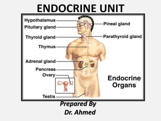
- 1. ENDOCRINE UNIT Prepared By Dr. Ahmed
- 2. ADRENAL GLAND (suprarenal gland) 5th Lecture
- 3. Adrenal gland LOCATION paired , retroperitoneal related to upper pole of kidney Yellowish in colour SHAPE AND MEASUREMENTS Shape: Each gland is flattened A-P 1. RT. : pyramidal in shape 2LT.: crescentric in shape like Measurements: Length 50 mm. Breadth 30 mm. Thickness 10 mm. Weight= About 5 g. At birth the gland is 1/3 of the size of kidney, in adults it is only 1/30th of the size of kidney
- 4. Adrenal gland LOCATION retroperitoneal in the epigastric region of abdomen, anterosuperior to the upper part of each kidney yellowish in color SHAPE AND MEASUREMENTS Shape: Each gland is flattened A-P 1. RT. : pyramidal in shape 2. LT.: crescentric in shape Measurements: Length 50 mm. Breadth 30 mm. Thickness 10 mm. Weight= About 5 g. At birth the gland is 1/3 of the size of kidney, in adults it is only 1/30th of the size of kidney
- 6. Adrenal gland Sheath surrounded by two sheaths : 1. Surrounded by fat called perirenal fat. 2. Outer to this, renal fascia encloses the suprarenal gland together with the kidney 3. but the gland is separated from the kidney by a septum.
- 7. Adrenal gland ARTERIAL SUPPLY Each gland is supplied by three arteries from three different sources : 1. Superior suprarenal artery: A branch of the inferior phrenic artery. 2. Middle suprarenal artery: A branch of the abdominal aorta. 3. Inferior suprarenal artery: A branch of the renal artery.
- 8. Adrenal gland VENOUS DRAINAGE Each gland is drained by only single vein which emerges from the hilus of the gland : 1. Right suprarenal vein drains into the inferior vena cava. 2. Left suprarenal vein drains into the left renal vein.
- 9. Adrenal gland Parts divided into outer cortex 80% inner medulla 20% outer cortex contains parenchymal cells synthesize & secrete but do not store various steroid hormones. Intracellular lipid droplets characteristic cytologic features inner medulla contains two populations of parenchymal cells, called chromaffin cells which synthesize, store, and secrete the catecholamine
- 10. Adrenal gland Endocrine Cells are generally characterized according to hormones they produce: 1. Nitrogenous-hormone secreting cell: RER. Golgi. Secretory granules. 2. Steroid-hormone secreting cell: SER. Lipid droplet: raw materials. Mitochondria: with tubular- vesicular cristae.
- 11. Adrenal gland
- 12. Adrenal gland
- 13. Adrenal gland Zona glomerulosa Occupy 15% of the cortex Immediately beneath the capsule cells arranged in closely packed, rounded cluster like glomeruli cells are smaller , Columnar or pyramidal with dark stain cytoplasm
- 14. Adrenal gland Zona Fasciculata Occupy 65% of the cortex Intermediate zone Arranged in one or two – cell thick straight cords(perpendicular to surface of the gland) cells are Polyhedral, binucleated light staining ( a lot of lipid droplets in their cytoplasm) Cells are also called spongyocytes (foamy cell) due to vacuolization
- 15. Adrenal gland Zona Reticularis occupy 7% of the cortex Innermost layer - lies between zona fasciculata and medulla Small cells arranged in irregular cords forming network Cells are darkly stained, characterize by presence of lipofuscin pigment granules ( with age)
- 16. Adrenal gland Medulla Lies in the center of the adrenal gland Clear line of demarcation Composed of polyhedral cells arranged in clusters surrounded by an extensive capillary network& , supported by reticular fiber network Composed of : Chromaffin cells of 2types A – CELLS N – CELLS
- 17. Adrenal gland
- 18. Adrenal gland Medulla A – CELLS Characterized by 80% containing small granules Store epinephrine N – CELLS Characterized by 20% containing large dense granules Store Norepinephrine
- 19. Adrenal gland Medulla has a dual blood supply that arises from (1) medullary arterioles that bypass the adrenal cortex (2) capillary sinusoids that perfuse the adrenal cortex (adrenocortical sinusoids). central adrenomedullary vein drains to IVC(Rt.) or Lt. renal V.(Lt.)
- 20. Adrenal gland Nerve supply of adrenal gland cortex is regulated by ACTH secreted by the anterior lobe of the pituitary gland Medulla is regulated by sympathetic NS (myelinated preganglionic sympathetic fibers ) derived from splanchnic nerves & distributed to the chromaffin cells (modified postganglionic sympathetic neurons) release of catecholamine from secretary granules stored in chromaffin cells by process called exocytosis
- 21. Adrenal gland
- 22. Adrenal gland Radiology of Suprarenal Gland deeply located difficult to visualize on plain X-ray. CT scan of abdomen inverted Y shape with medial & lateral limbs
- 23. Adrenal gland Adrenalectomy ( removal of the adrenal glands) Rt. suprarenal vein must be ligated before manipulating the gland 1. Short ,wide 2. To prevent surge of catecholamines to the circulation. surgical removal of Lt. gland is easier because the identification & clamping of the left suprarenal vein is easy Rt. gland is more difficult to approach than the left because part of it lies posterior to IVC
- 24. Adrenal gland
- 25. Adrenal gland
- 26. Adrenal gland
- 27. Adrenal gland
- 28. Adrenal gland
- 29. Adrenal gland
- 30. Adrenal gland
- 31. Adrenal gland Conn’s disease HYPERALDOSTERONISM usually caused by adrenal tumor Na and water retention K+ (hypokalemia) Hypertension
- 32. Adrenal gland Cushing disease / syndrome secretion of cortisol from Adrenal hyperplasia 4X more frequent in females Primary-tumor on the adrenal cortex Secondary-tumor on the anterior pituitary gland - truncal obesity - buffalo hump - “moon face” Adrenal hyperplasia with excessive secretion of sex steroids leads to feminization of male and masculanization of female.
- 33. Adrenal gland Cushing disease / syndrome secretion of cortisol from Adrenal hyperplasia 4X more frequent in females Primary-tumor on the adrenal cortex Secondary-tumor on the anterior pituitary gland - truncal obesity - buffalo hump - “moon face” Adrenal hyperplasia with excessive secretion of sex steroids leads to feminization of male and masculanization of female.
- 34. Adrenal gland
- 35. Adrenal gland Addison’s disease hypofunction of adrenal cortex What hormones will you have too little of??? - glucocorticoids or _______ - mineralocorticoids or _______ - sex steroid or ____________
- 36. Adrenal gland Addison’s disease hypofunction of adrenal cortex What hormones will you have too little of??? - glucocorticoids or _______ - mineralocorticoids or _______ - sex steroid or ____________
- 37. Adrenal gland Pheochromocytoma rare, benign tumor of the adrenal medulla Hallmark is hypertension-200/150 or greater
- 38. Adrenal gland Embryologically : adrenal gland consists of two parts develops from two different sources at 5th week : (a) a large outer part called cortex : is mesodermal in origin and develops from celomic epithelium (b) a small inner part called medulla. develops from neural crest.
- 39. Adrenal gland A. Cortex 1. forms from two episodes of mesoderm proliferation that occur between the root of the dorsal mesentery and the gonad. first episode forms the inner fetal cortex. second episode forms the outer adult cortex 2. During the fetal period , the suprarenal glands are very large due to the size of the fetal cortex( 10–20 times larger than adult adrenal gland). 3. suprarenal glands become smaller as the fetal cortex involutes rapidly during the first 2 weeks after birth and continues to involute during the first year of life. 4. zona glomerulosa and zona fasciculata of the adult cortex are present at birth, but the zona reticularis is not formed until age 3 years. B. Medulla 1. forms when neural crest cells aggregate at the medial aspect of the fetal cortex and become surrounded by the fetal and adult cortex. 2. The neural crest cells differentiate into chromaffin cells
- 40. Adrenal gland Congenital anomalies 1. Ectopic adrenal tissue/adrenal gland: The adrenal tissue or complete adrenal gland may be found fused to kidney deep in its capsule or in the right lobe of the liver. 2. adrenal hyperplasia a) Congenital adrenal hyperplasia: It is most commonly caused by mutation of genes for enzymes involved in adrenocortical steroid biosynthesis (e.g., 21-hydroxylase deficiency), b) Adrenogenital syndrome: It occurs due to congenital hyperplasia of the cells of the adrenal cortex, which secrete androgen. C/F differ in male and female : (a) In male: (adrenogenital syndrome) It leads to a very early development of secondary sexual characters. (b) In female: (pseudohermaphroditism) female child may be mistaken as a male.
- 41. Abdominopelvic splanchnic nerves Notes related to lecture chromaffin cells celomic epithelium
- 42. chromaffin cells
- 44. Neural Crest cells Derivatives of neural crest: 1. C.T. and bones of the face and skull. 2. ganglia of the cranial nerves. 3. C-cells of the thyroid gland. 4. conotruncal septum of the heart. 5. odontoblast cells. 6. dermis of the face and neck. 7. spinal ganglia. 8. autonomic ganglia. 9. Chromaffin cells (adrenal medulla). 10. schwan cells. 11. glial cells of the brain. 12. melanocytes. 13. arachnoid and pia matter
- 45. Adrenal gland Sites of chromaffin tissue : 1. para-aortic bodies 2. Along sympathetic chain near sympathetic ganglia 3. Along sympathetic plexuses 4. Near splanchnic nerves. chromaffin tissue 1. stain intensely with chromium salts yellow-brown . 2. Neural crest derivatives Chromaffin cells can be found in extrasuprarenal sites at birth, but these sites normally regress completely by puberty. In a normal adult, chromaffin cells are found only in the suprarenal medulla Pheochromocytoma: tumor arising from chromaffin cells usually from extra-adrenal
- 50. Sympathetic Chain Extension: from the base of the skull to the coccyx. bears a number of ganglia along its length On either side of V. column (paravertebral ganglion) Parts cervical, thoracic, lumbar and sacral Ganglion Cervical :3 usually one ganglion less than the number of nerves: 11 thoracic; 4 lumbar; and 4 sacral 2 neurons (pre & postganglionic ) Preganglionic N : T1- L2 in lat. horn Communication with spinal nerves T1-L2 : by grey &white communicants Above & below this level: only grey communicants NO white communicants Why?
- 51. Fate of preganglionic fibres preganglionic fibres arise from the lateral horns of spinal segments with ventral root 4 options Sympathetic ganglion of sympathetic trunk (paravertebral ganglion prevertebral ganglion
- 52. preganglionic fibres arise from the lateral horns of spinal segments with ventral root preganglionic fibres enter the sympathetic ganglion via white rami communicantes of the spinal nerve. Rely (synopsis) in the ganglion Postganglionic fibres from the ganglion re-enter the spinal nerve via grey rami communicantes supply the corresponding dermatome of the upper limb and the body wall.
- 53. BRANCHES of thoracic part Visceral & somatic Efferent Fibers divided into two groups: medial & lateral Medial branches(visceral) 1) postganglionic fibres from 1st to 5th ganglia distributed to the heart, great vessels, lungs, and esophagus through the following plexuses: a) Pulmonary plexus bronchodilatation b) b) Cardiac plexus. vasoconstriction c) Aortic plexus. Secretion d) Esophageal plexus HR & dilate coronary art. 2) preganglionic fibres 5th to 12th thoracic ganglia form three splanchnic nerves : a) Greater splanchnic nerve(5-9th) b) Lesser splanchnic nerve(10 &11th ) c) Least (lowest) splanchnic nerve(12th (renal)
- 54. BRANCHES of thoracic part Lateral branches(somatic) supply: • erector pili of hair follicle ( pilomotor) • Sweat gland (sudomotor) • Bl. V of the skin ( vasomotor) dilate Cutaneous Bl. V of limbs and body wall