
Recomendados
Más contenido relacionado
La actualidad más candente
La actualidad más candente (20)
Destacado
Destacado (19)
Similar a Chemistry of vision
Similar a Chemistry of vision (20)
Último
Último (20)
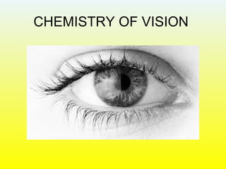
Chemistry of vision
- 2. Agenda • Eyes and its components. • Types of cells and how they work. • Vision in dim light. • Vision in coloured light. • Coloured Blindness. • Night Blindness.
- 3. ‘Light’
- 4. Eyes(Photoreceptor organs) • Vision is such an everyday occurrence that we seldom stop to think and wonder how we are able to see the objects that surround us. Yet the vision process is a fascinating example of how light can produce molecular changes. • The retina contain the molecules that undergo a chemical change upon absorbing light, but it is the brain that actually makes sense of the visual information to create an image. • The light image is mapped on the surface of the retina by activating a series of light-sensitive cells known as RODS and CONES or photoreceptors. The rods and cones convert the light into electrical impulses which are transmitted to the brain via nerve fibers. The brain then determines, which nerve fibers carried the electrical impulse activate by light at certain photoreceptors, and then creates an image. • Single eye gathers light and focuses it to many receptor cells. Extraordinary sensitive instrument • Wavelength range (400-800nm)
- 5. Vertebrate eye • RETINA • RODS • CONES Lens Light Optic Nerve
- 6. Rods (Dim Vision) • High sensitivity • Low resolution • Black & White vision • Can see only in shades of grey • Found at the periphery of retina
- 7. Cones (Colour Vision) • High resolution • Lower sensitivity • Centre of retina
- 8. • Have rods • Have cones • Can see only in bright light • Can see only in Dim light Some animals do not possess both rods and cones
- 9. Single Ommatidium Photoreceptor cell Rhabdomere Extension of photoreceptor cell that contains visual pigment Lens Photoreceptor axon Light
- 10. Rod cells contain Rhodopsin • First discovered by German physiologist Franz Boll. • Known as visual purple because of absorption maxima=500nm • Human rhodopsin has mol. wt.=41000 and has 348 amino residues. • This works at night time. • Cannot distinguish colour.
- 11. • Rhodopsin chromophore is polyunsaturated aldehyde, 11-cis retinal (A). • Rhodopsin is the product of the reaction of this aldehyde and protein called Opsin (B). • Reaction is between the aldehyde group of A and alpha amino group of the chain of the protein by the loss of a water molecule. • Other interactions of the –SH bonds of protein keeps the aldehyde on place.
- 13. Irradiation of rhodopsin leads to a series of conformational changes When rhodopsin absorbs a photon of light the 11-cis retinal isomerizes to all trans form
- 15. Rhodopsin (Red) Opsin all-trans-Retinal 11-cis-Retinal Light isomerase Opsin + 11-cis-Retinal Activated form Light converts 11-cis-Retinal to all-trans-Retinol Photochemistry of Pigment molecules all-trans-Retinol (Vitamin A) Thus precursor of 11-cis retinal is vitamin A Reduction
- 16. Photochemical changes • Absorption of one quantum does not result in vision. • Several quanta(2-6) should reach the rod in short time. • The absorption of light leads to chain reaction and it takes energy from the metabolism. • From the retina, amplification of the visual signal takes place with high gain and low noise ratio. • Photoreceptor act as photomultiplier. • Visual pigments are integral membrane proteins that reside in photoreceptors outer segment. • Photoisomerisation of retinal triggers many conformational changes in attached proteins that creates an enzyme site. • Which ultimately produces signal.
- 17. Introduction: Phototransduction A photon is absorbed by Rhodopsin which causes a conformational change in retinal 11-Cis Retinal
- 18. Introduction: Phototransduction The conformational change allows a G-protein to replace GDP with GTP - initiates transduction pathway. “All-Trans” Retinal
- 19. Colour Vision • Occurs due to cone cells. • Photosensitive pigment is Iodopsin. • The chromophore of Iodopsin is also 11-cis retinal. • 3 types of Iodopsin in cones. Blue (440nm) Green (535nm) Red (565nm) • Less sensitive to light than rod cells, so in dim light objects appear in shade of grey.
- 20. Color Vision: Cone Cells
- 21. Color Blindness • Missing or defective pigment proteins for certain cone cell types – ~8% of men, rare in women. • Types of Color Blindness – Achromatopsia - black and white vision. – Dichromacy - 2 functioning cone types. – Anomalous Trichromacy - shifted cone absorption. • Non-Genetic Causes – Disease, Accidents, Medication.
- 22. Colour Blindness Everyone should see number 12 ‘normal’ see number 8 Red-green deficiency see number 3 Total colour blind see no number Take care of your eyes
- 23. Night Blindness (Nyctalopia) • The most common cause of nyctalopia is retinitis pigmentosa, a disorder in which the rod cells in the retina gradually lose their ability to respond to the light. • Patients suffering from this genetic condition have progressive nyctalopia and eventually their daytime vision may also be affected.
- 25. Thank You